. 2017 Feb 10;355(6325):597-602.
doi: 10.1126/science.aal3316.

Redox-based reagents for chemoselective methionine bioconjugation

Affiliations

Redox-based reagents for chemoselective methionine bioconjugation

Shixian Lin et al. Science. .

Abstract

Cysteine can be specifically functionalized by a myriad of acid-base conjugation strategies for applications ranging from probing protein function to antibody-drug conjugates and proteomics. In contrast, selective ligation to the other sulfur-containing amino acid, methionine, has been precluded by its intrinsically weaker nucleophilicity. Here, we report a strategy for chemoselective methionine bioconjugation through redox reactivity, using oxaziridine-based reagents to achieve highly selective, rapid, and robust methionine labeling under a range of biocompatible reaction conditions. We highlight the broad utility of this conjugation method to enable precise addition of payloads to proteins, synthesis of antibody-drug conjugates, and identification of hyperreactive methionine residues in whole proteomes.


Figures

Fig. 1
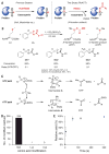
Fig. 1. The ReACT strategy for chemoselective methionine bioconjugation
(A) (Left) Acid-base conjugation strategies for cysteine-based protein functionalization. Iodoacetamide (IAA) and maleimide reagents are representative electrophiles for selective cysteine bioconjugation. (Right) ReACT strategies for methionine-based protein functionalization. Oxaziridine (Ox) compounds serve as oxidant-mediated reagents for direct functionalization by converting methionine to the corresponding sulfimide conjugation product. During this redox process, the Ox ReACT reagents are reduced to benzaldehyde. (B) Model redox conjugation reaction with 25 μM of N-acetyl-L-methionine methyl ester (S1) and 27.5 μM of various oxaziridine compounds as substrates in cosolvent (CD3OD/D2O = 1:1). The reactions were monitored by detecting the chemical shift of the methionine methyl group with 1H NMR (fig. S1). The reaction time was 10 min in 100% CD3OD solution due to slow reaction rate and 20 min in 5% CD3OD /D2O solution due to poor solubility of substrate in aqueous solution. (C) The proposed reaction mechanism between methionine and oxaziridine compound proceeds by nucleophilic attack of sulfide at N atom or O atom of oxaziridine ring, followed by N–O bond cleavage to generate reaction intermediate A or B, respectively. The NTP or OTP is generated, along with the corresponding aldehyde or imine as side product, through an intramolecular rearrangement. (D) Number of unique ReACT-sensitive Met, Lys, and Cys residues detected in HeLa cell lysates when treated with 1 mM Ox4 for 10 min. (E) Yield of conjugation reaction was performed with 15 μM of BSA carrying four methionines per protein and 100 μM Ox4 at the indicated time point as measured by in-gel fluorescence imaging. Error bars, mean ± SD from three independent experiments. Representative fluorescent gel is shown in fig. S4.
Fig. 2
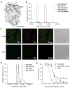
Fig. 2. The ReACT strategy for protein functionalization
(A) General two-step procedure for methionine-specific protein functionalization is a combination of ReACT and click reactions. Various pay-loads (red sphere) can be installed through methionine conjugation at a directed position on a given protein. (B) Redox conjugation of a CaM model protein (100 μM) with various Ox compounds (1 mM). The chemical structures of oxaziridine probes are shown with molecular weight changes (ΔM) listed for the corresponding modifications. The deconvoluted MS data of full protein peaks are plotted in the same figure. The major peaks correspond to CaM protein carrying nine sulfimide modifications (ΔM). For Ox2-labeled protein: expected mass 17,564 Da, found 17,565 Da; Ox4-labeled protein: expected mass 17,654 Da, found 17,654 Da; Ox5-labeled protein: expected mass 18,050 Da, found 18,051 Da; Ox6-labeled protein: expected mass 18,059 Da, found 18,060 Da. The minor peaks correspond to CaM protein bearing eight sulfimide modifications (ΔM) and one sulfoxide modification (O).
Fig. 3
Fig. 3. ReACT for synthesis of methionine-targeted antibody conjugates
(A) Crystal structure of Her-Fab [Protein Data Bank (PDB) 1n8z] with three native methionine residues shown as green sticks and sulfur atoms shown as yellow spheres. The sulfur atoms appear to be buried in the pocket of Fab on the crystal structure. (B) The ratio of probe/methionine for Fab labeling is 10, with 10-min labeling time at room temperature. The deconvoluted MS data of GFP-Fab constructs with or without ReACT labeling. GFP-Fab-CM: expected mass 47158 Da, found 47158 Da; GFP-Fab-N3 (labeled by Ox6): expected mass 47299 Da, found 47299 Da; biotin-functionalized GFP-Fab-N3 (GFP-Fab-Biotin): expected mass 48019 Da, found 48019 Da; Cy3-functionalized GFP-Fab-N3 (GFP-Fab-Cy3): expected mass 48282 Da, found 48282 Da. MMAE-functionalized GFP-Fab-N3 (GFP-Fab-MMAE): expected mass 48949 Da, found 48949 Da. (C) Fluorescence colocalization imaging of GFP-Fab-Cy3 with cell surface–targeted GFP in HEK-293T cells. The GFP was inducibly expressed on the cell surface with addition of 1 μM/mL of Dox before adding GFP-Fab-Cy3. Cells without addition of Dox were used as a control and show no antibody staining. All images use the same scale bar: 20 μm. (D) The de-convoluted MS data of Her-Fab constructs with or without ReACT labeling. Her-Fab carries one C-terminal methionine (Her-Fab1): expected mass 47544 Da, found 47544 Da; Ox6-labeled Her-Fab1 (Her-Fab1-N3): expected mass 47685 Da, found 47686 Da; Her-Fab carries two C-terminal methionine (Her-Fab2): expected mass 47676 Da, found 47677 Da; Ox6-labeled Her-Fab2 (Her-Fab2-N3): expected mass 48958 Da, found 47958 Da. (E) In vitro cytotoxicity of Her-Fab2 [median effective concentration (EC50) = 0.086 ± 0.02 μg/mL], noncovalent mixture of Her-Fab2 and free MMAE (EC50 = 0.096 ± 0.04 μg/mL), and the ReACT-derived ADC from Her-Fab2 and MMAE (EC50 = 0.015 ± 0.007 μg/mL). Error bars, mean ± SD from three independent experiments. EC50 values and EC50 ± SDs were determined using four-parameter logistic fitting.
Fig. 4
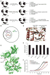
Fig. 4. Chemoproteomic methionine profiling with ReACT
(A) Reactive methionine profiling with ReACT involves treatment of proteomes with low, medium, and high doses of Ox4 probe, followed by CuAAC-based installation of acid cleavable biotin-azide tag, enrichment with streptavidin magnetic beads, and sequential on-bead trypsin digestions to afford probe-labeled peptides for LC-MS/MS analysis. (B) The number of peptides carrying the desired ReACT modification on methionine and appearing in two independent runs from the low-, medium-, and high-dose groups is shown. (C) Reactive methionine map on actin (PDB 3byh), with hyperreactive methionines colored in red, including Met44 and Met47, medium-reactive methionines colored in purple; and less-reactive methionines colored in blue. The methionines colored in yellow represent residues identified by LC-MS/MS that do not carry redox modification. The domains carrying these yellow-colored methionines are involved in actin polymerization. Because of this activity, no desired modification is detected on these residues even when these are surface accessible on the protein x-raycrystal structure. (D) Protein structure alignment of human alpha enolase (yellow; PDB 2psn) and yeast enolase 1 (red; PDB 2AL1), with conserved methionine residues shown in stick representation. (E) The relative activity of yeast enolase 1 variants with or without treatment of NaClO (100 μM). Error bars, mean ± SD from four independent experiments. P values indicated in the figure represent results of an unpaired t test. (F) Growth curve of wild-type (WT) (ENO2 null) and ENO1-M171L (ENO2 null) strains, with or without treatment of NaClO (100 μM). Knockout and mutation strains were generated by clustered regularly interspaced short palindromic repeats (CRISPR)–Cas9–mediated genome editing. Error bars, mean ± SD from three independent experiments.

References

    1. Stipanuk MH. Annu Rev Nutr. 2004;24:539–577. - PubMed
    1. Levine RL, Mosoni L, Berlett BS, Stadtman ER. Proc Natl Acad Sci USA. 1996;93:15036–15040. - PMC - PubMed
    1. Giles NM, et al. Chem Biol. 2003;10:677–693. - PubMed
    1. Paulsen CE, Carroll KS. Chem Rev. 2013;113:4633–4679. - PMC - PubMed
    1. Spicer CD, Davis BG. Nat Commun. 2014;5:4740. - PubMed

Publication types

MeSH terms