. 2017 Jun;15(6):753-764.
doi: 10.1158/1541-7786.MCR-16-0350. Epub 2017 Feb 9.

Phosphatidylserine Sensing by TAM Receptors Regulates AKT-Dependent Chemoresistance and PD-L1 Expression

Affiliations

Phosphatidylserine Sensing by TAM Receptors Regulates AKT-Dependent Chemoresistance and PD-L1 Expression

Canan Kasikara et al. Mol Cancer Res. 2017 Jun.

Abstract

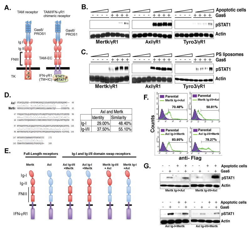
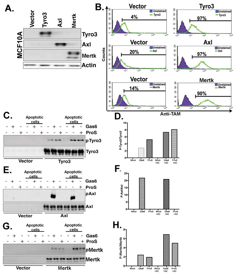
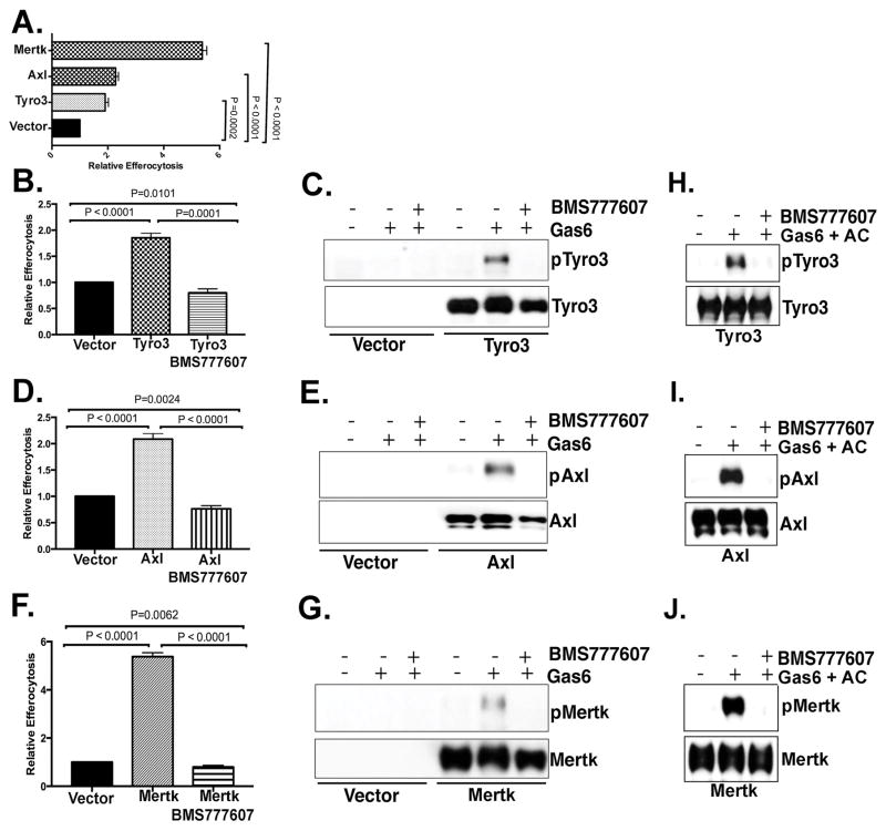
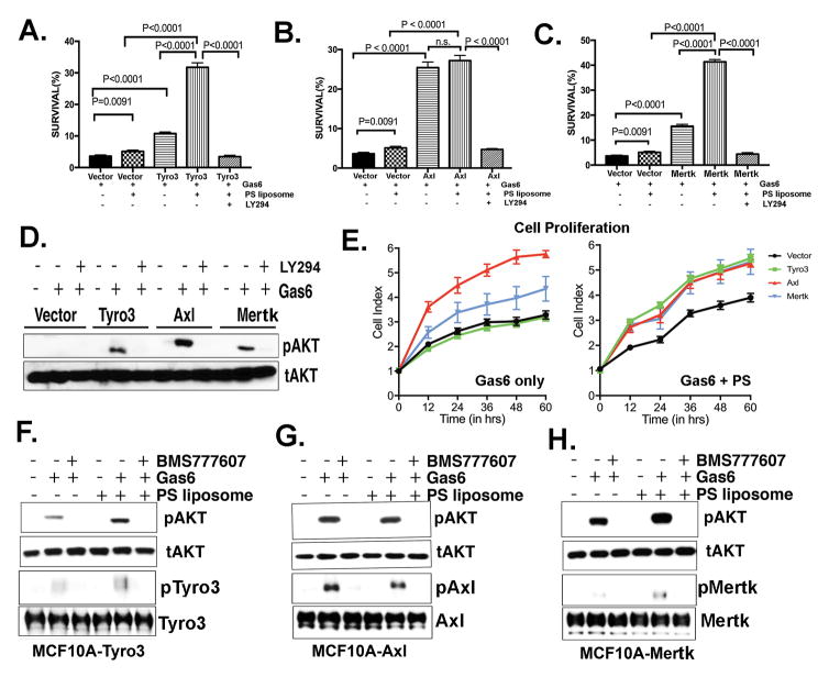
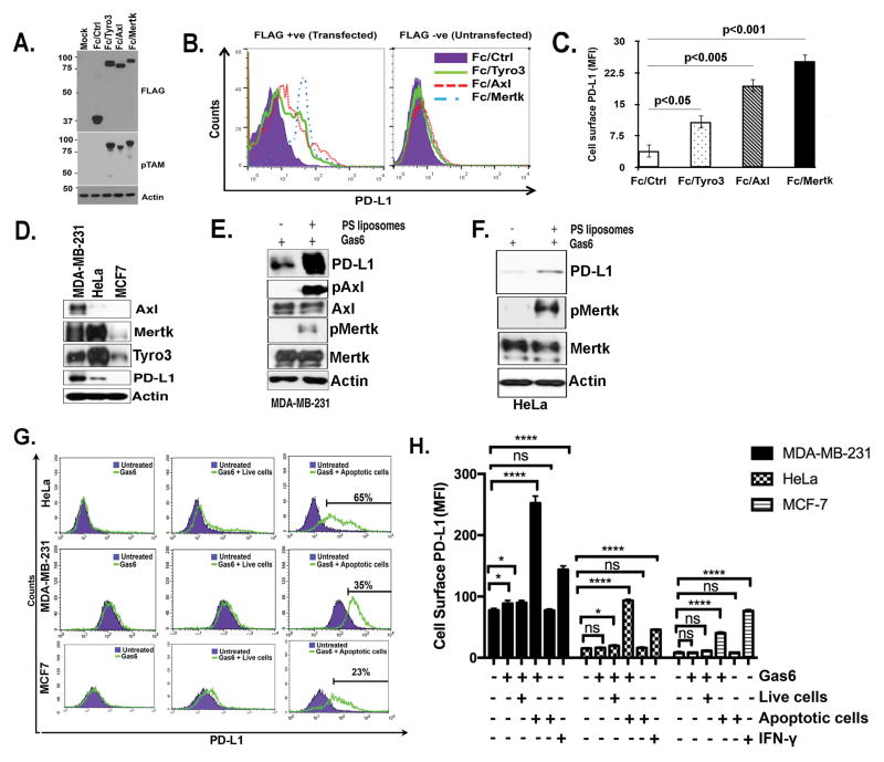
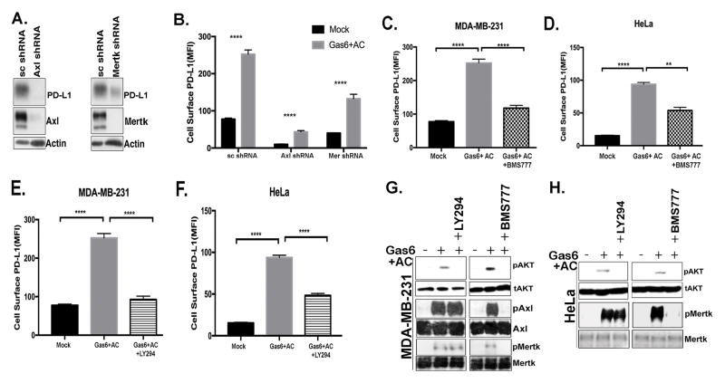
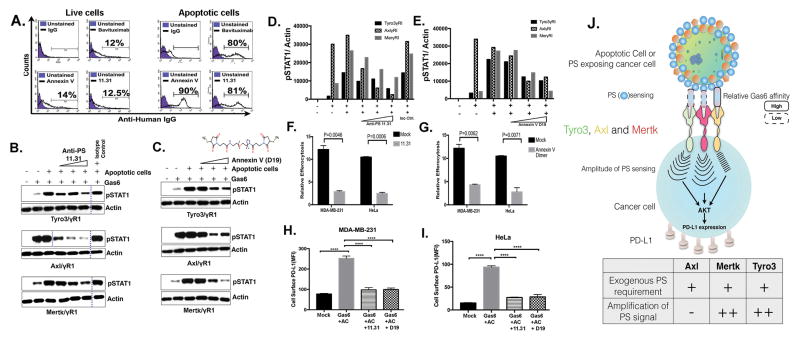
Tyro3, Axl, and Mertk (collectively TAM receptors) are three homologous receptor tyrosine kinases that bind vitamin K-dependent endogenous ligands, Protein S (ProS), and growth arrest-specific factor 6 (Gas6), and act as bridging molecules to promote phosphatidylserine (PS)-mediated clearance of apoptotic cells (efferocytosis). TAM receptors are overexpressed in a vast array of tumor types, whereby the level of expression correlates with the tumor grade and the emergence of chemo- and radioresistance to targeted therapeutics, but also have been implicated as inhibitory receptors on infiltrating myeloid-derived cells in the tumor microenvironment that can suppress host antitumor immunity. In the present study, we utilized TAM-IFNγR1 reporter lines and expressed TAM receptors in a variety of epithelial cell model systems to show that each TAM receptor has a unique pattern of activation by Gas6 or ProS, as well as unique dependency for PS on apoptotic cells and PS liposomes for activity. In addition, we leveraged this system to engineer epithelial cells that express wild-type TAM receptors and show that although each receptor can promote PS-mediated efferocytosis, AKT-mediated chemoresistance, as well as upregulate the immune checkpoint molecule PD-L1 on tumor cells, Mertk is most dominant in the aforementioned pathways. Functionally, TAM receptor-mediated efferocytosis could be partially blocked by PS-targeting antibody 11.31 and Annexin V, demonstrating the existence of a PS/PS receptor (i.e., TAM receptor)/PD-L1 axis that operates in epithelial cells to foster immune escape. These data provide a rationale that PS-targeting, anti-TAM receptor, and anti-PD-L1-based therapeutics will have merit as combinatorial checkpoint inhibitors.Implications: Many tumor cells are known to upregulate the immune checkpoint inhibitor PD-L1. This study demonstrates a role for PS and TAM receptors in the regulation of PD-L1 on cancer cells. Mol Cancer Res; 15(6); 753-64. ©2017 AACR.


Conflict of interest statement

Disclosure of potential conflicts of interest:

Cyril Empig, Bruce Freimark, Michael Gray and Jeff Hutchins are employees of Peregrine Pharmaceuticals, Inc. and have financial interest in the company. Allen Krantz and Andrzej Wilczynski are employees of Advanced Proteome Therapeutics Corporation. Allen Krantz has a financial interest in the company. Raymond B. Birge reports receiving a commercial research grant from Peregrine Pharmaceuticals, Inc. No potential conflicts of interest were disclosed by the other authors.

Figures

Figure 1
Figure 1. Differential PS sensing of chimeric TAM receptors is dependent on the TAM Ig-I/II domains
A. Schematic representation of TAM/IFN-γR1 chimeric receptors that exhibit pSTAT1 as a “read-out” for receptor activation. Effect of PS-positive apoptotic cells (B) PS-positive liposomes (C) on Gas6-inducible activation of Tyro3, Axl, and Mertk. D. Alignment of Ig-I/II domains of Axl and Mertk receptors and summary of the conservation between their Ig-I and Ig-II domains. E. Schematic representation of Ig-I or Ig-I/II swaps of Axl and Mertk chimeric receptors. F. Expression profiling by flow cytometry for cells expressing Axl and Mertk Ig-I/II domain swaps. Numbers indicate MFI of chimeric receptor expression using FACS staining with an anti-Flag antibody that is engineered into the extracellular domain of each chimeric receptor G. Effects of Ig-I or Ig-I/II swap on apoptotic cells and Gas6 mediated Axl and Mertk activation.
Figure 2
Figure 2. Differential PS sensing of native TAM receptors towards apoptotic cells
A. Generation of stable native human Tyro3, Axl and Mertk expressing MCF10A cell lines. B. Flow cytometric analysis of TAM expressing cells show comparable receptor expression in stable MCF10A-expressing cells. C. TAM receptors phosphorylation levels were evaluated by Western blotting after Tyro3-MCF10A cells (C), Axl-MCF10A cells (E), or Mertk-MCF10A cells (G) were treated with Gas6 or ProS in the presence or absence of apoptotic cells. Densitometric analysis of the Western blots shown in panels C, E and G respectively (D, F & H) are indicated to show expression levels of phospho TAMs normalized to total TAMs expression.
Figure 3
Figure 3. TAM mediated epithelial cell efferocytosis is dependent on the tyrosine kinase activity of TAMs
A. TAM-MCF10A cells, as shown in Fig. 2, were tested for epithelial efferocytosis of apoptotic Jurkat cells. MCF10A and apoptotic cells were co-cultured at a ratio of 1:5 (phagocyte/apoptotic cell ratio) for 5 hr. Effect of 300 nM BMS777607 (pan-TAM kinase inhibitor) on epithelial cell efferocytosis in MCF10A cells induced by Tyro3 (B), Axl (D) and Mertk (F). Effect of BMS777607 on TAM phosphorylation induced by Gas6 (C, E, G) and TAM phosphorylation induced by Gas6 in the presence of apoptotic cells (H–J).
Figure 4
Figure 4. TAM expression in MCF10A cells induces chemo-resistance, proliferation, and the activation of Akt; Effects of Gas6 and PS liposomes
A. Effect of Gas6 and/or Gas6 PS liposomes on camptothecin-mediated cell death in Tyro3-MCF10A cells (A), Axl-MCF10A cells (B) and Mertk-MCF10A cells (C) (see Materials and methods). After 16 hr, cells were evaluated by PI and Annexin V-conjugated FITC to quantify apoptosis. Bar 5 represents a control in which cells were treated in the presence of 50 μM Akt inhibitor LY-294002. D. Effect of Akt inhibitor LY294002 on TAM inducible pAkt. E. Real-time Xcelligence assay to assess cell proliferation induced by Gas6 alone (left panel) and Gas6 with PS liposomes (right panel) on Tyro3-MCF10A, Axl-MCF10A, and Mertk-MCF10A cells. Effect of BMS777607 pretreatment on the Gas6 inducible phosphorylation of Akt (pAkt) and phosphorylation of TAMs; pTyro3 (F), pAxl (G) and pMertk (H) is shown.
Figure 5
Figure 5. Role of TAMs and TAM-mediated efferocytosis on PD-L1 expression in cancer cells
A-B. Expression of constitutively active Fc-TAM receptors up-regulate surface PD-L1 expression in HEK293 cells. Data in panel B are quantified in panel C. D. Expression levels of endogenous PD-L1, Axl, Mertk and Tyro3 in MDA-MB-231, HeLa, and MCF7 cancer cell lines. Effect of PS liposomes on PD-L1 expression and phosphorylation of Mertk and Axl in MDA-MB-231 cells (E) and HeLa cells (F). G. Flow cytometric analysis for efferocytosis mediated changes in surface expression of PD-L1 on HeLa, MDA-MB-231 and MCF-7 cells by treatment with Gas6 and apoptotic cells. H. Quantification of cells surface expression of PD-L1 in multiple replicates samples (n=3). P values for * and **** are <0.01.
Figure 6
Figure 6. Axl and Mertk regulate PD-L1 expression by the activation of Akt
A. Effect of Axl and Mertk knockdown on PD-L1 expression in MDA-MB-231 cells by Western blotting. B. Quantification of cells surface expression of PD-L1 by Axl and Mertk knockdown and Gas6 and apoptotic cell stimulation in multiple replicates (n=3). Effect of BMS777607 (pan-TAM kinase inhibitor) on Gas6 and apoptotic cell induced PD-L1 surface expression in MDA-MB-231 (C) and HeLa cells (D). Effect of LY294 (Akt inhibitor) treatment on Gas6 and apoptotic cells induced PD-L1 surface expression MDA-MB-231 (E) and HeLa cells (F). Effects of LY294 (Akt inhibitor) and BMS777607 (pan-TAM kinase inhibitor) on phosphorylation of Akt, Mertk and Axl shown by western blot in MDA-MB-231 cells (G) and HeLa cells (H). P values **** are <0.01.
Figure 7
Figure 7. PS targeting agents 11.31 and Dimerized Annexin V suppress epithelial cell efferocytosis and PD-L1 expression
A. Flow cytometry based binding studies of PS targeting agents to PS on live and apoptotic cells. UV treatment mediated apoptosis and PS exposure was measured by Annexin-V staining by flow cytometry. Anti-human PS targeting antibody 11.31 or Bavituximab were incubated with thus produced apoptotic or live cells using Annexin V as control. Percentage of PS targeting antibodies bound with apoptotic cells were measured with conjugated flow antibodies by flow cytometry. (B–E) PS targeting agents affect Gas6 and apoptotic cells induced TAM receptor activation. Effects of PS targeting 11.31 antibody (B–D) and dimerized Annexin V D19 (C–E) on Tyro3, Axl and Mertk receptor activation by apoptotic cells and Gas6 in TAM-IFN-YR1 chimeric receptor system. Effects of 11.31 PS targeting antibody (F) and Annexin V dimer D19 (G) on relative efferocytosis by MDA-MB-231 and HeLa epithelial cells. Role of 11.31 and Annexin V dimer D19 treatments on cell surface expression of PD-L1 by flow cytometric analysis in Gas6 and apoptotic cells induced MDA-MB-231 (H) and HeLa (I) cells. Western blots bands that are cropped are separated with dotted lines. (J) Schematic model representing different PS sensing and PS signal amplifications paradigms by TAM receptors.

Similar articles

Cited by

References

    1. Robinson DR, Wu YM, Lin SF. The protein tyrosine kinase family of the human genome. Oncogene. 2000;19:5548–57. - PubMed
    1. Graham DK, Dawson TL, Mullaney DL, Snodgrass HR, Earp HS. Cloning and mRNA expression analysis of a novel human protooncogene, c-mer. Cell growth & differentiation : the molecular biology journal of the American Association for Cancer Research. 1994;5:647–57. - PubMed
    1. Kumar S, Birge RB. Efferocytosis. Current biology : CB. 2016;26:R558–r9. - PubMed
    1. Linger RM, Keating AK, Earp HS, Graham DK. TAM receptor tyrosine kinases: biologic functions, signaling, and potential therapeutic targeting in human cancer. Advances in cancer research. 2008;100:35–83. - PMC - PubMed
    1. Prasad D, Rothlin CV, Burrola P, Burstyn-Cohen T, Lu Q, Garcia de Frutos P, et al. TAM receptor function in the retinal pigment epithelium. Mol Cell Neurosci. 2006;33:96–108. - PubMed

MeSH terms