. 2017 Apr 15:53:260-267.
doi: 10.1016/j.actbio.2017.02.008. Epub 2017 Feb 7.

Customized biomaterials to augment chondrocyte gene therapy

Affiliations

Customized biomaterials to augment chondrocyte gene therapy

Izath Nizeet Aguilar et al. Acta Biomater. .

Abstract

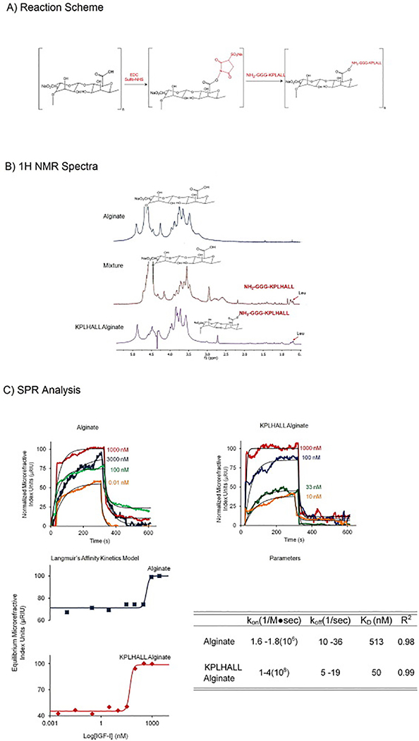
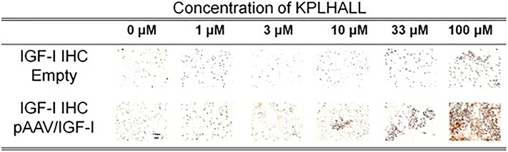
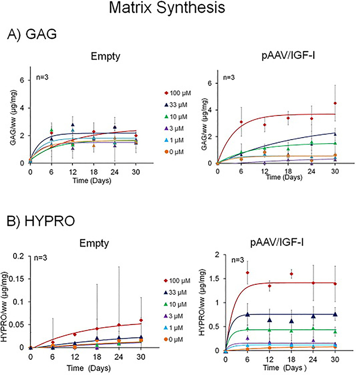
A persistent challenge in enhancing gene therapy is the transient availability of the target gene product. This is particularly true in tissue engineering applications. The transient exposure of cells to the product could be insufficient to promote tissue regeneration. Here we report the development of a new material engineered to have a high affinity for a therapeutic gene product. We focus on insulin-like growth factor-I (IGF-I) for its highly anabolic effects on many tissues such as spinal cord, heart, brain and cartilage. One of the ways that tissues store IGF-I is through a group of insulin like growth factor binding proteins (IGFBPs), such as IGFBP-5. We grafted the IGF-I binding peptide sequence from IGFBP-5 onto alginate in order to retain the endogenous IGF-I produced by transfected chondrocytes. This novel material bound IGF-I and released the growth factor for at least 30days in culture. We found that this binding enhanced the biosynthesis of transfected cells up to 19-fold. These data demonstrate the coordinated engineering of cell behavior and material chemistry to greatly enhance extracellular matrix synthesis and tissue assembly, and can serve as a template for the enhanced performance of other therapeutic proteins.

Statement of significance: The present manuscript focuses on the enhancement of chondrocyte gene therapy through the modification of scaffold materials to enhance the retention of targeted gene products. This study combined tissue engineering and gene therapy, where customized biomaterials augmented the action of IGF-I by enhancing the retention of protein produced by transfection of the IGF-I gene. This approach enabled tuning of binding of IGF-I to alginate, which increased GAG and HYPRO production by transfected chondrocytes. To our knowledge, peptide-based modification of materials to augment growth factor-targeted gene therapy has not been reported previously.

Keywords: Binding peptide; Growth factor; IGF-I; Osteoarthritis.


Figures

Figure 1
Figure 1
1A Reaction Scheme: EDC and Sulfo-NHS activate the alginate forming a carboxylate carbon intermediate. The carboxylate carbon intermediate is attacked by the primary amine nitrogen forming the amide bond to the alginate backbone. 1B NMR Spectra: First spectra shows the alginate sample with broad peaks for a polysaccharide, from 4.8 to 3.2, and distinct peak for the anomeric proton[59]. Second spectrum, mixture of the alginate and peptide, shows both different peaks for alginate and high leucine amino acids peaks. The third spectrum shows the modified alginate with KPLHALL where the chemical shift in the leucine peaks and the height of the peaks differs from the mixture. 1C SPR Spectra and Langmuir’s Affinity Kinetics Model: Representative examples of curve fits for the affinity kinetic analysis of control (alginate) and modified alginate (KPLHALL alginate). Concentrations of IGF-I for alginate varied from 3000 nM to 0 nM. Concentrations of IGF-I for KPLHALL alginate varied from 1000 nM to 0 nM. Langmuir’s Affinity Kinetics Model for alginate is the top one (blue) and KPLHALL alginate is on the bottom (red). KD shifted 10 times, where KD is 50 nM sin KPLHALL alginate (KD of 50 nM) when compared to alginate (KD of 513 nM). KPLHALL alginate had a kon ranging from 1 to 4(108/M●sec) and a koff from 5/s to 19/s (R2: 0.40 to 0.90).The parameters for alginate kon and koff ranged from 1.6 to 1.8(108/Ms) and koff 10/s to 29/s (R2: 0.74 to 0.9).
Figure 2
Figure 2. IGF-I Binding
Immunohistochemistry of constructs for IGF-I at Day 30. Scale bar = 100 μm. Alginate does not show differences in immunolocalization staining at day 30. Immunolocalization staining for IGF-I changes as the concentration of binding sites (KPLHALL) increases in the constructs. The constructs with 0 μM of binding sites barely show retention of f IGF-I; and constructs with 1 and 3 μM of binding sites show some retention of IGF-I. The highest immunolocalization of IGF-I is at 100 μM.
Figure 3
Figure 3
3A GAG matrix accumulation kinetic profiles: The production of GAG increases in both transfected and control groups. Control chondrocytes have smaller changes in kinetic profiles between the differences in KPLHALL concentrations. pAAV/IGF-I transfected chondrocytes have a greater effect (*p<0.001 by 2-way ANOVA) between the differences in KPLHALL concentrations. 3B HYPRO matrix accumulation kinetic profiles: The production of HYPRO increases in both pAAV/IGF-I transfected chondrocytes and control chondrocytes. The effect in chondrocytes that are transfected with pAAV/IGF-I is greater than the effect on those transfected with pAAV/MCS (Empty) (p<0.001 by 2-way ANOVA). This effect is greater in concentrations of 10, 33 and 100 μM binding sites (p<0.001 by 2-way ANOVA). When alginate is compared with the highest concentration of binding sites (100 μM) there is an increase from 0.08 μg/mg at 0 uM to 1.4 μg/mg at 100 μM (p<0.001 by 2-way ANOVA)in pAAV/IGF-I transfected chondrocytes.
Figure 4
Figure 4
4A GAG and HYPRO Dose Response: GAG at the steady state concentration of each concentration is plotted against the different concentrations of binding sites of KPLHALL covalently attached to alginate. Transfected chondrocytes with pAAV/IGF-I produce 600% GAG, than the control chondrocytes transfected with pAAV/ MCS (Empty) (*p<0.0001 by unpaired t test). pAAV/IGF-I transfected chondrocytes required 16.9 μM of binding sites in the alginate to produce an increase in GAG of 6.9 (Table 2). Control chondrocytes required almost twice the amount of KPLHALL modified alginate only to produce 60% (Table 2). *p<0.001 when compared to min. +p<0.001 when compared to max empty. 4B Dose Response: The production of HYPRO increases both in pAAV/IGF-I transfected chondrocytes and control chondrocytes. The effect in chondrocytes that are transfected with pAAV/IGF-I is greater than the effect in cells transfected with pAAV/ MCS (Empty). pAAV/IGF-I increased HYPRO accumulation by 20 fold at the maximum effective concentration of IGF-I binding sites(p<0.02 by unpaired t test). The parameters are summarized on table 2 where all R2 are higher than 0.93 (Table 2). %p<0.02 when compared to min. ^p<0.02 when compared to max empty.

Similar articles

Cited by

References

    1. Weisleder N, Takizawa N, Lin P, Wang X, Cao C, Zhang Y, Tan T, Ferrante C, Zhu H, Chen P-J, Yan R, Sterling M, Zhao X, Hwang M, Takeshima M, Cai C, Cheng H, Takeshima H, Xiao R-P, Ma J, Recombinant MG53 Protein Modulates Therapeutic Cell Membrane Repair in Treatment of Muscular Dystrophy, Sci. Transl. Med 4 (2012) 139ra85–139. doi:10.1126/scitranslmed.3003921. - DOI - PMC - PubMed
    1. Nagahara AH, a Merrill D, Coppola G, Tsukada S, Schroeder BE, Shaked GM, Wang L, Blesch A, Kim A, Conner JM, Rockenstein E, V Chao M, Koo EH, Geschwind D, Masliah E, a Chiba A, Tuszynski MH, Neuroprotective effects of brain-derived neurotrophic factor in rodent and primate models of Alzheimer’s disease., Nat. Med 15 (2009) 331–337. doi:10.1038/nm.1912. - DOI - PMC - PubMed
    1. Bartus K, James ND, Didangelos A, Bosch KD, Verhaagen J, Yáñez-Muñoz RJ, Rogers JH, Schneider BL, Muir EM, Bradbury EJ, Large-Scale Chondroitin Sulfate Proteoglycan Digestion with Chondroitinase Gene Therapy Leads to Reduced Pathology and Modulates Macrophage Phenotype following Spinal Cord Contusion Injury., J. Neurosci 34 (2014) 4822–36. doi:10.1523/JNEUROSCI.4369-13.2014. - DOI - PMC - PubMed
    1. Kafienah W, Al-Fayez F, Hollander AP, Barker MD, Inhibition of cartilage degradation: A combined tissue engineering and gene therapy approach, Arthritis Rheum. 48 (2003) 709–718. doi:10.1002/art.10842. - DOI - PubMed
    1. Evans CH, Gouze JN, Gouze E, Robbins PD, Ghivizzani SC, Osteoarthritis gene therapy., Gene Ther. 11 (2004) 379–89. doi:10.1038/sj.gt.3302196. - DOI - PubMed

Publication types

MeSH terms