Review
. 2017 Mar;13(3):171-191.
doi: 10.1038/nrneurol.2017.13. Epub 2017 Feb 10.

The far-reaching scope of neuroinflammation after traumatic brain injury

Affiliations
Review

The far-reaching scope of neuroinflammation after traumatic brain injury

Dennis W Simon et al. Nat Rev Neurol. 2017 Mar.

Erratum in

Abstract

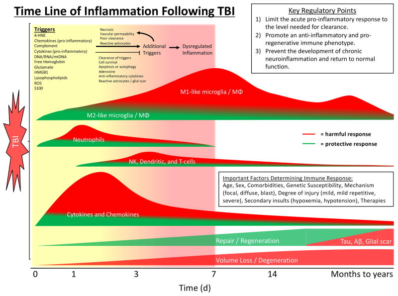
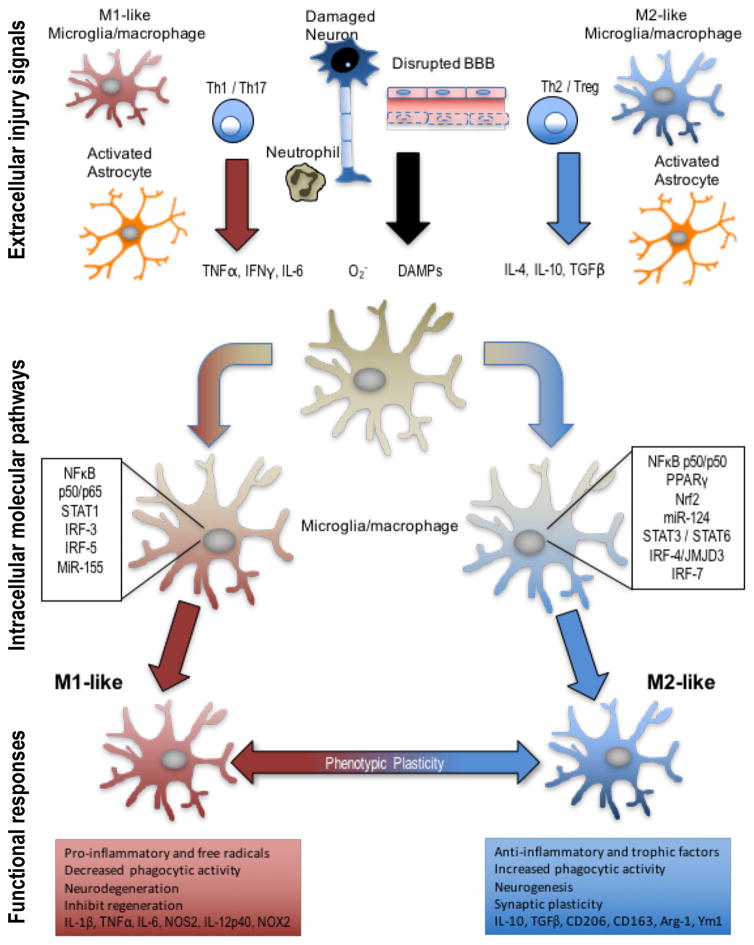
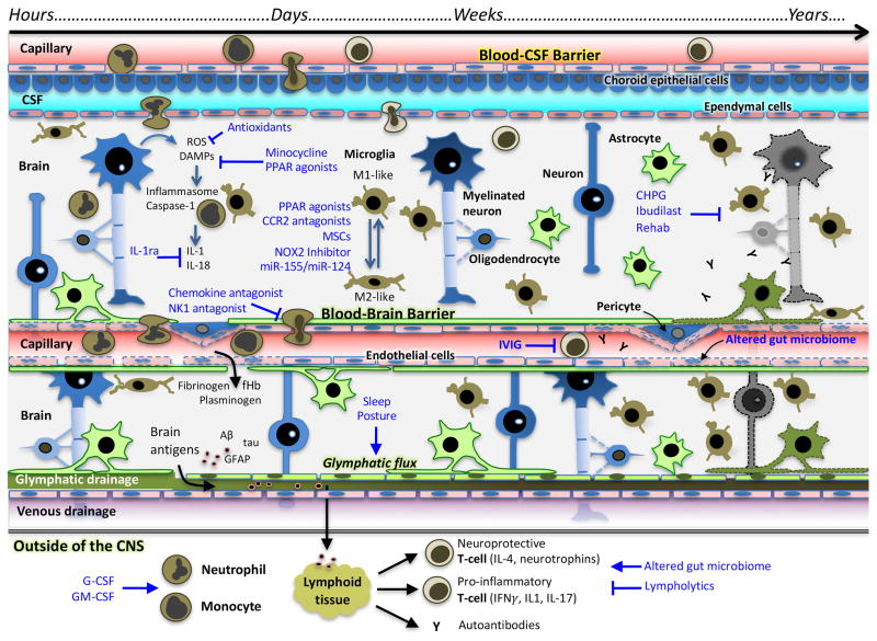
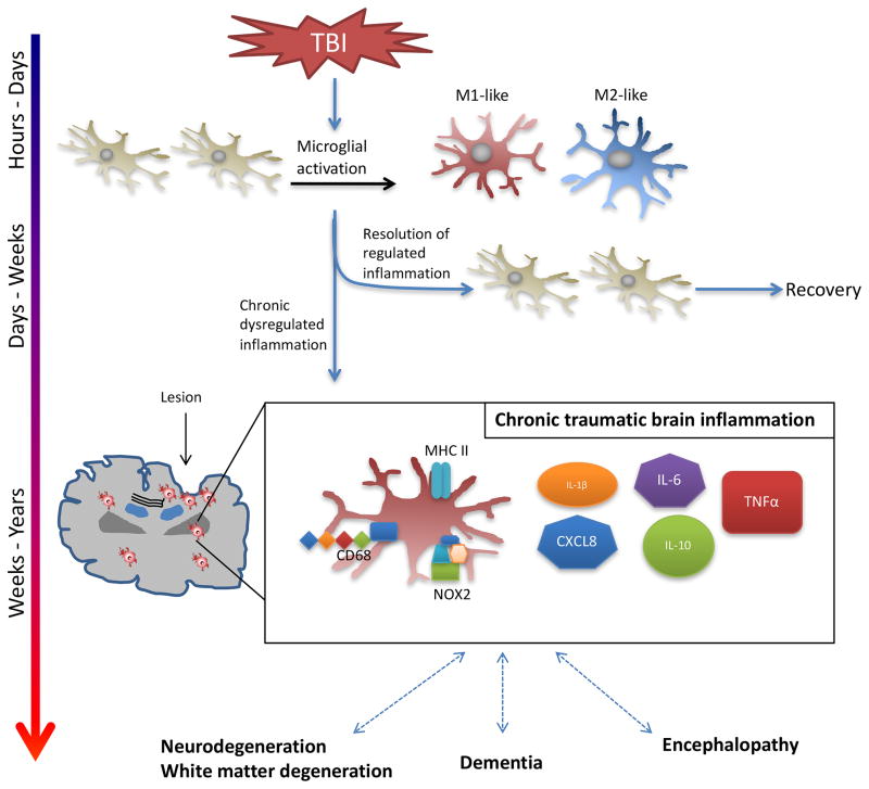
The 'silent epidemic' of traumatic brain injury (TBI) has been placed in the spotlight as a result of clinical investigations and popular press coverage of athletes and veterans with single or repetitive head injuries. Neuroinflammation can cause acute secondary injury after TBI, and has been linked to chronic neurodegenerative diseases; however, anti-inflammatory agents have failed to improve TBI outcomes in clinical trials. In this Review, we therefore propose a new framework of targeted immunomodulation after TBI for future exploration. Our framework incorporates factors such as the time from injury, mechanism of injury, and secondary insults in considering potential treatment options. Structuring our discussion around the dynamics of the immune response to TBI - from initial triggers to chronic neuroinflammation - we consider the ability of soluble and cellular inflammatory mediators to promote repair and regeneration versus secondary injury and neurodegeneration. We summarize both animal model and human studies, with clinical data explicitly defined throughout this Review. Recent advances in neuroimmunology and TBI-responsive neuroinflammation are incorporated, including concepts of inflammasomes, mechanisms of microglial polarization, and glymphatic clearance. Moreover, we highlight findings that could offer novel therapeutic targets for translational and clinical research, assimilate evidence from other brain injury models, and identify outstanding questions in the field.


Conflict of interest statement

DECLARATION OF INTERESTS

The authors declare no competing interests

Figures

Figure 1
Figure 1. Overview of Neuroinflammation after TBI
Primary mechanical injury to the CNS may cause cell membrane disruption, vascular rupture, and BBB damage followed by secondary reactions involving ionic imbalance, release of excitatory amino acids, calcium overload, and mitochondrial dysfunction - ultimately culminating in cell death pathways. Primary and secondary injury lead to release of DAMPs, cytokines, chemokines, activation of microglia and astrocytes, and recruitment of circulating immune cells. These immune responses largely overlap temporally. The inflammatory response is crucial to clearance of debris, repair, and regeneration after TBI. However, dysregulated inflammation can lead to additional acute and chronic brain injury. Abbreviations: CNS, central nervous system; BBB, blood brain barrier; DAMP, damage-associated molecular pattern; TBI, traumatic brain injury
Figure 2
Figure 2. Extracellular injury signals and intracellular molecular pathways control polarization of microglia and macrophages following TBI
Molecular signals from injured tissue can drive phenotypic and functional responses in microglia/macrophages after TBI. DAMPs released by injured neurons, pro-inflammatory or oxidative mediators released by infiltrating immune cells including TNFα, IFNγ, IL-6, and O2− can polarize cells towards an M1-like phenotype. M1-like populations are characterized by expression of proteins such as IL-1β, TNFα, IL-6, NOS2, IL-12p40, and NOX2. Molecular pathways that regulate the M1-phenotype include STAT1, IRF-3/5, NFκB p50/p65 and miR-155, among others. M1-like microglia and macrophages release pro-inflammatory factors and free radicals that promote chronic neuroinflammation, oxidative stress and neurodegeneration, and inhibit regeneration. In response to anti-inflammatory and neurotrophic signals microglia and macrophages can be polarized towards an M2-like phenotype, characterized by expression of proteins such as CD206, CD163, Arginase 1, FCγR, Ym1, IL-10, and TGFβ. Molecular pathways that regulate M2-like phenotypic transitions include STAT6/3, IRF-4/7, NF-κB p50/p50, Nrf2 and miR-124, among others. M2-like microglia and macrophages release anti-inflammatory and trophic factors that resolve inflammation. They also have increased phagocytic activity, and improve brain repair by modulating neurogenesis, axonal regeneration, synaptic plasticity, and angiogenesis. Microglia and macrophages demonstrate significant plasticity and can switch between M1- and M2-like phenotypes. Moreover, it is recognized that following TBI they present mixed phenotypes during the acute phase post-injury, and transitions to an M1-like dominant phenotype in the chronic phase after TBI. Abbreviations: TBI, traumatic brain injury; DAMP, damage-associated molecular pattern; TNF, tumor necrosis factor; IFN, interferon; IL, interleukin; NOS, nitric oxide synthase; NOX, nicotinamide adenine dinucleotide phosphate oxidase; STAT, signal transducer and activator of transcription; IRF, interferon regulatory factor; CD, cluster of differentiation; TGF, transforming growth factor; NF-κB, nuclear factor kappa-light-chain-enhancer of activated B cells
Figure 3
Figure 3. Novel therapies for TBI targeting inflammation at different time points from injury
Therapies targeting TBI-responsive inflammation may be effective at different time points depending on the therapeutic target(s). Similarly, design of pre-clinical and clinical trials of anti-inflammatory agents should note that inflammation causing secondary injury at one time-point may be protective at others. Initially, inflammation triggered by release of DAMPs and ROS generation can be blocked through the use of antioxidants, minocycline, and PPAR agonists, among others. Inflammasome activation will cause release of IL-1β, the action of which can be inhibited at IL-1 receptors with IL-1ra (Anakinra). Over the next several hours-days, invasion of CNS by circulating immune cells will contribute to neuroinflammation, and this process can be inhibited by therapies such as NK1 antagonism and chemokine antagonists. Microglial polarization to M2-like phenotype has been shown to be neuroprotective. M1-like phenotype, which peaks ~7 days from injury, is proinflammatory and associated with secondary injury. The M2-like phenotype can be promoted by MSC, PPAR agonists, and CCR2 antagonists, among other possibilities. The adaptive immune response peaks days after injury. T-cells must be primed to enter CNS – this may be inhibited by therapies such as IVIG. Additionally, alterations in gut microbiome may affect the relative number of pro- and anti-inflammatory T-lymphocytes. Glymphatic clearance may be impaired after TBI, which may lead to impaired clearance of pro-inflammatory mediators. Investigations are ongoing to determine ways to improve glymphatic flow, however it has been shown to be maximized during sleep. Chronic microglial activation may develop and lead to chronic neurodegeneration, encephalopathy, and dementia. Activation of the mGluR5 on microglia, such as with CHPG, attenuates M1-like microglial activation. Rehabilitation and exercise have also been shown to reduce M1-like microglial activation. Abbreviations: TBI, traumatic brain injury; CSF, cerebrospinal fluid; ROS, reactive oxygen species; DAMP, damage-associated molecular pattern; BBB, blood brain barrier; PPAR, peroxisome proliferator-activated receptor; IL, interleukin; MSC, mesenchymal stem cell; CHPG, (RS)-2-Chloro-5-hydroxyphenylglycine; IVIG, intravenous immunoglobulin; IFN, interferon; fHb, free hemoglobin; GFAP, glial fibrillary acidic protein
Figure 4
Figure 4. Chronic neuroinflammation contributes to chronic neurodegeneration, dementias, and encephalopathy after TBI
Neuroinflammation and microglial activation are key mediators of repair and recovery from TBI. However, recent clinical and laboratory data have shown that TBI can cause persistent neuroinflammation and microglial activation, in some cases lasting many years, and lead to chronic neurodegeneration, dementia, and encephalopathy. Prospective studies of TBI biomarkers in adults with severe TBI have shown that serum levels of IL-1β, IL-6, CXCL8, IL-10, and TNFα are chronically increased. Experiments in animal models have demonstrated persistently increased numbers of microglia expressing MHC II, CD68, and NOX2 at the margins of the lesion and in the thalamus at 1-year post-injury associated with oxidative stress and white matter disruption. These inflammatory findings have been correlated with chronic neurodegeneration, the development of dementia, and encephalopathies – which may subsequently cause additional inflammation in a self-perpetuating deleterious feedback mechanism. Abbreviations: TBI, traumatic brain injury; IL, interleukin; CD, cluster of differentiation; TNF, tumor necrosis factor

References

    1. Roozenbeek B, Maas AI, Menon DK. Changing patterns in the epidemiology of traumatic brain injury. Nature reviews. Neurology. 2013;9:231–236. doi: 10.1038/nrneurol.2013.22. - DOI - PubMed
    1. Feigin VL, et al. Incidence of traumatic brain injury in New Zealand: a population-based study. Lancet neurology. 2013;12:53–64. doi: 10.1016/s1474-4422(12)70262-4. - DOI - PubMed
    1. Goldstein LE, et al. Chronic traumatic encephalopathy in blast-exposed military veterans and a blast neurotrauma mouse model. Science translational medicine. 2012;4:134ra160. doi: 10.1126/scitranslmed.3003716. - DOI - PMC - PubMed
    1. Corps KN, Roth TL, McGavern DB. Inflammation and neuroprotection in traumatic brain injury. JAMA neurology. 2015;72:355–362. doi: 10.1001/jamaneurol.2014.3558. - DOI - PMC - PubMed
    1. Csuka E, et al. IL-10 levels in cerebrospinal fluid and serum of patients with severe traumatic brain injury: relationship to IL-6, TNF-alpha, TGF-beta1 and blood-brain barrier function. Journal of neuroimmunology. 1999;101:211–221. - PubMed