. 2017 Feb 10:8:14333.
doi: 10.1038/ncomms14333.

P113 is a merozoite surface protein that binds the N terminus of Plasmodium falciparum RH5

Affiliations

P113 is a merozoite surface protein that binds the N terminus of Plasmodium falciparum RH5

Francis Galaway et al. Nat Commun. .

Abstract

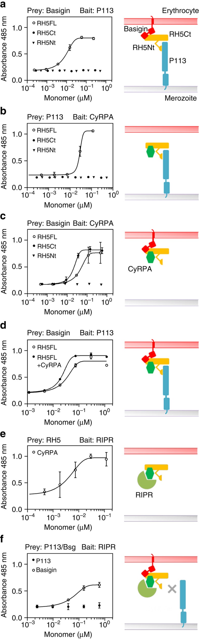
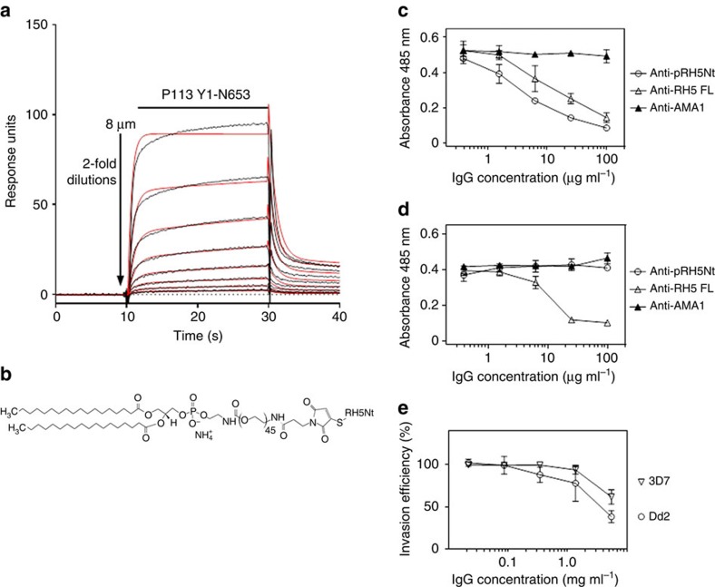
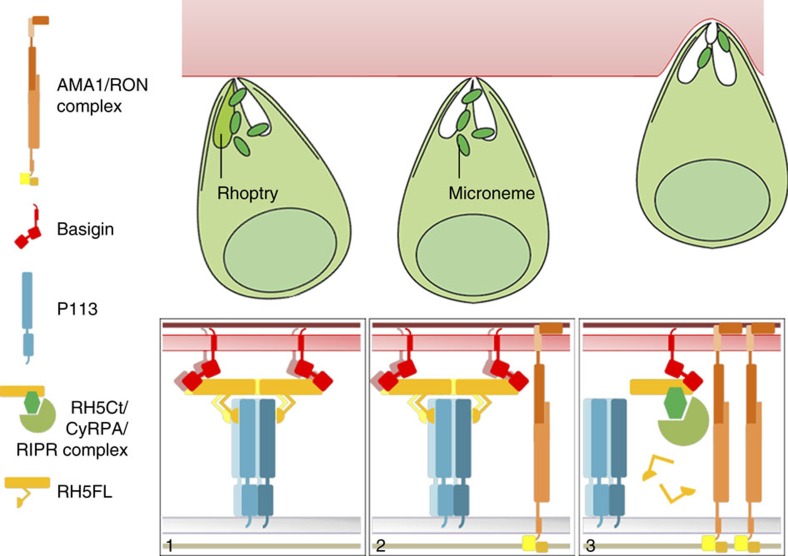
Invasion of erythrocytes by Plasmodium falciparum merozoites is necessary for malaria pathogenesis and is therefore a primary target for vaccine development. RH5 is a leading subunit vaccine candidate because anti-RH5 antibodies inhibit parasite growth and the interaction with its erythrocyte receptor basigin is essential for invasion. RH5 is secreted, complexes with other parasite proteins including CyRPA and RIPR, and contains a conserved N-terminal region (RH5Nt) of unknown function that is cleaved from the native protein. Here, we identify P113 as a merozoite surface protein that directly interacts with RH5Nt. Using recombinant proteins and a sensitive protein interaction assay, we establish the binding interdependencies of all the other known RH5 complex components and conclude that the RH5Nt-P113 interaction provides a releasable mechanism for anchoring RH5 to the merozoite surface. We exploit these findings to design a chemically synthesized peptide corresponding to RH5Nt, which could contribute to a cost-effective malaria vaccine.


Conflict of interest statement

The authors declare no competing financial interests.

Figures

Figure 1
Figure 1. The N-terminal domain of RH5 is not involved in basigin binding.
(a) Purified tagged recombinant RH5 expressed by HEK293 cells resolved as four species by SDS–PAGE under reducing conditions. The N-terminal sequence of each species was determined by Edman degradation and is shown. (b) The N-terminal region of RH5 is not involved in basigin binding. The indicated RH5 fragments were expressed as biotinylated bait proteins and probed for interactions with a highly avid β-lactamase-tagged basigin prey protein using the AVEXIS assay. The full length (FL) and the major processed form (RH5Ct) of RH5 bound basigin but the N-terminal region (RH5Nt) did not. Bars represent means±95% confidence intervals; n=3. (c) Biophysical analysis of the RH5Ct-basigin interaction. Serial dilutions of the indicated concentrations of purified RH5Ct protein were injected over the biotinylated ectodomain of basigin immobilized on a streptavidin-coated sensor chip. The data showed excellent fits to a simple (1:1) binding model (red line). The binding parameters of RH5Ct did not differ significantly from those obtained from RH5FL (Supplementary Table 1) demonstrating that the N-terminal region of RH5 does not contribute to basigin binding affinity.
Figure 2
Figure 2. The N-terminal domain of RH5 interacts with the merozoite surface protein P113.
(a) The N-terminal domain of RH5 (RH5Nt, F1-K116) was expressed as a pentameric, β-lactamase-tagged prey protein and systematically tested for interactions against a library of recombinant P. falciparum merozoite receptor ectodomains and secreted bait proteins using the AVEXIS assay; a single interaction with P113 was observed. (b) The P113-RH5Nt interaction detected in the reciprocal orientation using a P113 prey against RH5 baits. Bars in (a,b) represent means±95% confidence intervals; n=3; controls were RH5FL-basigin (+ve) and Cd4d3+4 tag alone (−ve). (c) Biochemical purification of native P113 from P. falciparum cultures with RH5Nt but not control RH5Ct-coated agarose beads. Parasite lysates were incubated with beads coated in either RH5Nt or RH5Ct protein, washed, and eluates resolved by SDS–PAGE under reducing conditions, blotted and probed with anti-P113 antibody. (d) P113 is expressed in schizonts and on the surface of free merozoites. Fixed blood-stage P. falciparum schizonts and free merozoites were probed with anti-P113 antibodies (green), the merozoite surface marker anti-MSP9 (red) and nucleic acid stained with DAPI (blue). Scale bars, 3 μm.
Figure 3
Figure 3. The P113-RH5 interaction interface involves a short linear sequence in RH5Nt and the cysteine-rich N-terminal domain of P113.
(a) The P113 interaction interface on RH5 was mapped to 19 amino acids located within the N terminus of RH5. Fragments of RH5Nt that could be expressed as prey proteins are depicted schematically and were tested for their ability to bind a P113 bait protein by AVEXIS. The K9-K27 fragment represented the minimal binding region. (b) The RH5 interaction interface on P113 was mapped to the N terminus corresponding to a cysteine-rich region encompassed by Y1-K197. Fragments of P113, including the entire ectodomain (EE), that could be expressed as bait proteins are depicted schematically as in a with the approximate locations of the 16 cysteines marked by vertical bars. The proteins were tested for their ability to bind an RH5 prey protein by AVEXIS. Bars represent means±95% confidence intervals n=3; the experiment shown is one of two independent experiments.
Figure 4
Figure 4. The interaction between P113 and the N-terminal region of RH5 is specific and 10-fold stronger in the context of full-length RH5.
(a) RH5Nt bound P113 specifically and with low affinity. Purified monomeric RH5Nt was serially diluted before being injected over 780 RU of monobiotinylated P113 immobilized on a streptavidin-coated sensor chip. The binding traces suggested the presence of a small amount of multimeric material (see Methods); consequently, binding equilibrium was not quite achieved even though long (>60 s) injection times were used; the dashed line (inset) marks the time point used for equilibrium binding. From these data, an equilibrium binding constant (KD) of 3.0±0.5 μM (mean±s.d., n=2) was calculated. (b,c) RH5FL binds P113 with a ten-fold higher affinity than RH5Nt. A kinetic analysis of P113 binding RH5Nt (b) or RH5FL (c) using SPR; inset gels demonstrate the homogeneity and integrity of each RH5 analyte preparation. Serially diluted RH5 analytes were injected over 780 RU of immobilized P113. Binding data (black lines) fitted a simple 1:1 binding model well (red lines), and were used to determine binding constants. (d) Gel filtration elution profiles of the entire ectodomain (EE) and an N-terminal subfragment (Y1-N653) of recombinant tagged P113 demonstrated that P113 EE can form multimers. The lettered dashed lines correspond to fractions collected over the resolved peaks; the elution volumes of gel filtration mass markers are indicated. (e) Native and denaturing (SDS–PAGE) gels corresponding to aliquots of the fractions indicated by the letters in d. P113 EE but not the Y1-N653 fragment resolves at an approximately fourfold higher mass under native when compared with denaturing conditions. The Y1-N653 fragment resolved as two species by native PAGE. Multimeric forms of P113 EE exhibited complex multivalent binding behaviour by SPR (f) compared with P113 Y1-N653 (g). Sensorgrams in (f,g) show a 120 s pulse of purified 8 μM P113 analyte injected over 510 RU of immobilized RH5Nt.
Figure 5
Figure 5. The RH5-CyRPA-RIPR complex can interact with basigin but not P113.
The binding interdependencies of the proteins within the RH5 invasion complex with basigin and P113 were determined using modified AVEXIS assays. The indicated purified monomeric components of the RH5 complex were titrated into binding reactions between the named baits and β-lactamase-tagged preys, which do not interact directly, and any resulting prey binding quantified by measuring the hydrolysis of a colorimetric β-lactamase substrate at 485 nm. Binding data are shown on the left panels with their interpretations shown schematically on the right. RH5FL can simultaneously bind P113 and basigin (a), CyRPA and P113 (b), and CyRPA and basigin (c). (d) The basigin-RH5FL-P113 complex is not overtly affected by the addition of CyRPA; here, the RH5FL monomer concentration was titrated with CyRPA held constant at 0.3 μM. (e) RH5FL prey could be captured on a RIPR bait by addition of purified CyRPA. (f) The RH5FL-CyRPA-RIPR complex interacted with basigin, but not P113 preys; here, the CyRPA monomer concentration was titrated with RH5FL held constant at 0.2 μM. Binding data points represent means±95% confidence interval (n=3); a representative experiment from at least two independent experiments is shown.
Figure 6
Figure 6. An ‘amph-vaccine' based on RH5Nt elicits antibodies that inhibit parasite growth in vitro.
(a) A synthetic 116 amino-acid peptide corresponding to RH5Nt interacts with P113. Serial dilutions of purified P113 (Y1-N653) were injected over the RH5Nt peptide immobilized on a streptavidin-coated sensor chip after the C-terminal cysteine was conjugated to biotin functionalized with maleimide. Reference-subtracted binding data are shown (black lines) which fit well to a 1:1 binding model (red lines). (b) Structure of an ‘amph-vaccine' based on RH5Nt created by conjugating the RH5Nt peptide to maleimide-functionalized 1,2-distearoyl-sn-glycero-3-phosphoethanolamine-N-[maleimide(polyethylene glycol)-2000] (PEG2000-DSPE). Antibodies raised against RH5Nt peptide (Anti-pRH5Nt) blocked RH5 binding to P113 (c), but not to basigin (d). The indicated concentrations of protein-G purified rabbit polyclonal antibodies were incubated with RH5 β-lactamase-tagged prey proteins before presenting them to immobilized P113 (c) or basigin (d) baits. Prey binding was quantified by nitrocefin hydrolysis at 485 nm; polyclonal antibodies to RH5FL and AMA1 were used as positive and negative controls respectively. (e) Polyclonal antibodies elicited against the RH5Nt amph-vaccine inhibited erythrocyte invasion of both 3D7 and Dd2 strain of P. falciparum. Data points represent means±95% confidence interval, n=3; a representative experiment from two independent experiments is shown.
Figure 7
Figure 7. A model describing the role of the RH5 complex and its interaction with P113 during erythrocyte invasion.
Before invasion, the different components of the RH5 invasion complex are segregated within different subcellular locations of the merozoite and therefore purposefully prevented from interacting: RH5 is located in the rhoptries, CyRPA and RIPR in the micronemes, and P113 on the surface of the merozoite. Following engagement of the erythrocyte and release of the rhoptry contents, RH5 is tethered via its N-terminal region at the merozoite membrane by the surface-localized multimeric P113, enabling direct presentation to the basigin receptor on the erythrocyte surface and leading to the formation of an open connection for other invasion ligands to be secreted (1). The AMA1/RON complex initiates the formation of the moving junction (2). The localized secretion of CyRPA and RIPR from the micronemes leads to the formation of the RH5-CyRPA-RIPR-basigin invasion complex which, either because of the cleavage of RH5 to remove the N-terminal region, and/or because P113 and RIPR cannot simultaneously bind the RH5 complex would release RH5 from the P113 tether at the merozoite surface to licence invasion (3). The P113-RH5 complex would therefore only be fleetingly formed during the rapid invasion process resulting in a soluble post-invasion RH5Ct-CyRPA-RIPR complex.

References

    1. Murray C. J. et al.. Global malaria mortality between 1980 and 2010: a systematic analysis. Lancet 379, 413–431 (2012). - PubMed
    1. Cowman A. F. & Crabb B. S. Invasion of red blood cells by malaria parasites. Cell 124, 755–766 (2006). - PubMed
    1. Cowman A. F., Berry D. & Baum J. The cellular and molecular basis for malaria parasite invasion of the human red blood cell. J. Cell Biol. 198, 961–971 (2012). - PMC - PubMed
    1. Weiss G. E. et al.. Revealing the sequence and resulting cellular morphology of receptor-ligand interactions during Plasmodium falciparum invasion of erythrocytes. PLoS Pathog. 11, e1004670 (2015). - PMC - PubMed
    1. Hadley T. J. Invasion of erythrocytes by malaria parasites: a cellular and molecular overview. Annu. Rev. Microbiol. 40, 451–477 (1986). - PubMed

Publication types

MeSH terms