. 2017 Feb 10:7:42159.
doi: 10.1038/srep42159.

Inhibition of Receptor-Interacting Protein Kinase 1 with Necrostatin-1s ameliorates disease progression in elastase-induced mouse abdominal aortic aneurysm model

Affiliations

Inhibition of Receptor-Interacting Protein Kinase 1 with Necrostatin-1s ameliorates disease progression in elastase-induced mouse abdominal aortic aneurysm model

Qiwei Wang et al. Sci Rep. .

Abstract

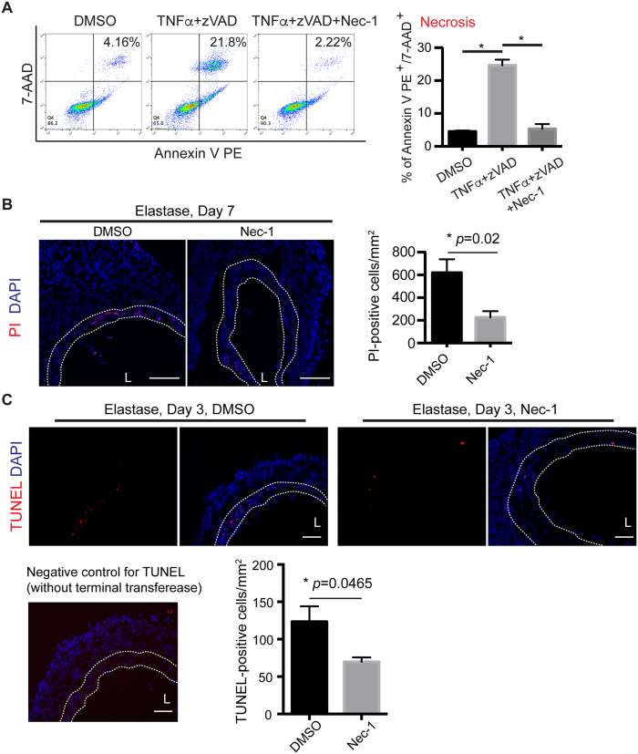
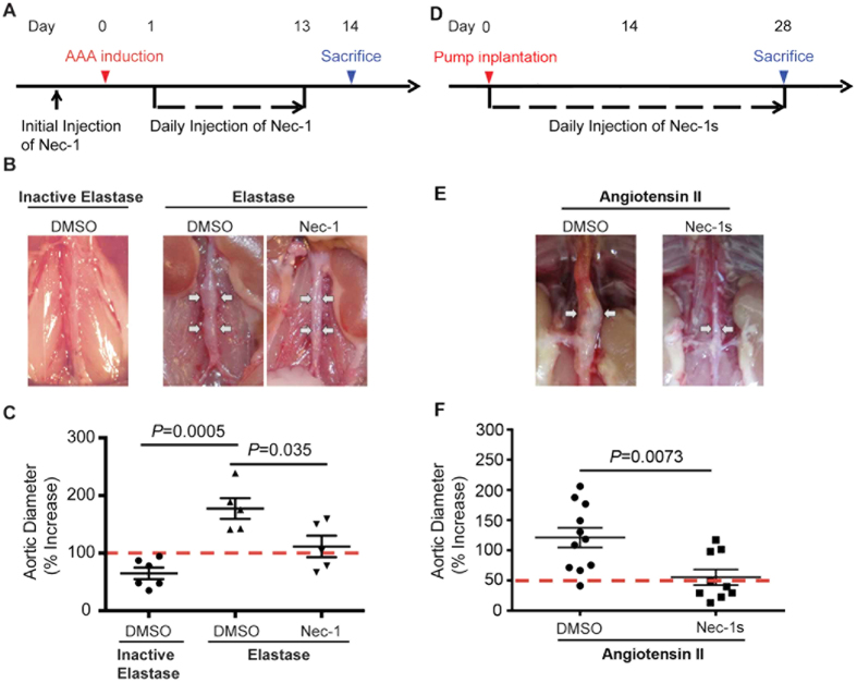
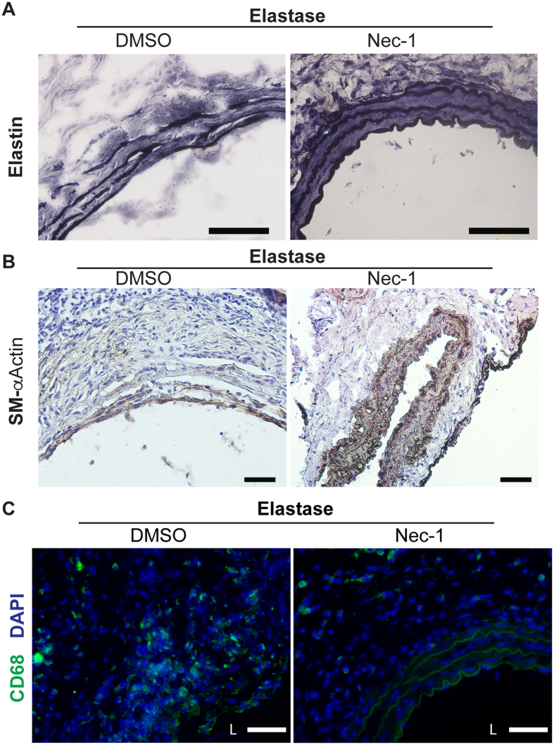
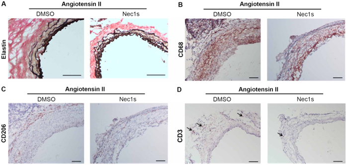
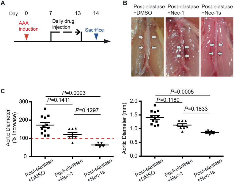
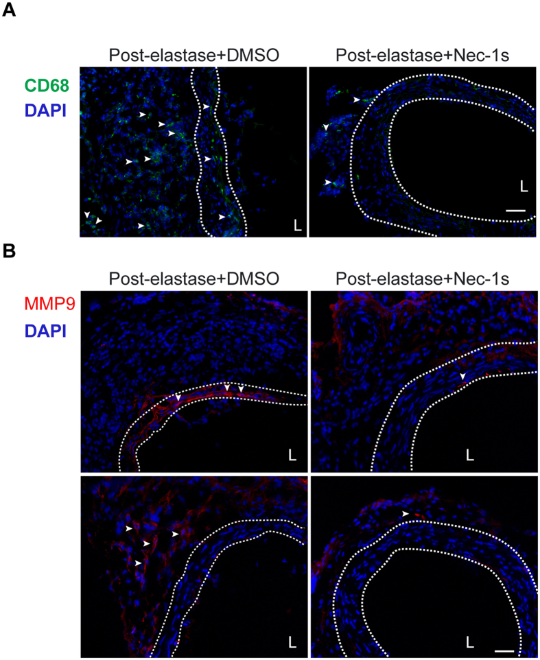
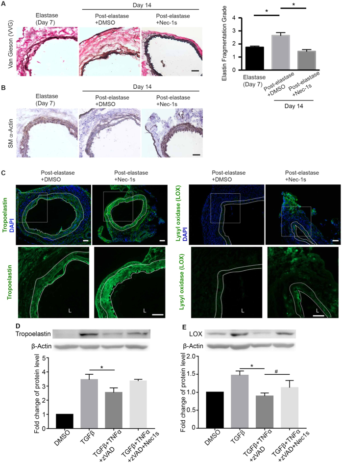
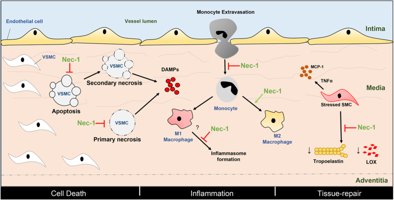
Abdominal aortic aneurysm (AAA) is a common aortic disease with a progressive nature. There is no approved pharmacological treatment to effectively slow aneurysm growth or prevent rupture. Necroptosis is a form of programmed necrosis that is regulated by receptor-interacting protein kinases (RIPs). We have recently demonstrated that the lack of RIP3 in mice prevented aneurysm formation. The goal of the current study is to test whether perturbing necroptosis affects progression of existing aneurysm using the RIP1 inhibitors Necrostatin-1 (Nec-1) and an optimized form of Nec-1, 7-Cl-O-Nec-1 (Nec-1s). Seven days after aneurysm induction by elastase perfusion, mice were randomly administered DMSO, Nec-1 (3.2 mg/kg/day) and Nec-1s (1.6 mg/kg/day) via intraperitoneal injection. Upon sacrifice on day 14 postaneurysm induction, the aortic expansion in the Nec-1s group (64.12 ± 4.80%) was significantly smaller than that of the DMSO group (172.80 ± 13.68%) (P < 0.05). The mean aortic diameter of Nec-1 treated mice appeared to be smaller (121.60 ± 10.40%) than the DMSO group, though the difference was not statistically significant (P = 0.1). Histologically, the aortic structure of Nec-1s-treated mice appeared normal, with continuous and organized elastin laminae and abundant αActin-expressing SMCs. Moreover, Nect-1s treatment diminished macrophage infiltration and MMP9 accumulation and increased aortic levels of tropoelastin and lysyl oxidase. Together, our data suggest that pharmacological inhibition of necroptosis with Nec-1s stabilizes pre-existing aneurysms by diminishing inflammation and promoting connective tissue repair.


Conflict of interest statement

The authors declare no competing financial interests.

Figures

Figure 1
Figure 1. Necrostatin-1 (Nec-1) inhibits SMC necroptosis in vitro and attenuates cell death in the elastase-injured aortae.
(A) MOVAS cells (a mouse aortic smooth muscle cell line) were treated with TNF-α (50 ng/ml), zVAD (40 μΜ), Nec-1 (40 μΜ) or DMSO (vehicle control) as indicated for 6 hours. Cells were then stained with PE Annexin V and 7-AAD and analyzed by flow cytometry. Necrotic cells were identified as PE Annexin V+/7-AAD+. Data represent mean ± SEM. n = 6. *P < 0.05. (B,C) Representative photographs of aortic sections from DMSO or Nec-1 (3.2 mg/kg/day) treated mice. (B) Mice were injected with propidium iodide (PI) 2 hours before euthanization. DAPI was used to stain nuclei. Quantification of PI-positive cells in the media layer (indicated by white dashed line) is shown on the right. (C) Apoptotic cells were stained by terminal deoxynucleotidyl transferase dUTP nick end labeling (TUNEL). Bottom left: a representative image of TUNEL staining negative control in which the aortic section was incubated with label solution only without terminal transferase. Bottom right: a bar graph showing quantification of apoptosis (TUNEL-positive) in the media layer (indicated by white dashed line). At least five non-serial cross-sections per aorta were analyzed (n = 3 aortae per treatment). Data represent mean ± SEM. L indicates lumen. Scale bars = 100 μm (B), 50 μm (C).
Figure 2
Figure 2. Necrostatin-1 (Nec-1) prevents aneurysm formation in mouse models of aneurysms.
(A) Experimental design for the elastase model. Mice were treated with vehicle (DMSO) or Nec-1 at 3.2 mg/kg/day starting 30 minutes before elastase perfusion. Mice were euthanized 14 days after. (B) Representative photos of perfused abdominal aortae with indicated treatments. Arrows indicate aneurysm formation. (C) Percentage increase of maximal external aortic diameter between pre-perfusion and on day 14 after perfusion. In the elastase model, an AAA is defined as a percentage increase in aortic diameter ≥100% (red dashed line). (D) Experimental design for the Angiotensin II (AngII) model. ApoE−/− mice received osmotic pumps that disbursed Ang II at 1000 ng/kg/min. Daily injection of DMSO or Nec-1s (1.6 mg/kg/day) started immediately following the pump implantation. Mice were euthanized 28 days after. (E) Representative photos of abdominal aortae from Ang II-treated mice on day 28. Arrows indicate aneurysm formation. (F) Aortic dilatation was expressed as the percentage increase of suprarenal diameter over infrarenal diameter. An AAA is defined in the Ang II model as a ≥50% increase in aortic diameter (red dashed line). Data represent mean ± SEM. One-Way ANOVA and unpaired student’s t test were used for C and F, respectively.
Figure 3
Figure 3. Necrostatin-1 (Nec-1) prevents aortic degradation and inflammation associated with the elastase model.
Elastase-perfused aortae were harvested from mice treated with DMSO or 3.2 mg/kg/day Nec-1 on day 14 after surgery. (A) Aortic sections were stained with Verhoeff’s working elastic stain solution for elastic fibers which appear black. (B,C) Representative photographs of immunostaining for a smooth muscle cell marker SM-αActin (B) or for a macrophage marker CD68 (C) in the elastase-perfused aortae. DAPI was used to stain the nuclei (C). L indicates lumen. Scale bars = 50 μm.
Figure 4
Figure 4. Necrostatin-1s (Nec-1s) prevents angiotensin II induced elastin degradation and aortic inflammation.
Aneurysm-prone aortae were harvested from mice treated with DMSO or 1.6 mg/kg/day Nec-1s 28 days after pump implantation. (A) Aortic sections were stained with Verhoeff’s working elastic stain solution for elastic fibers (black). (B–D) Representative photographs of immunostaining with antibodies specific to CD68 (B), CD206 (C) or CD3 (D). Arrows indicate representative staining of CD3. Scale bars = 100 μm.
Figure 5
Figure 5. Inhibition of RIP1 kinase with Necrostatin-1s (Nec-1s) blocks progression of existing aneurysms.
(A) Mice were subjected to elastase perfusion. Starting on Day 7 post-elastase perfusion, mice received daily intraperitoneal injection of DMSO, Necrostatin-1 (Nec-1, 3.2 mg/kg/day), or Nec-1s (1.6 mg/kg/day), respectively. Mice were euthanized on Day 14. (B) Representative photos of abdominal aortae with indicated treatments. (C) Aortic diameters are expressed (left) as the percentage increase of maximal external aortic diameter between pre-perfusion and on Day 14 and (right) in millimeter measured on Day 14. An AAA is defined as a percentage increase in aortic diameter ≥100% (red dashed line). Kruskal–Wallis nonparametric test. Data represent mean ± SEM.
Figure 6
Figure 6. Necrostatin-1s (Nec-1s) treatment decreases macrophage infiltration and MMP9 accumulation in the elastase-injured aortae.
Mice were subjected to elastase perfusion and started to receive daily intraperitoneal injection of DMSO or Nec-1s (1.6 mg/kg/day) from day 7 post-elastase perfusion. Mice were euthanized on Day 14. Representative photographs of immunostaining for the macrophage marker CD68 (A) and MMP9 (B) in the elastase-perfused aortae with indicated treatments. In (B), the top and bottom panels exemplify MMP9 expression in the media and adventitial aortic layers, respectively. Arrowheads indicate cells with positive CD68 or MMP9 staining. DAPI was used to stain the nuclei. Medial layer is highlighted by white dashed line. L indicates lumen. Scale bars = 50 μm.
Figure 7
Figure 7. Necrostatin-1s (Nec-1s) treatment promotes aortic tissue repair.
(A) Cross sections of harvested aortae were stained with Verhoeff-Van Gieson in which elastin fibers are stained in black (representative photos are shown in left panel). Scale bar = 50 μm. Fragmentation of elastin was graded according to an arbitrary scale of 1–4 as described in methods. Higher grading indicates more severe fragmentation. Quantification of the grading is shown on the right. Data are mean ± SEM. n = 5. *P < 0.05. (B,C) Representative photographs of immunostaining for the smooth muscle cell marker SM-αActin (B), tropoelastin (C, left) and lysyl oxidase (C, right) in the elastase-perfused aortae (Day 14) with indicated treatments. In C, bottom images represent increased magnification of highlighted region in the upper images. DAPI was used to stain the nuclei. Medial layer is highlighted by white dashed line. L indicates lumen. Scale bars = 50 μm. (D,E) Primary mouse aortic SMCs were starved in 0.5% FBS low glucose DMEM for 24 h, then treated with TGFβ (10 ng/ml) for 48 h, and challenged with TNFα (20 ng/ml) plus zVAD (40 μΜ), Nec-1s (20 μM) for 6 h. Protein levels of tropoelastin and lysyl oxidase were detected by Western blot. n = 3. One-way ANOVA. *P < 0.05; #P < 0.05.
Figure 8
Figure 8. Proposed actions of Necrostatin-1 and its optimized form Nec-1s during the development and progression of abdominal aortic aneurysm.
The common features of aneurysms– cell-death, inflammation and degradation of the extracellular matrix– are depicted in the vessel wall that consists of the intima, media and adventitia. VSMC in the media undergo apoptosis, primary or secondary (post-apoptotic) necrosis causing the release of DAMPs that subsequently stimulate inflammation. Nec-1/Nec-1s prevents apoptosis and necrosis, thereby reducing inflammation indirectly. Nec-1/Nec-1s may also directly reduce inflammation by preventing monocyte migration, inflammasome formation and increasing monocyte to M2 macrophage differentiation. Nec-1/Nec-1s may also promote tissue-repair through rescuing the reduction in the tropoelastin and LOX normally observed in the stressed VSMCs and therefore preserve extracellular matrix. Red arrows depict inhibition whereas green arrows show activation. VSMC: vascular smooth muscle cell, DAMP: damage-associated molecular patterns, LOX: lysyl oxidase, MCP-1: monocyte chemoattractant protein-1, TNFα: tumor necrosis factor alpha.

References

    1. Baxter B. T., Terrin M. C. & Dalman R. L. Medical management of small abdominal aortic aneurysms. Circulation 117, 1883–1889 (2008). - PMC - PubMed
    1. Weintraub N. L. Understanding abdominal aortic aneurysm. The New England journal of medicine 361, 1114–1116 (2009). - PMC - PubMed
    1. Powell J. T. & Greenhalgh R. M. Clinical practice. Small abdominal aortic aneurysms. The New England journal of medicine 348, 1895–1901 (2003). - PubMed
    1. Kent K. C. Clinical practice. Abdominal aortic aneurysms. The New England journal of medicine 371, 2101–2108 (2014). - PubMed
    1. Lederle F. A. et al.. Prevalence and associations of abdominal aortic aneurysm detected through screening. Aneurysm Detection and Management (ADAM) Veterans Affairs Cooperative Study Group. Annals of internal medicine 126, 441–449 (1997). - PubMed

Publication types

MeSH terms

LinkOut - more resources