. 2017 Feb 10:7:42546.
doi: 10.1038/srep42546.

Gene Transfer of Prolyl Hydroxylase Domain 2 Inhibits Hypoxia-inducible Angiogenesis in a Model of Choroidal Neovascularization

Affiliations

Gene Transfer of Prolyl Hydroxylase Domain 2 Inhibits Hypoxia-inducible Angiogenesis in a Model of Choroidal Neovascularization

Anna Takei et al. Sci Rep. .

Abstract

Cellular responses to hypoxia are mediated by the hypoxia-inducible factors (HIF). In normoxia, HIF-α proteins are regulated by a family of dioxygenases, through prolyl and asparagyl hydroxylation, culminating in proteasomal degradation and transcriptional inactivation. In hypoxia, the dioxygenases become inactive and allow formation of HIF transcription factor, responsible for upregulation of hypoxia genes. In ocular neoangiogenic diseases, such as neovascular age-related macular degeneration (nAMD), hypoxia seems pivotal. Here, we investigate the effects of HIF regulatory proteins on the hypoxia pathway in retinal pigment epithelium (RPE) cells, critically involved in nAMD pathogenesis. Our data indicates that, in ARPE-19 cells, prolyl hydroxylase domain (PHD)2 is the most potent negative-regulator of the HIF pathway. The negative effects of PHD2 on the hypoxia pathway were associated with decreased HIF-1α protein levels, and concomitant decrease in angiogenic factors. ARPE-19 cells stably expressing PHD2 impaired angiogenesis in vitro by wound healing, tubulogenesis, and sprouting assays, as well as in vivo by iris-induced angiogenesis. Gene transfer of PHD2 in vivo resulted in mitigation of HIF-mediated angiogenesis in a mouse model of nAMD. These results may have implications for the clinical treatment of nAMD patients, particularly regarding the use of gene therapy to negatively regulate neoangiogenesis.


Conflict of interest statement

The authors declare no competing financial interests.

Figures
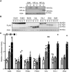
Figure 1
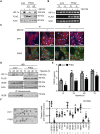
Figure 1. PHDs inhibit hypoxia-mediated transcriptional activation in RPE cells.
(A) ARPE-19 cells were transfected with pFLAG-HIF-1α or -HIF-2α, or an empty plasmid (CMX), and exposed to normoxia (N) or CoCl2 (C). Immunoblots demonstrated endogenous levels of HIF-1α, but not HIF-2α, when compared to transfected controls. (B) Expression analysis of HIF regulatory proteins in ARPE-19. Cells were transfected with plasmids encoding FLAG-tagged PHD1, PHD2, PHD3, VHL, FIH-1, IPAS, or empty CMX and exposed to normoxia (N), hypoxia (H), or treated with CoCl2 (C). (C) ARPE-19 cells were transfected with an HRE-driven luciferase reported plasmid and expression vectors for PHD1, PHD2, PHD3, VHL, FIH-1, or IPAS, and exposed to normoxia (N), hypoxia (H), or CoCl2 (C). Data are presented as relative luciferase activity to cells transfected with an empty plasmid (CMX) and kept at normoxia. All PHD-expressing plasmids significantly reduced hypoxia-mediated HIF-1α transactivation, and PHD2 and PHD3 also abrogate HIF-1α transactivation in response to CoCl2 (mean + SEM, n = 6; *P < 0.05 vs corresponding CMX).
Figure 2
Figure 2. PHD2 overexpression reduces HIF-1α lifetime in RPE cells.
Endogenous HIF-1α was stabilized by exposing ARPE-19 cells to hypoxia and the protein lifetime was determined by relative densitometry (RD) of anti-HIF-1α immunoblot in cells transfected with empty CMX plasmid (A) under reoxygenation. Analysis of ARPE-19 cells transfected with plasmids encoding FLAG-tagged PHD1 (B), PHD3 (D), VHL (E), FIH-1 (F), or IPAS (G) did not display reduction of HIF-1α lifetime. Cells overexpressing pFLAG-PHD2 (C) showed a considerable reduction on endogenous HIF-1α protein lifetime and expression.
Figure 3
Figure 3. RPE cells stably expressing PHD2 display reduced levels of endogenous HIF-1α and VEGF proteins.
(A) ARPE-19 cells were transfected and selected to stably express FLAG-tagged PHD2 or an empty puromycin-resistance control (puro). Two clones of RPE-PHD2 cells (cl1 and cl2) were analyzed for HIF-1α and PHD2 protein expression (FLAG), under proteasome blockage with MG132 (+). RPE-PHD2 cells clone 1 displayed high levels of PHD2 expression and considerably lower levels of endogenous HIF-1α protein when compared to the puro control. Clone 1 was used for all subsequent experiments. (B) RPE-puro and RPE-PHD2 were exposed to normoxia (N) or hypoxia (H), in the presence (+) or absence (−) of MG123, and HIF-1α transcript expression pattern was analyzed by RT-PCR. No discernable differences could be observed in HIF-1α transcripts in PHD2-overexpressing RPE cells. (C) ARPE-19 cells stably expressing FLAG-PHD2 (green) showed reduced immunostaining for endogenous HIF-1α protein (white) in both normoxia (N) and hypoxia (H), even in the presence of MG123, as compared to the RPE-puro control cells. Cells were counter stained to visualize nuclei (blue) and cytoskeleton (red), respectively. Scale bar = 100 μm. (D) RPE-puro and RPE-PHD2 cells were exposed to increasing times of hypoxia versus normoxia (N). Immunoblots display a considerable reduction in HIF-1α and intracellular VEGF protein levels in RPE-PHD2 cells. VEGF-capturing assay using Bevacizumab also revealed lower levels of soluble VEGF produced by RPE-PHD2 cells, when compared to the RPE-puro cells. (E) VEGF soluble factor was quantified by ELISA in conditioned media from ARPE-19 stably expressing puro control or PHD2 exposed to increasing times of hypoxia. Significantly lower levels of VEGF soluble factor were quantified in media from RPE-PHD2 cells when compared to the RPE-puro control cells (mean + SEM, n = 6; *P < 0.05). (F) Angiogenesis-related protein expression profile in media conditioned to 24 h of hypoxia from RPE-puro and RPE-PHD2 cells. Densitometric analysis of predominantly expressed proteins (alphabetized) was preformed against positive controls (+) and presented as expression relative to control (dashed line). A general reduction of angiogenic factors was observed in RPE-PHD2 cells as compared to RPE-puro control cells (mean ± SEM, n = 3; *P < 0.05).
Figure 4
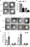
Figure 4. RPE-PHD2 cells impair angiogenesis in HUVE cells.
Confluent HUVE cell cultures were scratched to induce wounding or seeded onto matrigel for tubulogenesis, and cultured in media conditioned by RPE-puro or RPE-PHD2 cells exposed to 24 h hypoxia. (A) A delay in wound healing is noticeable, particularly at 12 h post-exposure to RPE-PHD2 cells conditioned media. Scale bar = 200 μm. (B) Wound marginal distances were measured in HUVE cell cultures. Data is presented as percentage decrease in average marginal distance. RPE-PHD2 cells conditioned medium significantly delays HUVE cells wound healing at 12 h post-exposure, when compared to RPE-puro control cells (mean ± SEM, n = 6; *P < 0.05). (C) A considerable reduction in both cell number and tube-like cells was observed in HUVE cells exposed to RPE-PHD2 conditioned medium, when compared to the RPE-puro control cell line. Sale bar = 100 μm. (D) Tube-like cells and nuclei were counted in HUVE cell cultures exposed to RPE conditioned media and evaluated through time. Data is displayed as tubes/nuclei ratio. A statistically significant lower tubulogenesis was observed in HUVE cells exposed to RPE-PHD2 cell conditioned media as compared to its correspondent RPE-puro control (mean ± SEM, n = 6; *P < 0.05).
Figure 5
Figure 5. RPE-PHD2 cells reduce angiogenesis in ocular endothelial cells.
(A) RE or CE spheroids were embedded in matrigel and cultured in media conditioned by RPE-puro or RPE-PHD2 cells exposed to 24 h hypoxia. Scale bar = 100 μm. (B) A significant reduction in the number of sprouts were determined in RE and CE spheroids exposed to RPE-PHD2 hypoxia conditioned media when compared to RPE-puro control (mean + SEM, n = 6; *P < 0.05). Data is presented as number of sprouts per spheroid. (C) 3D cultures were obtained by mixing cell suspensions of RE or CE cells with either RPE-puro or RPE-PHD2 prior to formation of spheroids, and embedding in matrigel. Cultures where allowed to create sprouts by exposure to normoxia (N) or hypoxia (H) for 36 h. Scale bar = 100 μm. (D) 3D cultures containing RPE-puro displayed increased sprouting in hypoxic (H) when compared to normoxic (N) conditions. 3D cultures containing RPE-PHD2 failed to induce sprouts, both in normoxia or hypoxia. A critical reduction of sprouts was observed in 3D cultures containing RPE-PHD2 cells. Data is presented as number of sprouts per culture (mean + SEM, n = 6; *P < 0.05 N vs H; #P < 0.05 PHD2 vs puro).
Figure 6
Figure 6. Hypoxic RPE-PHD2 conditioned media mitigates iris angiogenesis.
(A) Schematic representation of iris angiogenesis protocol. P12.5 mice were IVt injected with RPE-puro or RPE-PHD2 media conditioned by 24 h of hypoxia. Injections were repeated every fourth day posteriorly to the iris (red). At P27.5 mice vasculature was labeled with Dextran-FITC (green). (B) Irises were isolated and prepared for whole mount fluorescence. Scale bar = 100 μm. (C) Fluorescence densitometry (intensity per area) was determined and presented as percentage of vehicle (dashed line). Media conditioned by hypoxic RPE-puro cells showed an increase in iris angiogenesis, while hypoxic RPE-PHD2 conditioned media presented mitigated angiogenesis (mean + SEM, n = 6; *P < 0.05 vs vehicle; #P < 0.05 vs puro).
Figure 7
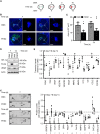
Figure 7. Gene transfer of PHD2 reduces angiogenesis in vivo.
(A) Schematics of laser-induced mouse CNV model with DNA subretinal injection and electroporation. CNV lesions (green) were induced nasally and temporally. Four days post-laser induction the lesions initiate angiogenesis (red) and mice were subretinally injected (dashed circle) with plasmid DNA encoding FLAG-PHD2 or empty CMX. Gene transfer was achieved by electroporation. The creation of a subretinal bleb during DNA injection leads to an increase in the lesion area, while the fellow-lesion (opposing the large DNA bleb) undergoes canonical CNV progression. On days 7 and 14 after laser (equivalent of 3 and 10 days of DNA expression), the posterior eye segments were analyzed. (B) Whole-mounts of posterior eye segments were stained with isolectin (blue) and with a FLAG antibody to detect exogenous PHD2 (green). Expression of FLAG-PHD2 expression was observed surrounding the site of injection (dashed circle indicates DNA bleb) on the RPE-side of the posterior eye segments. Fellow-lesion (dotted square) is magnified. Scale bar = 100 μm. (C) Quantitative analysis of the fellow-lesion area. A significant reduction of CNV area was observed in fellow-lesions of CNV mice overexpressing PHD as compared to CMX control (mean + SEM, n = 6; *P < 0.05). (D) Expression analysis for HIF-1α, VEGF and PHD2 proteins in posterior eye segments of CNV mice expressing control CMX or overexpressing FLAG-PHD2. Increased PHD2 expression correlated with decreased expression of both HIF-1α and VEGF proteins. (E) HIF-mediated transcript levels were quantified by qPCR in posterior eye segments of CNV mice overexpressing PHD2. Data is presented as fold (2−ΔΔCt) normalized to time-corresponding CMX control (dashed line). Analyzed transcript levels displayed reduced expression in posterior eye segments from PHD2 overexpressing CNV mice (mean ± SEM, n = 3; *P < 0.05). (F) Angiogenesis-related factors were profiled in whole-tissue from posterior eye segments from CNV mice expressing CMX or PHD2. Densitometric analysis of proteins of interest (alphabetized) was preformed against positive controls (+) and presented as expression relative to time-corresponding control (dashed line). Angiogenic-factors were reduced in posterior eye segments from CNV mice overexpressing PHD2 (mean ± SEM, n = 3; *P < 0.05).

Similar articles

Cited by

References

    1. Ruas J. L. & Poellinger L. Hypoxia-dependent activation of HIF into a transcriptional regulator. Semin. Cell Dev. Biol. 16, 514–522 (2005). - PubMed
    1. Weidemann A. & Johnson R. S. Biology of HIF-1α. Cell Death Differ 15, 621–627 (2008). - PubMed
    1. Semenza G. L., Nejfelt M. K., Chi S. M. & Antonarakis S. E. Hypoxia-inducible nuclear factors bind to an enhancer element located 3′ to the human erythropoietin gene. Proc. Natl. Acad. Sci. USA 88, 5680–5684 (1991). - PMC - PubMed
    1. Wang G. L. & Semenza G. L. Purification and characterization of hypoxia-inducible factor 1. J. Biol. Chem. 270, 1230–1237 (1995). - PubMed
    1. Wang G. L., Jiang B. H., Rue E. A. & Semenza G. L. Hypoxia-inducible factor 1 is a basic-helix-loop-helix-PAS heterodimer regulated by cellular O2 tension. Proc. Natl. Acad. Sci. USA 92, 5510–5514 (1995). - PMC - PubMed

Publication types

MeSH terms

Substances

LinkOut - more resources