. 2017 Apr;24(4):660-671.
doi: 10.1038/cdd.2016.162. Epub 2017 Feb 10.

STAT1 mediates transmembrane TNF-alpha-induced formation of death-inducing signaling complex and apoptotic signaling via TNFR1

Affiliations

STAT1 mediates transmembrane TNF-alpha-induced formation of death-inducing signaling complex and apoptotic signaling via TNFR1

Yaping Jiang et al. Cell Death Differ. 2017 Apr.

Abstract

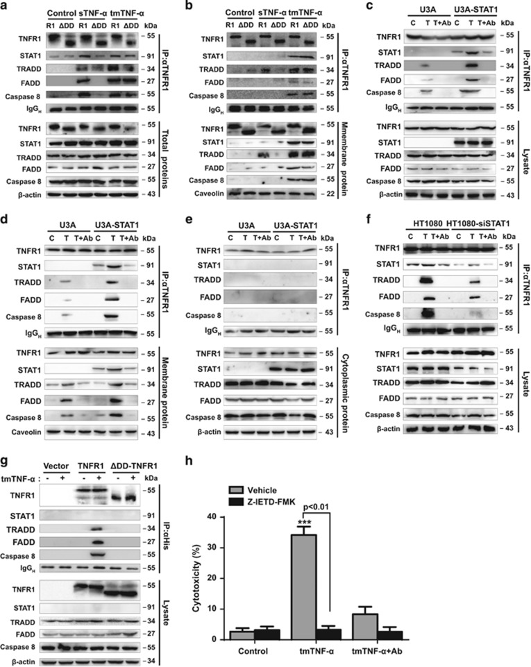
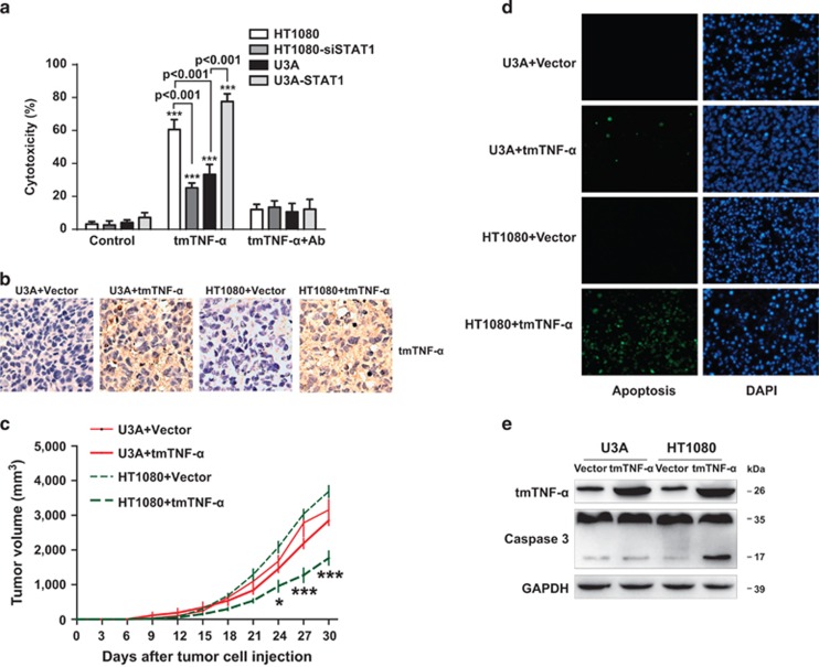
Tumor necrosis factor-alpha (TNF-α) exists in two forms: secretory TNF-α (sTNF-α) and transmembrane TNF-α (tmTNF-α). Although both forms of TNF-α induce tumor cell apoptosis, tmTNF-α is able to kill tumor cells that are resistant to sTNF-α-mediated cytotoxicity, indicating their differences in signal transduction. Here, we demonstrate that internalization of TNFR1 is crucial for sTNF-α- but not for tmTNF-α-induced apoptosis. sTNF-α induces binding of tumor necrosis factor receptor type 1-associated death domain protein (TRADD) to the death domain (DD) of TNFR1 and subsequent activation of nuclear factor kappa B (NF-κB), and the formation of death-inducing signaling complexes (DISCs) in the cytoplasm after internalization. In contrast, tmTNF-α induces DISC formation on the membrane in a DD-independent manner. It leads to the binding of signal transducer and activator of transcription 1 (STAT1) to a region spanning amino acids 319-337 of TNFR1 and induces phosphorylation of serine at 727 of STAT1. The phosphorylation of STAT1 promotes its binding to TRADD, and thus recruits Fas-associated protein with DD (FADD) and caspase 8 to form DISC complexes. This STAT1-dependent signaling results in apoptosis but not NF-κB activation. STAT1-deficiency in U3A cells counteracts tmTNF-α-induced DISC formation and apoptosis. Conversely, reconstitution of STAT1 expression restores tmTNF-α-induced apoptotic signaling in the cell line. Consistently, tmTNF-α suppresses the growth of STAT1-containing HT1080 tumors, but not of STAT1-deficient U3A tumors in vivo. Our data reveal an unappreciated molecular mechanism of tmTNF-α-induced apoptosis and may provide a new clue for cancer therapy.


Conflict of interest statement

The authors declare that they have no conflict of interest.

Figures

Figure 1
Figure 1
The cytotoxicity of tmTNF-α was independent of the TNFR1 internalization. U937 cells were pre-treated with 100 μM MDC for 1 h and then stimulated with either 100 ng/ml sTNF-α or tmTNF-α on fixed NIH3T3 cells at an effector/target ratio of 10:1 for 24 h. For neutralization of tmTNF-α, effector cells were treated with anti-TNF-α (Ab) for 30 min prior to the addition to the target cells. (a) Cytotoxicity of TNF-α detected by MTT assays. (b) Apoptosis induced by TNF-α determined by Annexin V-PI staining. (c) Western blotting of caspase 3 activation. (d) TNFR1 expression analyzed by flow cytometry. (e) Confocal images of the colocalization of TNFR1 (red) and EEA (green) after a 30 min-stimulation of U937 cells with both forms of TNF-α (magnification, × 600). All the quantitative data represent means±S.D. of at least three independent experiments. *P<0.05, ***P<0.001 versus corresponding treatment in the control group
Figure 2
Figure 2
tmTNF-α-mediated cytotoxicity via TNFR1 was independent of DD and NSD. HEK 293T cells were transfected with empty vector, wild-type TNFR1, ΔDD-TNFR1 or ΔNSD-TNFR1 containing plasmids. (a and c) The cell surface expression of TNFR1 and its mutants (a), and TNFR2 (c) detected by flow cytometry. (b) The transfected HEK 293T cells were stimulated for 24 h with 100 ng/ml sTNF-α or tmTNF-α on fixed NIH3T3 cells at an effector/target ratio of 10:1. For neutralization of tmTNF-α, effector cells were treated with anti-TNF-α (Ab) for 30 min prior to the addition to the target cells. The cytotoxicity was detected by MTT assays. (d) HEK 293T cells were co-transfected with control or TNFR2 siRNA and expression vectors for TNFR1 or its mutants as indicated. After 48 h, the cells were stimulated with tmTNF-α on fixed NIH3T3 for 24 h. The cytotoxicity of tmTNF-α was analyzed by MTT assays. (e) The 24 h-cytotoxicity of R32W-tmTNF-α expressed on COS-7 cells towards HEK 293T cells expressing TNFR1 or mutants thereof. (f) The 24 h-cytotoxicity of sTNF-α to HEK 293T cells co-treated with indicated concentrations of S1P. (g) The 24 h-cytotoxicity of tmTNF-α to HEK 293T cells expressing TNFR1 or its mutants in the presence of 10 μM S1P. All the quantitative data represent means±S.D. of at least three independent experiments. *P<0.05, **P<0.01, ***P<0.001 versus corresponding treatment in the control group
Figure 3
Figure 3
tmTNF-α induced formation of DISC via TNFR1 at the plasma membrane. U937 cells were pre-treated with 100 μM MDC for 1 h and then stimulated with 100 ng/ml sTNF-α or R32W-tmTNF-α on fixed COS-7 cells at an E/T ratio of 10:1 for 30 min. For neutralization of tmTNF-α, effector cells were treated with anti-TNF-α (Ab) for 30 min prior to the addition to the target cells. The total (a), cytoplasmic (c) and membrane (d and f) protein was immunoprecipitated with an anti-TNFR1 antibody and analyzed by immunoblotting with the indicated antibodies. (e) Confocal images of the cellular distribution of FADD or caspase 8 (green) in U937 cells in response to sTNF-α or R32W-tmTNF-α (magnification, × 400). (g) Degradation of Iκ-B was analyzed by western blotting. All immunoprecipitation (IP) or/and western data are representative of three independent experiments. (b and h) The 24 h-cytotoxicity of tmTNF-α (E/T: 10:1) or sTNF-α (100 ng/ml) towards U937 cells transfected with control or FADD siRNA (b) or pre-treated with PDTC (100 μM) for 1 h (h). The data represent means±S.D. of at least three independent experiments. ***P<0.001 versus corresponding treatment in the control group
Figure 4
Figure 4
STAT1 was necessary for tmTNF-induced DISC formation. (a and b) HEK 293T cells were transfected to express wild-type TNFR1, or ΔDD-TNFR1 and then stimulated with 100 ng/ml sTNF-α or R32W-tmTNF-α for 30 min. For neutralization, R32W-tmTNF-α-expressing cells were treated with anti-TNF-α (Ab) for 30 min prior to the addition to the target cells. The total (a) and membrane (b) protein was immunoprecipitated with an anti-TNFR1 antibody and analyzed by immunoblotting. R1: TNFR1; ΔDD: ΔDD-TNFR1. U3A (c–e) or HT1080 (f) cells were transfected by STAT1-containing plasmid or siRNA against STAT1, respectively, followed by stimulation with R32W-tmTNF-α for 30 min. Immunoprecipitation (IP)/western blotting was performed in total (c and f), membrane (d) and cytoplasmic (e) protein with an antibody to TNFR1. Ab, anti-TNF-α antibody; C, Control; T, tmTNF-α. (g) U3A cells were transfected with His-tagged empty vector, His-TNFR1 or His-ΔDD-TNFR1 containing plasmids and stimulated with R32W-tmTNF-α for 30 min. DISC formation was analyzed by IP/western blotting using an anti-His antibody. All the IP/western data are representative of three independent experiments. (h) The 24 h-cytotoxicity of R32W-tmTNF-α to U3A cells pre-treated for 30 min with caspase 8 inhibitor Z-IETD-FMK (4 μM). The data represent means±S.D. of three independent experiments. ***P<0.001 versus corresponding treatment in the control group
Figure 5
Figure 5
STAT1 was required for tmTNF-induced apoptosis. (a) The 24 h-cytotoxicity of R32W-tmTNF-α to U3A, STAT1-expressing U3A, STAT1 knockdown HT1080 or parental HT1080 cells. The data represent means±S.D of at least three independent experiments. ***P<0.001 versus corresponding treatment in the control group. (b–e) 5 × 10⁶ HT1080 or U3A cells were subcutaneously injected into the armpit of forelimb of nude mice. At day 5 after tumor cell inoculation, 100 μg of expression vector for R32W-tmTNF-α or empty vector was injected into the tumor site. (b) The expression of tmTNF-α was analyzed by immunohistochemistry (magnification, × 200). (c) Tumor growth curves (n=8 each group). *P<0.05, ***P<0.001 versus HT1080+vector. (d) Apoptosis in situ detected by terminal deoxynucleotidyl transferase-mediated dUTP nick end labeling assay (magnification, × 100). (e) Caspase 3 activation analyzed by western blotting that is representative of three independent experiments
Figure 6
Figure 6
STAT1 as an adaptor recruited DISC to TNFR1 in tmTNF-α-mediated apoptotic signaling. (a) The scheme of three TNFR1 truncations used in the following experiments. (b) HEK 293T cells were transfected to express wild-type TNFR1, Δ338-455-TNFR1, Δ319-455-TNFR1 or Δ266-455-TNFR1, and then treated with R32W-tmTNF-α for 30 min. Total protein was immunoprecipitated with an anti-TNFR1 antibody and analyzed by immunoblotting. (c) Bacterially expressed different GST fusion proteins and GST alone were incubated with His-tagged STAT1 bound to Ni-NTA resin, respectively, and then western blot was performed using antibodies against TNFR1 or STAT1. (d) HEK 293T cells transfected to express Δ338-455-TNFR1 were treated with 100 ng/ml sTNF-α or R32W-tmTNF-α for 30 min. STAT1 was detected by immunoprecipitation (IP)/western blotting. (e) Western blot of STAT1 phosphorylation in U937 cells treated with 100 ng/ml sTNF-α or R32W-tmTNF-α for 30 min. (f) U3A cells expressing STAT1, S727A-, Y701I- or DM-STAT1 were stimulated with R32W-tmTNF-α for 24 h. The cytotoxicity was detected by an MTT assay. Data represent means±S.D. of at least three independent experiments. ***P<0.001 versus corresponding treatment in the control group. (g) U3A cells expressing STAT1, S727A- or Y701I-STAT1 were treated for 30 min with R32W-tmTNF-α. STAT1 and DISC were visualized by IP/western blotting. For neutralization, R32W-tmTNF-α- expressing cells were treated with anti-TNF-α (Ab) for 30 min prior to addition to the target cells (df). All IP/western data are representative of three independent experiments
Figure 7
Figure 7
tmTNF-α-induced STAT1-dependent apoptotic signaling via TNFR1. sTNF-α induces binding of TRADD to the DD of TNFR1. TNFR1-bound TRADD further recruits TRAF2 and cIAP1/2 at the plasma membrane to activate NF-κB pathway. However, after TNFR1 internalization, STAT1 binds to TRADD and is phosphorylated at tyrosine 701, which excludes the binding of TRAF2 and cIAP1 to TRADD and attenuates NF-κB activation. Meanwhile, TRADD recruits FADD and caspase 8 to form DISC in the cytoplasm to mediate apoptosis. In contrast, tmTNF-α induces binding of STAT1 to the SD of TNFR1 and STAT1 serine phosphorylation at 727. The S727 phosphorylated STAT1 further recruits TRADD to form DISC at the plasma membrane to trigger apoptosis, but not NF-κB activation

References

    1. Kriegler M, Perez C, DeFay K, Albert I, Lu SD. A novel form of TNF/cachectin is a cell surface cytotoxic transmembrane protein: ramifications for the complex physiology of TNF. Cell 1988; 53: 45–53. - PubMed
    1. Black RA, Rauch CT, Kozlosky CJ, Peschon JJ, Slack JL, Wolfson MF et al. A metalloproteinase disintegrin that releases tumour-necrosis factor-alpha from cells. Nature 1997; 385: 729–733. - PubMed
    1. Grell M, Douni E, Wajant H, Lohden M, Clauss M, Maxeiner B et al. The transmembrane form of tumor necrosis factor is the prime activating ligand of the 80 kDa tumor necrosis factor receptor. Cell 1995; 83: 793–802. - PubMed
    1. Ardestani S, Li B, Deskins DL, Wu H, Massion PP, Young PP. Membrane versus soluble isoforms of TNF-alpha exert opposing effects on tumor growth and survival of tumor-associated myeloid cells. Cancer Res 2013; 73: 3938–3950. - PMC - PubMed
    1. Zhang H, Yan D, Shi X, Liang H, Pang Y, Qin N et al. Transmembrane TNF-alpha mediates 'forward' and 'reverse' signaling, inducing cell death or survival via the NF-kappaB pathway in Raji Burkitt lymphoma cells. J Leuk Biol 2008; 84: 789–797. - PMC - PubMed

Publication types

MeSH terms

Substances