

Phenotype- and genotype-specific structural alterations in spasmodic dysphonia
- PMID: 28186656
- PMCID: PMC5578762
- DOI: 10.1002/mds.26920
Phenotype- and genotype-specific structural alterations in spasmodic dysphonia
Abstract
Background: Spasmodic dysphonia is a focal dystonia characterized by involuntary spasms in the laryngeal muscles that occur selectively during speaking. Although hereditary trends have been reported in up to 16% of patients, the causative etiology of spasmodic dysphonia is unclear, and the influences of various phenotypes and genotypes on disorder pathophysiology are poorly understood. In this study, we examined structural alterations in cortical gray matter and white matter integrity in relationship to different phenotypes and putative genotypes of spasmodic dysphonia to elucidate the structural component of its complex pathophysiology.
Methods: Eighty-nine patients with spasmodic dysphonia underwent high-resolution magnetic resonance imaging and diffusion-weighted imaging to examine cortical thickness and white matter fractional anisotropy in adductor versus abductor forms (distinct phenotypes) and in sporadic versus familial cases (distinct genotypes).
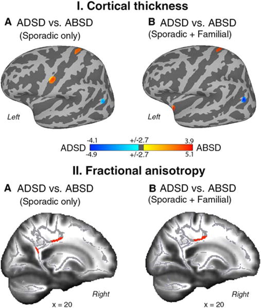
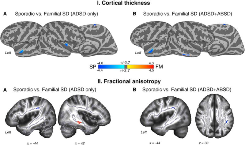
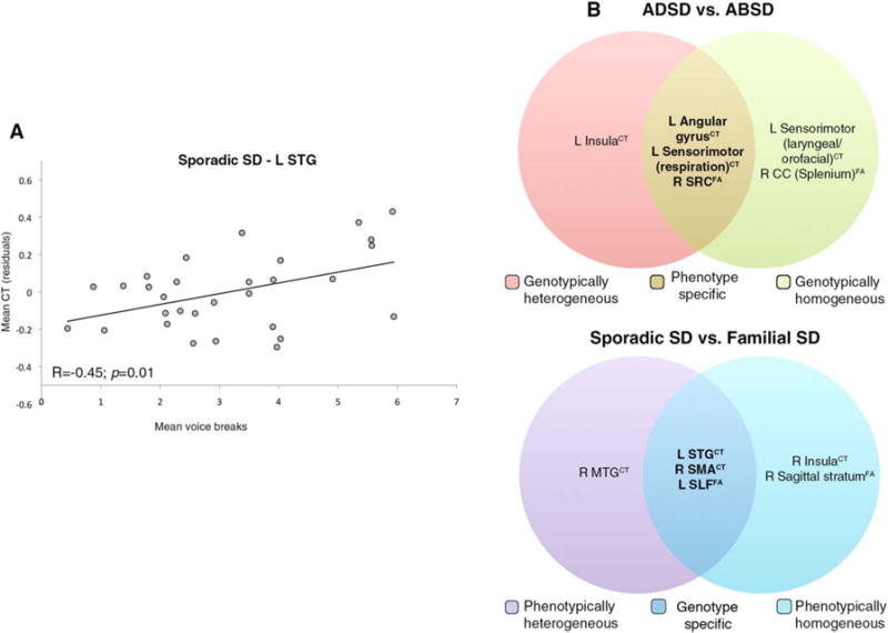
Results: Phenotype-specific abnormalities were localized in the left sensorimotor cortex and angular gyrus and the white matter bundle of the right superior corona radiata. Genotype-specific alterations were found in the left superior temporal gyrus, supplementary motor area, and the arcuate portion of the left superior longitudinal fasciculus.
Conclusions: Our findings suggest that phenotypic differences in spasmodic dysphonia arise at the level of the primary and associative areas of motor control, whereas genotype-related pathophysiological mechanisms may be associated with dysfunction of regions regulating phonological and sensory processing. Identification of structural alterations specific to disorder phenotype and putative genotype provides an important step toward future delineation of imaging markers and potential targets for novel therapeutic interventions for spasmodic dysphonia. © 2017 International Parkinson and Movement Disorder Society.
Keywords: cortical thickness; diffusion-weighted imaging; genetics; laryngeal dystonia.
© 2017 International Parkinson and Movement Disorder Society.
Conflict of interest statement
Figures
References
-
- Blitzer A, Brin MF, Stewart C. Botulinum toxin management of spasmodic dysphonia (laryngeal dystonia): a 12 year experience in more than 900 patients. Laryngoscope. 1998;108:1435–1441. - PubMed
Publication types
MeSH terms
Substances
Grants and funding
LinkOut - more resources
Full Text Sources
Other Literature Sources
Medical
