. 2017 Mar;6(3):788-798.
doi: 10.1002/sctm.16-0162. Epub 2017 Feb 6.

Skin-Derived Precursors as a Source of Progenitors for Corneal Endothelial Regeneration

Affiliations

Skin-Derived Precursors as a Source of Progenitors for Corneal Endothelial Regeneration

Emi Inagaki et al. Stem Cells Transl Med. 2017 Mar.

Abstract

Corneal blindness is the fourth leading cause of blindness in the world. Current treatment is allogenic corneal transplantation, which is limited by shortage of donors and immunological rejection. Skin-derived precursors (SKPs) are postnatal stem cells, which are self-renewing, multipotent precursors that can be isolated and expanded from the dermis. Facial skin may therefore be an accessible autologous source of neural crest derived cells. SKPs were isolated from facial skin of Wnt1-Cre/Floxed EGFP mouse. After inducing differentiation with medium containing retinoic acid and GSK 3-β inhibitor, SKPs formed polygonal corneal endothelial-like cells (sTECE). Expression of major corneal endothelial markers were confirmed by Reverse transcription polymerase chain reaction (RT-PCR) and quantitative Real time polymerase chain reaction (qRT-PCR). Western blots confirmed the expression of Na, K-ATPase protein, the major functional marker of corneal endothelial cells. Immunohistochemistry revealed the expression of zonular occludens-1 and Na, K-ATPase in cell-cell junctions. In vitro functional analysis of Na, K-ATPase pump activity revealed that sTECE had significantly high pump function compared to SKPs or control 3T3 cells. Moreover, sTECE transplanted into a rabbit model of bullous keratopathy successfully maintained corneal thickness and transparency. Furthermore, we successfully induced corneal endothelial-like cells from human SKPs, and showed that transplanted corneas also maintained corneal transparency and thickness. Our findings suggest that SKPs may be used as a source of autologous cells for the treatment of corneal endothelial disease. Stem Cells Translational Medicine 2017;6:788-798.

Keywords: Cell culture; Cornea; Corneal endothelium; Neural crest; Skin progenitors.


Figures

Figure 1
Figure 1
Isolation of SKPs and corneal endothelial induction. (A): SKPs were successfully isolated and maintained in floating culture. (B): Morphological changes without inducing factors exhibit a fibroblastic phenotype (upper panel), in contrast to sTECE that have a cobblestone appearance (lower panel). (C): RT‐PCR showed that sTECE expressed a series of corneal endothelial markers, Atp1a1, Cdh2, Pitx2, Slc4a4, Col4a4, and Col8a2 (upper panel), similar to positive controls of murine corneal endothelial cells (lower panel). (D): Comparison of qRT‐PCR analysis of corneal endothelial markers before (left) and after (right) corneal endothelial induction. qRT‐PCR showed that all markers were upregulated after endothelial induction. (E): Western blots of ATP1a1, PITX2, and CDH2. Semiquantitative analysis shows significant upregulation at the protein level. (F): Immunohistochemistry of ZO1 (green) and Na, K‐ATPase (red) in sTECE (upper panel) was similar to mCE control (lower panel). Data expressed as mean ± SD of three replicate experiments. (D, E; Student's t test) Scale bars = 100 μm (A, F). Abbreviations: CDH2, cadherin type 2; mCE, mouse corneal endothelium; SKPs, skin‐derived precursors; sTECE, SKPs derived tissue engineered corneal endothelium; ZO‐1, zonular occluding‐1.
Figure 2
Figure 2
Immunofluorescence staining of Wnt1‐Cre/Floxed‐EGFP derived skin‐derived precursors (SKPs) and tissue corneal endothelial‐like cells (TECE). (A): Wnt1‐Cre/Floxed‐EGFP mice show fluorescence in neural crest‐derived tissue. (B): Magnified view of the cornea shows EGFP positive cells (green) in facial skin as well as the corneal endothelium and stroma. (C): Cryosection of Wnt1‐Cre/Floxed‐EGFP (day14) mouse cornea reveals positive staining (green) in corneal endothelium. (D): SKPs derived from Wnt1‐Cre/Floxed EGFP mice were maintained by sphere culture. Spheres were EGFP positive, confirming that facial skin‐derived SKPs were of neural crest‐origin. (E): SKPs spheres expressed neural crest markers p75NTR (red) and CDH2 (red). (F): TECE derived from spheres were all EGFP‐positive, and expressed ZO‐1 (red) along the cell boarders. Nuclei were labeled with DAPI (blue). Scale bar: 100 μm (C, F). Scale bar: 50 μm (D, E). Abbreviations: DAPI, 4′,6‐diamidino‐2‐phenylindole; CDH2, cadherin type 2 N‐cadherin; mCE, mouse corneal endothelium; p75NTR, p75 neurotrophin receptor; ZO‐1, zonular occluding‐1.
Figure 3
Figure 3
Measurement of pump function by the Ussing chamber system. (A): Representative tracings of SCC in cultured mice CE, 3T3, SKPs, and sTECE. (Aa): Represents the measurement of loaded voltage to calculate TER, (Ab): Represents the influence of repeated ouabain addition until 10 mM. The arrow represents SCC changes before and after final Ouabain addition. (B): Total SCC change. (C): Ouabain dependent SCC calculated before and after ouabain addition. (D): Total PD. (E): Ouabain‐dependent PD. (E) TER of cultured cell sheets. (B–F) Multiple t‐test with BonFerroni correction after ANOVA. (**, p < .01; *, p < .05). Abbreviations: PD, potential difference; SKPs, skin‐derived precursors; sTECE, SKPs derived tissue engineered corneal endothelium; SCC, short circuit current; TER, trans‐epithelial resistance.
Figure 4
Figure 4
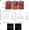
In vivo transplantation of mouse sTECE to rabbit cornea. (A): Anterior segment photographs of rabbit cornea 1, 5, 8 days after transplantation. Control eyes transplanted with grafts without endothelium showed severe edema (upper panel), while grafts with sTECE remained transparent (lower panel). (B): Change in corneal thickness after transplantation shows significant decrease in thickness due to edema in the sTECE group. (C): IOP was similar in all groups indicating that the difference in corneal thickness was not due to differences in IOP. (Normal eyes: open triangles n = 6), (control eyes: open squares n = 6), (sTECE transplanted eyes: open circles, n = 6), **, p < .01, multiple t test with Bonferroni correction after ANOVA). (D): Host‐graft junction of harvested cornea 8 days after transplantation. EGFP positive cells showed fluorescence in the host cornea. Scale bar: 100 μm (D). Abbreviations: IOP, intraocular pressure; sTECE, SKPs derived tissue engineered corneal endothelium.
Figure 5
Figure 5
Functional TECE induced from human SKPs. (A): Photograph of eyelid skin samples acquired during oculoplastic surgery. (B): Human SKPs were successfully isolated in sphere culture. SKPs sphere derived from a 44‐year‐old female (left panel), and from an 82‐year‐old male (right panel). (C): Real time RT‐PCR show that corneal endothelial markers (ATP1a1, CDH2, Pitx2) were upregulated after endothelial induction (Left bar: before induction, right bar: after induction) Student's t test (**, p < .01). (D): Immunohistochemistry of ZO1 (green) and Na, K‐ATPase (red) in hTECE (upper panel) was similar to control human corneal endothelial cell line (B4G12) (lower panel). Nuclei were labeled with DAPI (blue). (E): Ouabain‐dependent PD. hTECE showed significantly higher PD compared to hSKPs or B4G12 cells (Student's t test **, p < .01). (F): In vivo transplantation of hTECE to rabbit cornea. Anterior segment photographs of rabbit cornea 1, 2, 5, 8 days after transplantation. Control eyes transplanted with grafts without endothelium showed severe edema (upper panel), while grafts with sTECE remained transparent (lower panel). (G): Change in corneal thickness after transplantation shows significant decrease in thickness in the hTECE group (normal eyes: open triangles n = 4), (control eyes: open squares n = 4), (hTECE transplanted eyes: open circles, n = 4), **, p < .01, multiple t test with Bonferroni correction after ANOVA). (H): Change in IOP was similar in all groups indicating that the difference in corneal thickness was not due to difference in IOP. (I): Host‐graft junction of harvested cornea 8 days after transplantation of hTECE. Anti‐human Nuclei antibody (EGFP) was stained only in the transplantation site. Scale bar: 50 μm (B), Scale bar: 100 μm (C), Scale bar: 200 μm (I). Abbreviations: hTECE, TECE from human SKPs; IOP, intraocular pressure; hSKPs, human SKPs; ZO1, zonular occluding‐1; hCE, human corneal endothelium; SKPs, skin‐derived precursors; TESE, tissue engineered corneal endothelium.

References

    1. Bourne WM. Clinical estimation of corneal endothelial pump function. Trans Am Ophthalmol Soc. 1998;96:229–239; discussion 239–242. - PMC - PubMed
    1. Joyce NC. Proliferative capacity of the corneal endothelium. Prog Retin Eye Res 2003;22:359–389. - PubMed
    1. Hollingsworth J, Perez‐Gomez I, Mutalib HA et al. A population study of the normal cornea using an in vivo, slit‐scanning confocal microscope. Optom Vis Sci 2001;78:706–711. - PubMed
    1. Hatou S, Shimmura S, Shimazaki J et al. Mathematical projection model of visual loss due to fuchs corneal dystrophy. Invest Ophthalmol Vis Sci 2011;52:7888–7893. - PubMed
    1. Peh GS, Beuerman RW, Colman A et al. Human corneal endothelial cell expansion for corneal endothelium transplantation: An overview. Transplantation 2011;91:811–819. - PubMed

Publication types

MeSH terms