. 2017 Apr 25;8(17):27772-27785.
doi: 10.18632/oncotarget.15115.

An oral quinoline derivative, MPT0B392, causes leukemic cells mitotic arrest and overcomes drug resistant cancer cells

Affiliations

An oral quinoline derivative, MPT0B392, causes leukemic cells mitotic arrest and overcomes drug resistant cancer cells

Min-Wu Chao et al. Oncotarget. .

Abstract

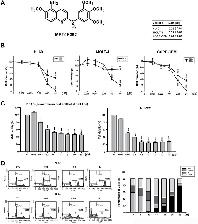
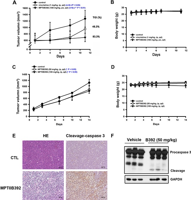
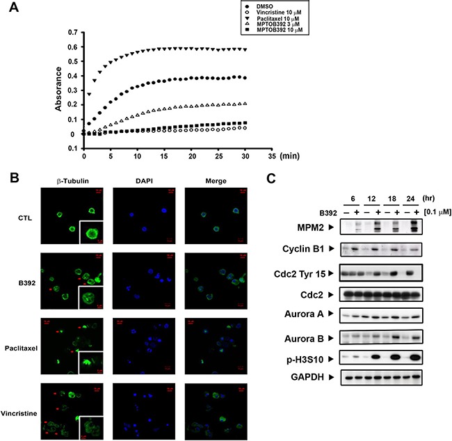
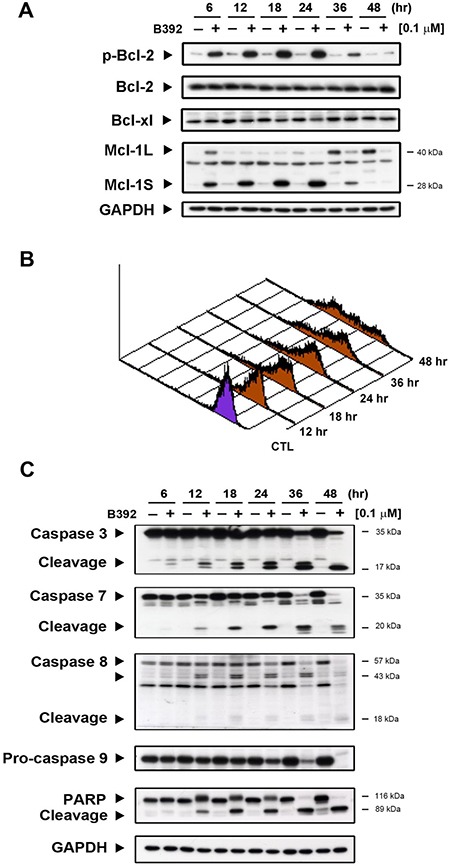
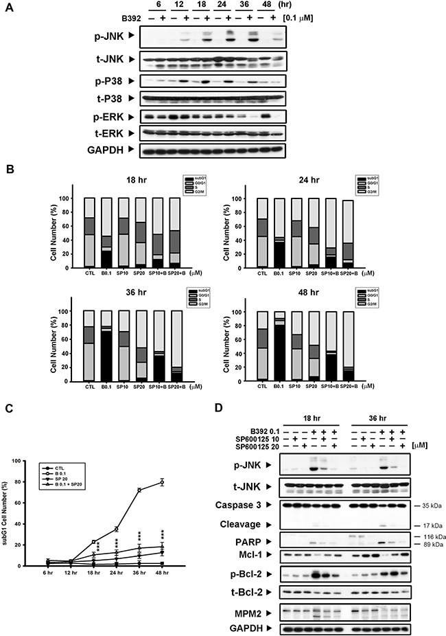
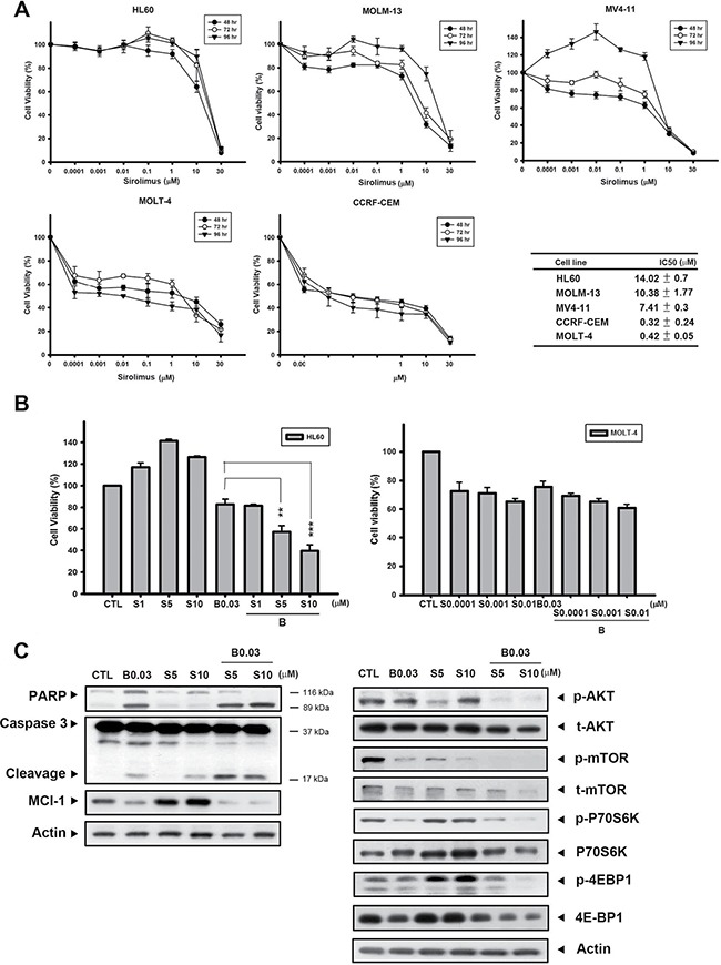
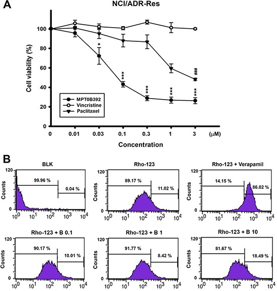
Despite great advances in the treatment of acute leukemia, a renaissance of current chemotherapy needs to be improved. The present study elucidates the underlying mechanism of a new synthetic quinoline derivative, MPT0B392 (B392) against acute leukemia and its potential anticancer effect in drug resistant cells. B392 caused mitotic arrest and ultimately led to apoptosis. It was further demonstrated to be a novel microtubule-depolymerizing agent. The effects of oral administration of B392 showed relative potent anti-leukemia activity in an in vivo xenograft model. Further investigation revealed that B392 triggered induction of the mitotic arrest, followed by mitochondrial membrane potential loss and caspases cleavage by activation of c-Jun N-terminal kinase (JNK). In addition, B392 enhanced the cytotoxicity of sirolimus in sirolimus-resistant acute leukemic cells through inhibition of Akt/mTOR pathway and Mcl-1 protein expression, and also was active in the p-glycoprotein (p-gp)-overexpressing National Cancer Institute/Adriamycin-Resistant cells with little susceptibility to p-gp. Taken together, B392 has potential as an oral mitotic drug and adjunct treatment for drug resistant cancer cells.

Keywords: MPT0B392; acute leukemia; drug resistance; mitotic arrest.


Conflict of interest statement

CONFLICTS OF INTEREST

There are no competing interests in this article.

Figures

Figure 1
Figure 1. Chemical structure of B392 and its potential anticancer effect in vitro
(A) The chemical structure of B392. (B) Cell viabilities of B392 in HL60 (acute promyelocytic leukemia), MOLT-4 (acute lymphoblastic leukemia), CCRF-CEM (acute lymphoblastic leukemia) and (C) normal cells (BEAS, human bronchial epithelial and HUVEC, human umbilical vein endothelial cell) were determined by MTT assay at 48 h. *P < 0.05, ***P < 0.001, #P < 0.05, ###P < 0.001 compared with the control group. (D) Time (right panel) and concentration-dependent (left panel) of B392 on cell cycle progression. HL60 cells were treated with vesicle (0.1% DMSO) or 0.01, 0.03, 0.1 μM of B392 for 24 and 48 h (left panel) and treated with vesicle (0.1% DMSO) or 0.1 μM of B392 for 6, 12, 18, 24, 36, 48 h (right panel). Cell cycle distribution was performed by flow cytometry. IC50 values were calculated by sigmoidal dose-response equation and expressed as mean plus minus SD at 48 h treatment.
Figure 2
Figure 2. The effect of B392 in MOLT-4 and HL60 xenograft models
SCID mice were ectopically implanted with MOLT-4 cells (A, B) or HL60 cells (C, D). Vincristine 1 mg/kg (i.p.), B392 50 mg/kg or 100 mg/kg (p.o. or i.p.) were treated. (A and C) The curves show the effect of B392 on tumor volume and percentage of tumor growth delay (TGD), which was calculated for treatment groups relative to control group. (B and D) The body weight of mice after indicated drugs treatment. (E) Immunohistochemical staining for the MOLT-4 tumors’ sections. Upper panel is the control, and lower panel is the B392 treatment group. Left panel was stained with hematoxylin and eosin; and right panel with cleavage caspase 3, which represents cells under apoptosis. Each tumor sample is under 160 ×magnification. (F) HL60 xenograft tumour homogenates were used to analyse cleavage caspase 3 protein expressions to determine apoptosis.
Figure 3
Figure 3. B392 is a depolymerizing agent and caused mitotic arrest
(A) In vitro tubulin polymerization assay was performed to evaluate the effect of B392 on microtubule dynamics. In cell-free condition, tubulin proteins were in reaction buffer in the presence or absence of B392 (3 or 10 μM), paclitaxel (10 μM) or vincristine (10 μM). Assembly of microtubules was determined by measuring absorbance at 340 nm. (B) HL60 cells were exposed to 0.1 μM B392, 10 μM vincristine and 10 μM paclitaxel for 24 h. The changes of microtubule network were visualized by staining β-tubulin as arrows indicated. Nuclear DNA was stained by DAPI. 10 μM vincristine and 10 μM paclitaxel were used as positive controls for tubulin depolymerization and tubulin polymerization, respectively. (C) HL60 cells were treated 0.1 μM B392 time-dependently to detect the expressions of G2/M related proteins.
Figure 4
Figure 4. The effect of B392 on apoptosis
(A) HL60 cells were treated with 0.1 μM B392 for indicated time then were harvested for detection of p-Bcl-2, Bcl-xl, Bcl-2, and Mcl-1 by western blot analysis. (B) The phenomena of mitochondria potential loss was measured by flow cytometry analysis with rhodamine-123. HL60 cells were treated with B392 (0.1 μM) for indicated time and then incubated with rhodamine-123 (10 μM) at 37°C for 30 min. The horizontal axis shows the relative fluorescence intensity, when the right curve shift to the left cure represents a loss of mitochondrial membrane potential. (C) HL60 cells were treated with vehicle (0.1% DMSO) or B392 (0.1 μM) for indicated times. The expressions of cleavage caspases and PARP were detected by western blot analysis. GAPDH used as an internal control.
Figure 5
Figure 5. JNK activation was involved in B392-caused cell apoptosis
(A) HL60 cells were treated with vehicle (0.1% DMSO) or B392 (0.1 μM) for time course. The cells were harvested for detection of indicated proteins by western blot analysis. (B) HL60 cells were exposed to B392 (0.1 μM) in the absence or presence of SP600125 (10, 20 μM) for 18, 24, 36 and 48 hours. Cell cycle distribution was analyzed by flow cytometry as well as determining (C) the percentage of subG1 cell number. (***P < 0.001) (D) The expression of indicated proteins was determined after treatment of HL60 with 0.1 μM B392 in the absence or presence of SP600125 (10, 20 μM) pretreatment for 30 min. SP: SP600126, p-JNK inhibitor.
Figure 6
Figure 6. B392 sensitized sirolimus anticancer activity in sirolimus-resistant cell line
(A) Acute myeloid leukemia cell lines (HL60, MV4-11, MOLM-13) and lymphoblastic cell lines (MOLT-4, CCRF-CEM) were treated with indicated concentration of sirolimus for 48, 72, 96 h. The cell viability was determined by MTT assay. The right table summarizes the IC50 of sirolimus at 72 h. IC50 values were calculated by sigmoidal dose-response equation and expressed as mean plus minus SD. (B) The cell viability of B392 combined with sirolimus at 72 h. (C) The expression of caspase 3, PARP, p-AKT, t-AKT, p-mTOR, t-mTOR, p-P70S6K, P70S6K, p-4EBP1, 4EBP, Mcl-1 and actin was determined after 0.03 μM B392 combination with 5 μM or 10 μM sirolimus for 72 h. S: sirolimus **P < 0.01, ***P < 0.001.
Figure 7
Figure 7. The cell viability of B392 on NCI/ADR-RES cell line and the effect on p-gp activity
(A) NCI/ADR-RES cells were treated with indicated concentrations of B392, vincristine or paclitaxel for 48 h. The cell viability was determined by MTT assay. (*P < 0.05, **P < 0.01, ***P < 0.001) (B) NCI/ADR-RES cells were treated with or without indicated agents (Verapamil, 50 μM; B392, 0.1, 1, 10 μM) for 1 h. After treatment with indicated drugs for 30 min, 10 μM Rhodiamine-123 was added and incubation for 30 min at 37°C. The signal of fluorescence was detected by flow cytometry. BLK, blank; Rho-123, Rhodiamine 123; B, B392.

Similar articles

Cited by

References

    1. Rose-Inman H, Kuehl D. Acute leukemia. Emerg Med Clin North Am. 2014;32:579–96. doi: 10.1016/j.emc.2014.04.004. - DOI - PubMed
    1. Douer D. Efficacy and Safety of Vincristine Sulfate Liposome Injection in the Treatment of Adult Acute Lymphocytic Leukemia. Oncologist. 2016;21:840–7. doi: 10.1634/theoncologist.2015-0391. - DOI - PMC - PubMed
    1. Al Ustwani O, Gupta N, Bakhribah H, Griffiths E, Wang E, Wetzler M. Clinical updates in adult acute lymphoblastic leukemia. Crit Rev Oncol Hematol. 2016;99:189–99. doi: 10.1016/j.critrevonc.2015.12.007. - DOI - PubMed
    1. Paul S, Kantarjian H, Jabbour EJ. Adult Acute Lymphoblastic Leukemia. Mayo Clin Proc. 2016;91:1645–66. doi: 10.1016/j.mayocp.2016.09.010. - DOI - PubMed
    1. Dumontet C, Jordan MA. Microtubule-binding agents: a dynamic field of cancer therapeutics. Nat Rev Drug Discov. 2010;9:790–803. doi: 10.1038/nrd3253. - DOI - PMC - PubMed

MeSH terms