. 2017 Apr 4;8(14):22662-22673.
doi: 10.18632/oncotarget.15153.

Inhibiting DNA-PKcs in a non-homologous end-joining pathway in response to DNA double-strand breaks

Affiliations

Inhibiting DNA-PKcs in a non-homologous end-joining pathway in response to DNA double-strand breaks

Jun Dong et al. Oncotarget. .

Abstract

DNA-dependent protein kinase catalytic subunit (DNA-PKcs) is a distinct factor in the non-homologous end-joining (NHEJ) pathway involved in DNA double-strand break (DSB) repair. We examined the crosstalk between key proteins in the DSB NHEJ repair pathway and cell cycle regulation and found that mouse embryonic fibroblast (MEF) cells deficient in DNA-PKcs or Ku70 were more vulnerable to ionizing radiation (IR) compared with wild-type cells and that DSB repair was delayed. γH2AX was associated with phospho-Ataxia-telangiectasia mutated kinase (Ser1987) and phospho-checkpoint effector kinase 1 (Ser345) foci for the arrest of cell cycle through the G2/M phase. Inhibition of DNA-PKcs prolonged IR-induced G2/M phase arrest because of sequential activation of cell cycle checkpoints. DSBs were introduced, and cell cycle checkpoints were recruited after exposure to IR in nasopharyngeal carcinoma SUNE-1 cells. NU7441 radiosensitized MEF cells and SUNE-1 cells by interfering with DSB repair. Together, these results reveal a mechanism in which coupling of DSB repair with the cell cycle radiosensitizes NHEJ repair-deficient cells, justifying further development of DNA-PK inhibitors in cancer therapy.

Keywords: DNA-PKcs; NU7441; double-strand break; nasopharyngeal carcinoma; non-homologous end-joining.


Conflict of interest statement

CONFLICTS OF INTEREST

None.

Figures

Figure 1
Figure 1. Cellular radiosensitivity depends on NHEJ after exposure to ionizing radiation (IR)
(A) Western blot analysis showed expression of Ku70 and DNA-PKcs proteins in MEF, DNA-PKcs−/− MEF, and Ku70−/− MEF cells. (B) MEF, DNA-PKcs−/− MEF, and Ku70−/− MEF cells were treated with various X-ray doses. MEF (C), DNA-PKcs−/− (D), and Ku70−/− (E) cells were exposed to IR alone or in combination with NU7441 (1 μM) for 16 h before seeding for colony formation. Clonogenic survival data are means of three independent experiments ± SD.
Figure 2
Figure 2. γH2AX foci formation is detected in MEF, DNA-PKcs−/− MEF, and Ku70−/− MEF cells
γH2AX foci were formed immediately after 5 Gy X-ray and the resolution of it delayed in NHEJ repair-deficient cells. Representative photomicrographs (×1000 magnification) shown.
Figure 3
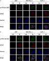
Figure 3. γH2AX is associated with phosphorylation of ATM and CHK1
MEF cells with knockdown of DNA-PKcs and Ku70 were irradiated with 5 Gy X-ray and co-immunofluorescence stained with γH2AX antibody and p-ATM (Ser1987) antibody (A), or with γH2AX antibody and p-CHK1 (Ser345) antibody (B) at 1 h post-irradiation. Nuclei were visualized with Hoechst 33342 staining. Representative photomicrographs (×1000 magnification) shown.
Figure 4
Figure 4. Deficiency in NHEJ repair results in cell cycle arrest
Cell cycle distribution in asynchronous MEF (A), DNA-PKcs−/− (B), and Ku70−/− (C) cells at indicated times post-IR (5 Gy) was assayed by flow cytometric analysis (FACS) using propidium iodide (PI) staining for DNA content. The proportion of G1, S, and G2/M cells (D) and G2/M phase arrest (E) post-IR was determined. Data are means ± SD of the percentages of cells of three independent experiments.
Figure 5
Figure 5. Inhibition of DNA-PKcs by NU7441 prolongs G2/M phase arrest
(A) Cells were pretreated with 2 μM NU7441 for 1 h and exposed to 5 Gy IR. Cell cycle progression was monitored. (B) Cell cycle distribution in asynchronous MEF was measured at 8, 16, and 24 h post-IR. (C) Percentage of cells in G2/M phase. Data are means ± SD of the percentages of cells of three independent experiments.
Figure 6
Figure 6. The ATM–CHK2 pathway interacts with the NHEJ repair pathway
Irradiation at 5 Gy up-regulated cell cycle checkpoint signaling in MEF (A), DNA-PKcs−/− (B), and Ku70−/− (C) cells. Whole cell extract was prepared from untreated cells or collected at various times after irradiation and subjected to western blot analysis. β-actin was used as the loading control. Representative image from three independent experiments is shown.
Figure 7
Figure 7. Activation of the ATR–CHK1 pathway is related to DSB repair
ATR–CHK1 pathway proteins were activated post-IR (5 Gy) in MEF (A), DNA-PKcs−/− (B), and Ku70−/− (C) cells. Whole cell extract was prepared from untreated cells or collected at various times after irradiation and subjected to western blot analysis. β-actin was used as the loading control. Representative image from three independent experiments is shown.
Figure 8
Figure 8. Inhibition of DNA repair induces radiosensitivity in nasopharyngeal carcinoma
(A) Sensitization of nasopharyngeal carcinoma to IR was determined by clonogenic survival of SUNE-1 cells. Data are means ± SD from three independent experiments. (B) Average number of γH2AX foci per cell after exposure to 5 Gy IR in SUNE-1 cells, calculated using more than 30 cells. Data are mean ± SEM. (C) Attenuation of DNA-PKcs interfered with DNA repair. The cells were fixed at 6 or 12 h after treatment with IR at 5 Gy or a combination of IR and NU7441 (2 μM). Representative photomicrographs (×1000 magnification) are shown. Average number of foci per cell (D) and average number of foci per nucleus area (mm2) (E) were calculated by counting at least 30 nuclei. Data are mean ± SEM. (F) Expression of cell cycle checkpoint proteins analyzed by western blot. Whole cell extract was prepared from untreated cells or collected at specific times after irradiation.

Similar articles

Cited by

References

    1. Liu P, Gan W, Guo C, Xie A, Gao D, Guo J, Zhang J, Willis N, Su A, Asara JM, Scully R, Wei W. Akt-mediated phosphorylation of XLF impairs non-homologous end-joining DNA repair. Mol Cell. 2015;57:648–661. - PMC - PubMed
    1. Barton O, Naumann SC, Diemer-Biehs R, Kunzel J, Steinlage M, Conrad S, Makharashvili N, Wang J, Feng L, Lopez BS, Paull TT, Chen J, Jeggo PA, et al. Polo-like kinase 3 regulates CtIP during DNA double-strand break repair in G1. J Cell Biol. 2014;206:877–894. - PMC - PubMed
    1. Felgentreff K, Du L, Weinacht KG, Dobbs K, Bartish M, Giliani S, Schlaeger T, DeVine A, Schambach A, Woodbine LJ, Davies G, Baxi SN, van der Burg M, et al. Differential role of nonhomologous end joining factors in the generation, DNA damage response, and myeloid differentiation of human induced pluripotent stem cells. Proc Natl Acad Sci USA. 2014;111:8889–8894. - PMC - PubMed
    1. Curtin NJ. DNA repair dysregulation from cancer driver to therapeutic target. Nat Rev Cancer. 2012;12:801–817. - PubMed
    1. Foulkes WD, Shuen AY. brief: BRCA1 and BRCA2. J Pathol. 2013;230:347–349. - PubMed

MeSH terms

LinkOut - more resources