. 2017 Apr 4;8(14):23507-23516.
doi: 10.18632/oncotarget.15183.

C-terminal truncated hepatitis B virus X protein regulates tumorigenicity, self-renewal and drug resistance via STAT3/Nanog signaling pathway

Affiliations

C-terminal truncated hepatitis B virus X protein regulates tumorigenicity, self-renewal and drug resistance via STAT3/Nanog signaling pathway

Rachel Hiu Ha Ching et al. Oncotarget. .

Abstract

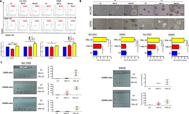
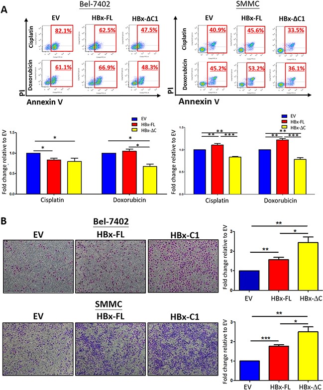
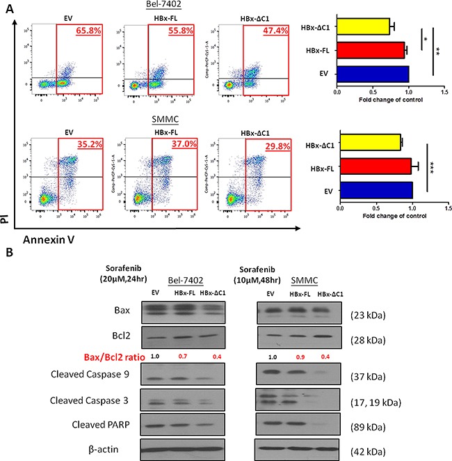
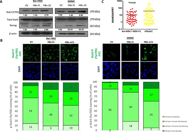
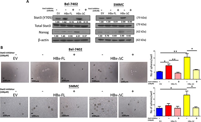
Hepatitis B virus (HBV) is a major risk factor of chronic liver disease and hepatocellular carcinoma (HCC). Random integration of HBV DNA into the host genome is frequent in HCC leading to truncation of the HBV DNA, particularly at the C-terminal end of the HBV X protein (HBx). C-terminally truncated HBx (HBx-ΔC) has been implicated in playing a pro-oncogenic role in hepatocarcinogenesis. However, the mechanism whereby HBx-ΔC1 contributes to hepatocarcinogenesis remains unclear. In this study, we investigated the functional role of HBx-ΔC1 in regulating liver cancer stem cell (CSC) properties. Using Tet-on inducible system, we found that HBx-ΔC1 enhanced CSC properties including self-renewal, tumorigenicity, chemoresistance, migration and expression of liver CSC markers, when compared with the full-length HBx counterpart and vector control. Interestingly, HBx-ΔC1 conferred resistance in HCC cells towards sorafenib treatment through suppression of apoptotic cascade. In addition, HBx-ΔC1 upregulated a panel of stemness genes, in which Nanog was found to be among the most significant one in both trasnfected cell lines. Consistently, Nanog was upregulated in human HCC samples which had HBx-ΔC1 expression. Furthermore, the induction of CSC properties by HBx-ΔC1 was via the Stat3/Nanog pathway, as administration of Stat3 inhibitor abolished the HBx-ΔC1-induced self-renewing capacity. In conclusion, our data suggest that HBx-ΔC1 enhances liver CSCs properties through Stat3/Nanog cascade, and provide a new insight for the therapeutic intervention for HBV-related HCC.

Keywords: HCC; Stat3; hepatocellular carcinoma; nanog; stemness; truncated HBx.


Conflict of interest statement

CONFLICTS OF INTEREST

The authors declare no conflicts of interest.

Figures

Figure 1
Figure 1. HBx-ΔC1 expressing cells increased expression of stemness-related genes
A. Direct protein lysates of control and HBx expressing Bel-7402 and SMMC-7721 cells were collected after adding doxycycline for 23 and 6 days respectively and confirmed that HBx-FL and HBx-ΔC1 proteins were successfully expressed in respective transfected Bel-7402 and SMMC cells, by indirect immunoblotting using c-myc antibodies to detect myc-tagged-Hbx-FL and HBx-ΔC1 protein. B. qPCR analysis revealed HBx-ΔC1 expressing Bel-7402 had increased expression of stemness-related genes including of Nanog and Sox2 after adding doxycyclines for 23 days when compared with control (EV) and HBx-FL expressing Bel-7402. After adding doxycycline for 6 days, HBx-ΔC1 expressing SMMC-7721 cells had increased mRNA expression of Nanog and Sox2 when compared with EV and HBx-FL expressing SMMC-7721. C. Western blot analysis showed that Nanog was found to be preferentially expressed in transfectants of HBx-ΔC1, when compared with those with EV and HBx-FL.
Figure 2
Figure 2. HBx-ΔC1 expressing cells possessed enhanced CSC properties
A. Flow cytometry analysis revealed increased expression of CSC markers including CD47 and CD133 in HBx-ΔC1 clones when compared with control (EV) and HBx-FL clones in both Bel-7402 and SMMC-7721 (*p<0.05, **p<0.01, Student's t test). B. Spheroid formation after 8-10 days (10X objective) showed that both HBx-ΔC1 expressing Bel-7402 and SMMC-7721 cells had increased number and size of hepatospheres formed when compared with respective control (EV) and HBx-FL expressing cells (*p<0.05, **p<0.01, ***p<0.001, Student's t test). P1 and P2 = passage 1 and 2, respectively C. Control, HBx-FL- or HBx-ΔC1 expressing Bel-7402 and SMMC-7721 cells were injected subcutaneously at 4 sites (2 in the right flank and 2 in the left flank) per NOD/SCID mouse in three different cell numbers (5000, 10000, 50000 cells). HBx-ΔC1 expressing cells exhibited increased tumor-forming incidence and size when compared with EV and HBx-FL expressing cells. For SMMC-7721, no tumor was formed in all three clones when 5000 cells were injected (data not shown).
Figure 3
Figure 3. HBx-ΔC1 conferred resistance to chemotherapeutic drugs and enhanced cell migration
A. HBx-ΔC1 overexpression conferred resistance to chemotherapeutic drugs including cisplatin and doxorubicin in both Bel-7402 and SMMC-7721, when compared to respective EV and HBx-FL, (Cisplatin: Bel-7402: 47.5% vs 82.1% and 62.5%; SMMC-7721: 33.5% vs 40.9% and 45.6%); and (Doxorubicin: Bel-7402: 48.3% vs 61.1% and 66.9%; SMMC-7721: 36.1% vs 45.2% and 53.2%) after treatment for 48hr at 0.25 and 2 μg/ml at 2% serum containing medium, 48hr at 4 and 10 μg/ml at 2% serum containing medium, respectively (*p<0.05, **p<0.01, ***p<0.001, Student's t test). B. Light micrograph of the cell migration assays, with staining of the cells in the lower chamber (4× objective), demonstrated that HBx-ΔC1 overexpression was more potent to enhance metastatic ability of cells in both Bel-7402 and SMMC-7721 clones (*p<0.05, **p<0.01, ***p<0.001, Student's t test).
Figure 4
Figure 4. HBx-ΔC1 enhanced the resistance to sorafenib-induced apoptosis
A. HBx-ΔC1 overexpression enhanced sorafenib resistance in both Bel-7402 and SMMC-7721, when compared to EV and HBx-FL expressing cells (Bel-7402: 47.4% vs 65.8% and 55.8%; SMMC-7721: 29.8% vs 35.2% and 37.0%), after treatment for 48hr at 10 and 20 μM at 5% serum containing medium respectively (*p<0.05, **p<0.01, ***p<0.001, Student's t test). B. HBx-ΔC1 overexpression reduced apoptotic signaling pathway by decreasing the Bax/Bcl2 ratio, indicator of apoptosis, the expression of activated cleaved form of caspase 9 and 3 and PARP when compared to control and HBx-FL expressing cells.
Figure 5
Figure 5. HBx-ΔC1 regulated liver CSC properties through Stat3-Nanog signaling
A. Western blot analysis revealed that HBx-ΔC1 expressing Bel-7402 and SMMC-7721 cells had increased Stat3 phosphorylation (Y705) and expression of Nanog, when compared to respective EV and HBx-FL expressing cells. B. Increased fluorescent nuclear staining of p-Stat3 (Y705) was observed in HBx-ΔC1 cells in both Bel-7402 and SMMC-7721when compared with respective EV and HBx-FL, as analyzed by immunefluorescence (IF) staining (40X objective). Stained cells were classified into 4 groups as per the fluorescent intensity measured by Photoshop CS5. C. qPCR analysis demonstrated that the expression of Nanog was up-regulated in HCC samples detected with HBx-ΔC1 (n=48) when compared with those with HBx-negative and HBx-FL (n=59) (*p=0.03, t test).
Figure 6
Figure 6. Effect of Stat3 inhibitor on HBx-induced self-renewal
A. Nanog expression was found to be down-regulated upon treatment with S3I-201 for 24hr, as analyzed by western blot analysis. B. Pre-treatment of S3I-201 at the concentration of 100 and 150μM led to abolishment of HBx-ΔC1-induced self-renewal indicated by sphere forming assay where the number and size of sphere formed (4X objective) were significantly reduced in HBx-ΔC1 expressing Bel-7402 and SMMC-7721 cells (*p<0.05, **p<0.01, Student's t test).

References

    1. Cervello M, McCubrey JA, Cusimano A, Lampiasi N, Azzolina A, Montalto G. Targeted therapy for hepatocellular carcinoma: novel agents on the horizon. Oncotarget. 2012;3:236–260. doi: 10.18632/oncotarget.466. - DOI - PMC - PubMed
    1. Tarocchi M, Polvani S, Marroncini G, Galli A. Molecular mechanism of hepatitis B virus-induced hepatocarcinogenesis. World Journal of Gastroenterolology. 2014;20:11630–11640. - PMC - PubMed
    1. Kew MC. Hepatitis B virus x protein in the pathogenesis of hepatitis B virus-induced hepatocellular carcinoma. Journal of Gastroenteroolgy and Hepatolology. 2011;26:144–152. - PubMed
    1. Yu DY, Moon HB, Son JK, Jeong S, Yu SL, Yoon H, Han YM, Lee CS, Park JS, Lee CH, Hyun BH, Murakami S, Lee KK. Incidence of hepatocellular carcinoma in transgenic mice expressing the hepatitis B virus X-protein. Journal of Hepatology. 1999;31:123–132. - PubMed
    1. Wang Y, Lau SH, Sham JS, Wu MC, Tang T, Guan XY. Characterization of HBV integrants in 14 hepatocellular carcinomas: association of truncated X gene and hepatocellular carcinogenesis. Oncogene. 2004;23:142–148. - PubMed

MeSH terms

LinkOut - more resources