. 2017 Mar 7;8(10):16851-16874.
doi: 10.18632/oncotarget.15170.

Interface between breast cancer cells and the tumor microenvironment using platelet-rich plasma to promote tumor angiogenesis - influence of platelets and fibrin bundles on the behavior of breast tumor cells

Affiliations

Interface between breast cancer cells and the tumor microenvironment using platelet-rich plasma to promote tumor angiogenesis - influence of platelets and fibrin bundles on the behavior of breast tumor cells

Sheila Siqueira Andrade et al. Oncotarget. .

Abstract

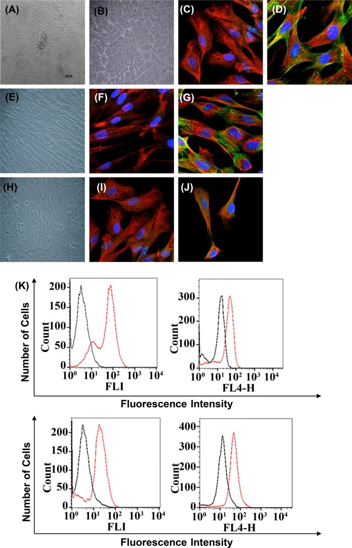
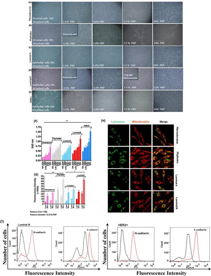
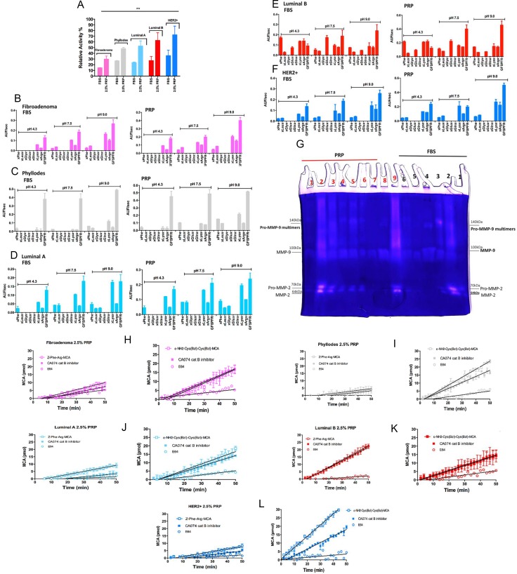
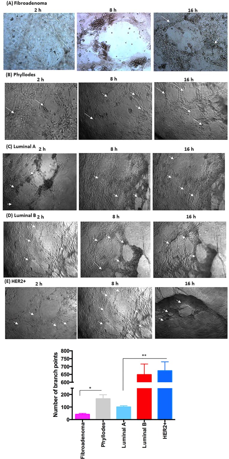
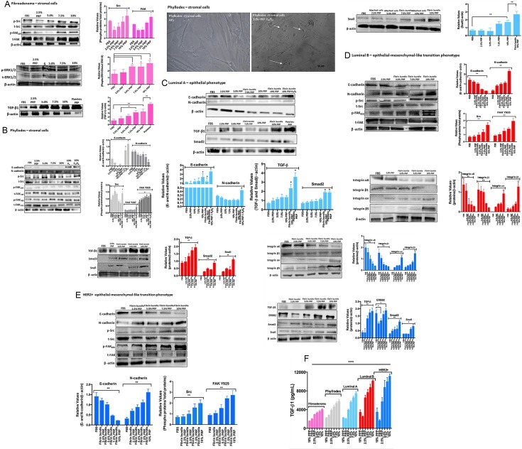
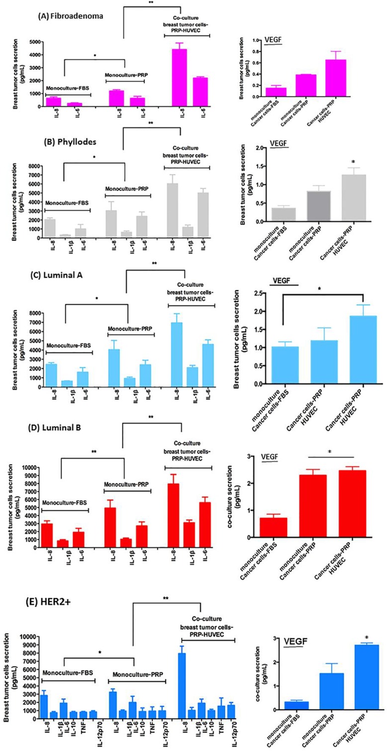
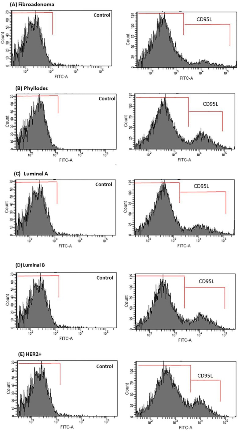
Cancer progression is associated with an evolving tissue interface of direct epithelial-tumor microenvironment interactions. In biopsies of human breast tumors, extensive alterations in molecular pathways are correlated with cancer staging on both sides of the tumor-stroma interface. These interactions provide a pivotal paracrine signaling to induce malignant phenotype transition, the epithelial-mesenchymal transition (EMT). We explored how the direct contact between platelets-fibrin bundles primes metastasis using platelet-rich plasma (PRP) as a source of growth factors and mimics the provisional fibrin matrix between actively growing breast cancer cells and the tumor stroma. We have demonstrated PRP functions, modulating cell proliferation that is tumor-subtype and cancer cell-type-specific. Epithelial and stromal primary cells were prepared from breast cancer biopsies from 21 women with different cancer subtypes. Cells supplemented with PRP were immunoblotted with anti-phospho and total Src-Tyr-416, FAK-Try-925, E-cadherin, N-cadherin, TGF-β, Smad2, and Snail monoclonal antibodies. Breast tumor cells from luminal B and HER2 subtypes showed the most malignant profiles and the expression of thrombin and other classes of proteases at levels that were detectable through FRET peptide libraries. The angiogenesis process was investigated in the interface obtained between platelet-fibrin-breast tumor cells co-cultured with HUVEC cells. Luminal B and HER2 cells showed robust endothelial cell capillary-like tubes ex vivo. The studied interface contributes to the attachment of endothelial cells, provides a source of growth factors, and is a solid substrate. Thus, replacement of FBS supplementation with PRP supplementation represents an efficient and simple approach for mimicking the real multifactorial tumor microenvironment.

Keywords: breast cancer; cancer; platelet-rich plasma; platelets; tumor microenvironment.


Conflict of interest statement

CONFLICTS OF INTEREST

The authors declare that they have no conflicts of interest to disclose.

Figures

Figure 1
Figure 1. Immunophenotype of breast cancer cells from patients with fibroadenoma and phyllodes tumors (non-cancerous breast condition), luminal A and B, and HER2+ breast cancer subtypes used in all experiments
Cells are shown under phase contrast microscopy and indirect immunofluorescence for vimentin, cytokeratin, E-cadherin, N-cadherin, PAI-1, claudin 1, phalloidin, and DAPI (blue for nuclei). (A) Phase contrast–confluent culture of organoid tumor cells after 3 days. (B) Confluent epithelial breast tumor cells after 2 days in culture. Mycoplasma contamination was not observed in any of the processed tissues. (CD) Analysis of epithelial and mesenchymal markers by confocal microscopy; cytokeratin, vimentin, and phalloidin. (E) Confluent stromal breast tumor cells after 2 days in culture. (FG) Positivity for the mesenchymal marker vimentin by confocal microscopy. We observed the cytoskeletal organization pattern when using phalloidin. (IJ) Breast cancer cells in epithelial-mesenchymal transition. Analysis of epithelial and mesenchymal markers by confocal microscopy; cytokeratin, vimentin, and phalloidin-cytoskeleton. (K) Flow cytometry histograms representative of E-cadherin, N-cadherin, PAI-1, and claudin 1 on tumor cells with EMT. The histogram on the left represents a control staining using an isotype-matched control antibody. These experiments were performed with cultured cells from all specimens collected from patients.
Figure 2
Figure 2. Morphology of stromal and epithelial cells during gradual PRP supplementation
(AB) The primary culture of stromal breast tumor cells from patients with fibroadenoma and phyllodes benign breast tumors developed more spindle-shaped morphology and proliferated faster than cells cultured in FBS. (CE) Primary culture of epithelial breast tumor cells from patients with luminal A and B and HER2+ breast cancer subtypes developed more prominent polygonal shape, proliferated faster, and migrated in higher amounts from the plastic surface to fibrin bundles than cells cultured in FBS. (F) Cell viability by the MTT assay. Stromal and epithelial breast tumor cells (≈2 × 10³ densities) were cultured in the appropriate medium without phenol red and supplemented with specifically 2.5% PRP for 24 h in 96-well microtiter plates. (G and H) MitoTracker Orange, LysoTracker Green, and DAPI fluorescent micrographs of stromal and epithelial breast tumor cells specifically supplemented with 5.0% PRP. Total lysosomal and mitochondrial amount. Scale bar = 20 μm. Quantification of fluorescence intensity of LysoTracker and MitoTracker in living cells to evaluate the number of lysosomes and mitochondria numerically in each cell. (I) Representative flow cytometry histograms of E-cadherin and N-cadherin on HER2+ epithelial breast tumor cells with EMT supplemented with PRP. The histogram on the left represents a control staining using an isotype-matched control antibody. These experiments were performed with cultured cells from all specimens collected from patients.
Figure 3
Figure 3. Detection of proteolytic activity on CM with 2.5% PRP supplementation of stromal and epithelial breast tumor cells
(A) Evaluation of degradation of the Benzoyl-Phe-Val-Arg-MCA substrate, a specific substrate for α-thrombin. Epithelial and stromal-CM with 2.5% PRP supplementation were compared with FBS supplementation (both conditions were incubated at 37°C for 16 h). (BF) Screening of endopeptidase activities presents in epithelial and stromal-CM with PRP supplementation. The rate of hydrolysis of the Abz-GXXZXXQ-EDDnp peptide sublibraries by epithelial and stromal-CM is shown under three pH conditions: 4.3, 7.5, and 9.0. The endopeptidase activity was measured for each sublibrary in which “Z” was either phenylalanine (XPheX), leucine (XLeuX), glycine (XGlyX), glutamate (XGluX), glutamine (XGlnX), lysine (XLysX), or arginine (XArgX). The velocity of hydrolysis was measured as Arbitrary Units of Fluorescence (AUF)/sec. (G) Zymography for MMP-2 and MMP-9 in epithelial and stromal-CM cells isolated from women with breast cancer. Cell culture in monolayers and co-cultures (stromal and epithelial breast tumor cells from the same patient). Samples: FBS (black) – 1) Fibroadenoma, 2) Phyllodes, 3) Luminal A, 4) Luminal B co-culture, 5) Luminal B, 6) HER2+; PRF (red) 1) Fibroadenoma, 2) Phyllodes, 3) Luminal A (2 × concentrated – 20 μg), 4) Luminal A (1 × concentrated – 10 μg), 5) Luminal A co-culture, 6) Luminal B co-culture, 7) Luminal B, 8) HER2+ and 9) Her2+ co-culture. MMP-9 multimers formation was previously described [32]. (HL) Cysteine protease activities in stromal and epithelial-CM cells from women with different breast cancer subtypes. Approximately 2 × 10⁵ cells were cultivated for 3 days; the enzymatic activity was measured in CM submitted to PRP supplementation (in the absence of phenol red) and removed from each well using stromal and epithelial cells; Z-FR-MCA (20 μM) (200 μL final volume) and stromal and epithelial cells; ε-NH2-caproyl-Cys(Bzl)- Cys(Bzl)-MCA (20 μM) in 200 μL final volume. A maximum of 10% substrate consumption was considered; each point represents the mean ± 95% confidence interval of two replicates. The presence of PMSF did not affect the total activity of proteases. Inhibitor concentrations: PMSF = 1 mM; CA074 = 1 μM; E64 = 5 μM. These experiments were performed with cultured cells from all specimens collected from patients.
Figure 5
Figure 5. Co-culture of breast tumor cells under PRP supplementation and the formation capillary-structure tubes induced by HUVEC
(A) Fibroadenoma stimulates minimally cluttered capillary tube formation. (B) Phyllodes showed enhanced angiogenesis-promoting ability. (C) Luminal A promoted a slight formation of angiogenic structures in HUVEC. (D and E) In luminal B and HER2+, the capillary tube structure was stable and defined, and 6.5-fold higher than luminal A. Arrows indicate the formation of angiogenic structures. Quantification of HUVEC branch points over an 8–16 hour time course (see Methods). (F) Measurement of the angiogenic structure formation (per well). These experiments were performed with cultured primary cells from all specimens collected from patients. Significant differences versus controls are presented (ANOVA; *p ≤ 0.05, **p ≤ 0.001). Inverted microscope images (× 40 magnification). Scale bars 20 μm.
Figure 4
Figure 4. Stromal and epithelial breast tumor cells under PRP supplementation (including cells attached to the plastic surface and cells attached to the network of fibrin bundles) were assessed by immunoblotting
(A) Fibroadenoma stromal cells under PRP supplementation showed increased phospho-Src, phosphos-FAK(Y)397, and phospho-ERK1/2, and upregulated TGF-β1 expression. The graph represents the densitometry analyses from immunoblotting results. (B) Phyllodes stromal cells under PRP supplementation showed EMT reversion and upregulation of E-cadherin and downregulation of N-cadherin expression after treatment with P4 and the E2+P4 combination. Phospho-Src and phospho-FAK(Y)925, but not FAK(Y)397, showed increased phosphorylation. Phase contrast–confluent culture of phyllodes stromal cells showed morphology alteration in the presence of steroid hormones under PRP supplementation. (CE) Luminal A and B and HER2+ epithelial cells under PRP supplementation showed EMT process with downregulation of E-cadherin and upregulation of N-cadherin expression. Phospho-Src and phospho-FAK(Y)925, but not FAK(Y)397, showed increased phosphorylation, and the α6β1 integrin subunits showed decreased expression. Conversely, the αvβ5 integrin subunits showed increased expression. The analysis of the TGF-β, Smad2, and Snail metastasis markers showed increased expression in the highest PRP concentrations. The results are represented as band intensities in arbitrary units relative to the respective total load of total proteins and control (β-actin). The statistical significance was evaluated using one-way ANOVA followed by the Tukey's test. These experiments were performed with cultured primary cells from specimens collected from patients, *p ≤ 0.05, **p ≤ 0.001. (F) Concentration of total TGF-β in CM from breast tumor cells. The conditioned medium was collected, centrifuged to remove platelets, and the presence of TGFβ1 in the supernatant measured by ELISA. Each bar represents the mean ± SEM of n = 2–6. ***p < 0.001 as determined by Student's t test.
Figure 6
Figure 6. Relative levels of indicated cytokines in CM of co-culture of breast tumor cells under PRP supplementation and HUVEC
(AE) The conditioned medium was recovered to measure protein secretion by flow cytometer in co-cultured cells in pg/ml). (FJ) VEGF concentration in CM after 8–16-hour culture measured by flow cytometry (human VEGF flex set’, BDTM cytometric bead array, CBA, BD Biosciences, USA). Values are the means ± SEM of duplicate determinations in co-cultures from all specimens collected from patients with fibroadenoma, phyllodes, luminal A and B, and HER2+ *p ≤ 0.05, **p ≤ 0.001.
Figure 7
Figure 7. CD95L secretion is enhanced in CM of co-cultured breast tumor cells under PRP supplementation-HUVEC
(AE) After 16 h, media were removed and analyzed for CD95L by flow cytometry in the Accuri C6 (BD Biosciences, USA). Values are the means ± SEM of duplicate determinations in co-cultures from all specimens collected from patients with fibroadenoma, phyllodes, luminal A and B, and HER2+*p ≤ 0.05.

Similar articles

Cited by

References

    1. Kuznetsov HS, Marsh T, Markens BA, Castaño Z, Greene-Colozzi A, Hay SA, Brown VE, Richardson AL, Signoretti S, Battinelli EM, McAllister SS. Identification of luminal breast cancers that establish a tumor-supportive macroenvironment defined by proangiogenic platelets and bone marrow-derived cell. Cancer Discov. 2012;2:1150–65. - PMC - PubMed
    1. Shen K, Luk S, Hicks DF, Elman JS, Bohr S, Iwamoto Y, Murray R, Pena K, Wang F, Seker E, Weissleder R, Yarmush ML, Toner M, et al. Resolving cancer-stroma interfacial signalling and interventions with micropatterned tumour-stromal assays. Nat Commun. 2014;5:5662. - PMC - PubMed
    1. Scheel C, Onder T, Karnoub A, Weinberg RA. Adaptation versus selection: the origins of metastatic behavior. Cancer Res. 2007;2:11476–9. - PubMed
    1. Labelle M, Begum S, Hynes RO. Direct signaling between platelets and cancer cells induces an epithelial-mesenchymal-like transition and promotes metastasis. Cancer Cell. 2011;20:576–590. - PMC - PubMed
    1. Angelucci C, Maulucci G, Lama G, Proietti G, Colabianchi A, Papi M, Maiorana A, De Spirito M, Micera A, Balzamino OB, Di Leone A, Masetti R, Sica G. Epithelial-stromal interactions in human breast cancer: effects on adhesion, plasma membrane fluidity and migration speed and directness. PLoS One. 2012;7:e50804. - PMC - PubMed