. 2017 Feb 10;5(1):20.
doi: 10.1186/s40168-017-0234-1.

Sputum DNA sequencing in cystic fibrosis: non-invasive access to the lung microbiome and to pathogen details

Affiliations

Sputum DNA sequencing in cystic fibrosis: non-invasive access to the lung microbiome and to pathogen details

Rounak Feigelman et al. Microbiome. .

Abstract

Background: Cystic fibrosis (CF) is a life-threatening genetic disorder, characterized by chronic microbial lung infections due to abnormally viscous mucus secretions within airways. The clinical management of CF typically involves regular respiratory-tract cultures in order to identify pathogens and to guide treatment. However, culture-based methods can miss atypical or slow-growing microbes. Furthermore, the isolated microbes are often not classified at the strain level due to limited taxonomic resolution.

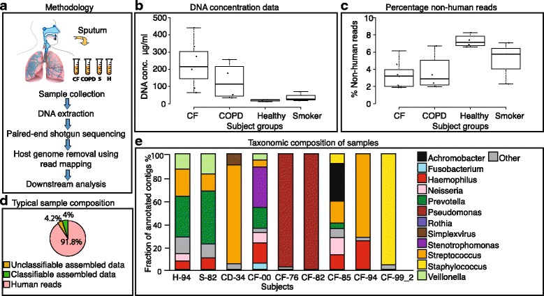
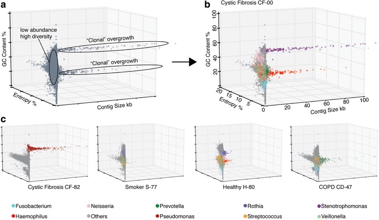
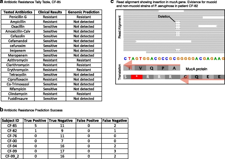
Results: Here, we show that untargeted metagenomic sequencing of sputum DNA can provide valuable information beyond the possibilities of culture-based diagnosis. We sequenced the sputum of six CF patients and eleven control samples (including healthy subjects and chronic obstructive pulmonary disease patients) without prior depletion of human DNA or cell size selection, thus obtaining the most unbiased and comprehensive characterization of CF respiratory tract microbes to date. We present detailed descriptions of the CF and healthy lung microbiome, reconstruct near complete pathogen genomes, and confirm that the CF lungs consistently exhibit reduced microbial diversity. Crucially, the obtained genomic sequences enabled a detailed identification of the exact pathogen strain types, when analyzed in conjunction with existing multi-locus sequence typing databases. We also detected putative pathogenicity islands and indicators of antibiotic resistance, in good agreement with independent clinical tests.

Conclusions: Unbiased sputum metagenomics provides an in-depth profile of the lung pathogen microbiome, which is complementary to and more detailed than standard culture-based reporting. Furthermore, functional and taxonomic features of the dominant pathogens, including antibiotics resistances, can be deduced-supporting accurate and non-invasive clinical diagnosis.

Keywords: COPD; Cystic fibrosis; Lung metagenome; Sputum; WGS metagenomic sequencing.


Figures

Fig. 1
Fig. 1
Sputum metagenomics workflow. a Overview of the procedure. b Concentration of extractable DNA in sputum, across subject groups. c Fraction of non-human DNA sequence reads across subject groups. d Fraction of DNA sequence reads of a representative healthy sample, further broken down according to taxonomic assignability to the assembled nucleotides from non-human fraction. e Taxonomic composition of all taxonomically assignable, non-human sequences, at genus level (for each of the control groups, only one representative sample is shown). All genera constituting at least 4.5% of the annotated fraction in each sample are assigned with a color code
Fig. 2
Fig. 2
Pathogen overgrowth can be separated from background diversity. Sequence contig feature plots (“entropy landscapes”), depicting at least one sample from every subject group. Each data point represents an assembled contig, with colors corresponding to genus level taxonomy annotations. The three axes show contig length (X-axis), contig sequence heterogeneity (entropy, Y-axis), and GC-content (Z-axis). a Magnified view of the plot of patient CF-00 without taxonomic annotation. b The same plot (CF-00) but with taxonomic annotation. c Representative plots of one subject from each group. Throughout, genera constituting less than 5% of the annotated fraction, as well as unannotated contigs are shown in gray color
Fig. 3
Fig. 3
High-precision strain typing from sputum sequences. Multi-locus sequence typing for two selected pathogen strains from CF samples. Yellow color highlights the phylogenetic position of the strains observed in this study, relative to previously typed strains deposited in MLST databases. a Patient CF-00 is colonized by a S. maltophilia strain that has close relatives in the database. Isolation sources of database strains are shown color-coded. b Patient CF-85 has a strain from the genus Achromobacter, for which no close relatives have been observed before (the strain likely does not belong to a named species). All monophyletic clades with 95% members from a single species have been collapsed
Fig. 4
Fig. 4
Prediction of antibiotic resistances and other phenotypes. a The Achromobacter strain isolated from patient CF-85 underwent routine clinical testing for antibiotic sensitivity; the compounds tested and the observed results are shown. This is contrasted with automated predictions based on the gene content of the sputum sequence data. b Summary table for all CF subjects, indicating the overlap between the resistance predictions and the clinical test results. c Read alignment against a section of the mucA gene from P. aeruginosa, from patient CF-82. Eleven reads show a wild-type sequence at this position, but 7 reads show a deletion event predicting a non-functional protein and a corresponding shift from a non-mucoid to a mucoid phenotype in this strain
Fig. 5
Fig. 5
Pathogen genome comparisons reveal patient-specific additions. Two public reference genomes of S. maltophilia are compared against assembled contigs from patient CF-00. a Genome-wide alignment showing blocks of colinearity, additions, and deletions. White stretches indicate unalignable, unique regions in each genome. Vertical red lines separate individual assembled contigs. b Magnified view centered on a genomic region that is unique to the strain in patient CF-00. Genes with homology to type six secretion system (T6SS) have been labeled with numerical IDs (see panel c below). Genes marked with an asterisk showed no detectable homology in sequence databases. c The core gene cluster of T6SS is depicted in yellow; additional accessory T6SS genes which are also observed in patient CF-00 are colored in gray. d Schematic model of the T6SS protein structure based on present knowledge

References

    1. Walters S, Mehta A. Epidemiology of cystic fibrosis. In: Hodson M, Geddes DM, Bush A, editors. Cystic fibrosis, 3rd edn. London: Edward Arnold Ltd; 2007. p. 21–45.
    1. Boucher RC. An overview of the pathogenesis of cystic fibrosis lung disease. Adv Drug Deliv Rev. 2002;54(11):1359–71. doi: 10.1016/S0169-409X(02)00144-8. - DOI - PubMed
    1. Mahenthiralingam E. Emerging cystic fibrosis pathogens and the microbiome. Paediatr Respir Rev. 2014;15:13–5. - PubMed
    1. Bell SC, De Boeck K, Amaral MD. New pharmacological approaches for cystic fibrosis: promises, progress, pitfalls. Pharmacol Ther. 2015;145:19–34. doi: 10.1016/j.pharmthera.2014.06.005. - DOI - PubMed
    1. Laura GAO, Filkins M. Cystic fibrosis lung infections: polymicrobial, complex, and hard to treat. 2015. pp. 1–8. - PMC - PubMed

Publication types