. 2017 Jun;91(6):1398-1409.
doi: 10.1016/j.kint.2016.12.010. Epub 2017 Feb 7.

The renal TRPV4 channel is essential for adaptation to increased dietary potassium

Affiliations

The renal TRPV4 channel is essential for adaptation to increased dietary potassium

Mykola V Mamenko et al. Kidney Int. 2017 Jun.

Abstract

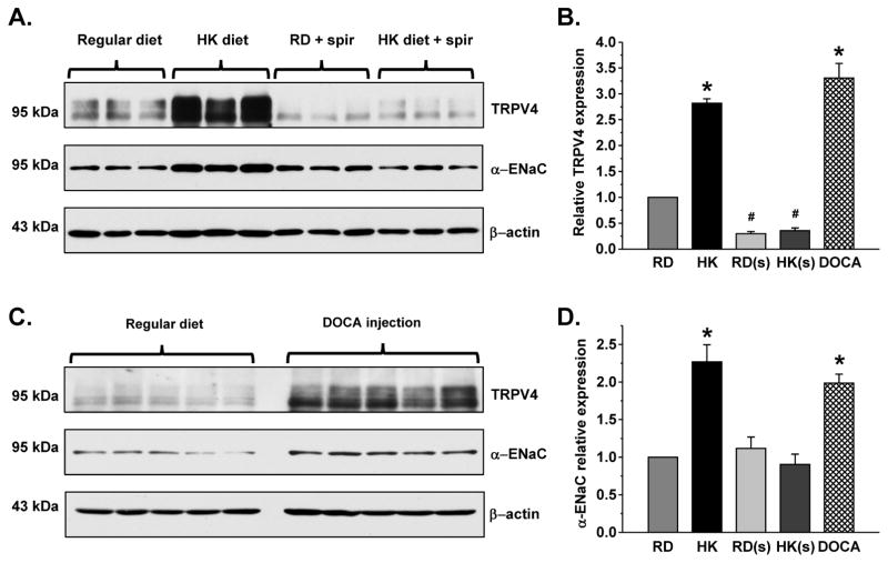
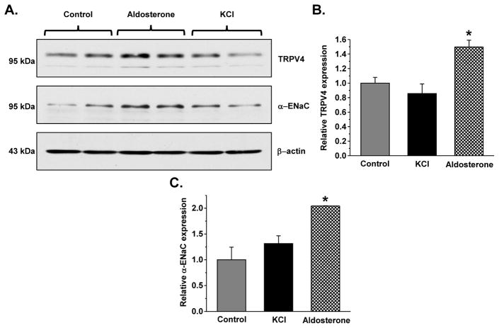
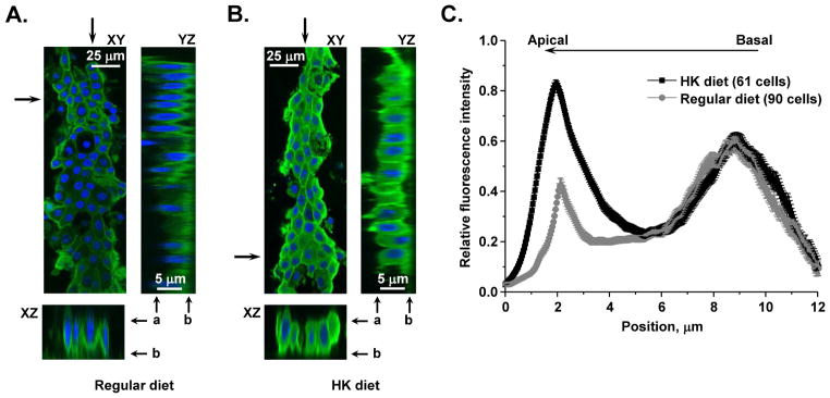
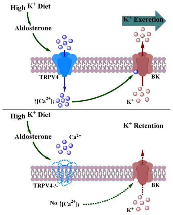
To maintain potassium homeostasis, kidneys exert flow-dependent potassium secretion to facilitate kaliuresis in response to elevated dietary potassium intake. This process involves stimulation of calcium-activated large conductance maxi-K (BK) channels in the distal nephron, namely the connecting tubule and the collecting duct. Recent evidence suggests that the TRPV4 channel is a critical determinant of flow-dependent intracellular calcium elevations in these segments of the renal tubule. Here, we demonstrate that elevated dietary potassium intake (five percent potassium) increases renal TRPV4 mRNA and protein levels in an aldosterone-dependent manner and causes redistribution of the channel to the apical plasma membrane in native collecting duct cells. This, in turn, leads to augmented TRPV4-mediated flow-dependent calcium ion responses in freshly isolated split-opened collecting ducts from mice fed the high potassium diet. Genetic TRPV4 ablation greatly diminished BK channel activity in collecting duct cells pointing to a reduced capacity to excrete potassium. Consistently, elevated potassium intake induced hyperkalemia in TRPV4 knockout mice due to deficient renal potassium excretion. Thus, regulation of TRPV4 activity in the distal nephron by dietary potassium is an indispensable component of whole body potassium balance.

Keywords: BK channels; [Ca(2+)](i) signaling; aldosterone; distal nephron; flow-induced potassium secretion.


Figures

Figure 1
Figure 1. TRPV4 expression in the kidney depends on dietary potassium intake
(A) Representative Western blots from whole kidney lysates of C57BL/6 mice kept on regular (0.9% K+) and high (5% K+, HK) potassium diet for one week. The lysates were probed with anti-TRPV4 and anti-β-actin antibodies, respectively. The channel appears as a duplet of upper glycosylated and lower non-glycosylated forms. (B) Summary graph comparing TRPV4 expression from Western blots similar to that shown in (A). Intensities of the TRPV4-reporting reporting bands were normalized to the intensities of the respective actin bands. (C) Summary graph of relative TRPV4 mRNA levels in the kidney as detected by RT q-PCR in mice treated with regular (0.9% K+) and high (5% K+) potassium diet for one week. Mean TRPV4 cycle threshold values were normalized to the respective HPRT cycle threshold values. * - significant increase versus regular diet.
Figure 2
Figure 2. Aldosterone-MR signaling is essential for regulation of TRPV4 in the kidney by dietary K+ intake
(A) Representative Western blots of whole kidney lysates from C57BL/6 mice kept on regular (0.9% K+) and high (5% K+) potassium (HK) diet for one week in the absence and presence of spironolactone (spir, 30 30 mg/kgBW) probed with anti-TRPV4 (upper), α-ENaC (middle), and β-actin (lower). (B) Summary graph comparing renal TRPV4 expression normalized to the respective intensity of β-actin in mice on regular diet (RD), high K+ diet (HK), regular diet + spironolactone (RD(s)), high K+ diet + spironolactone (HK(s)), and regular diet injected with Deoxycorticosterone acetate (DOCA). (C) Representative Western blots of whole kidney lysates from C57BL/6 mice kept on regular diet and injected with DOCA for 3 consecutive days (2.4 mg/injection/animal) probed with anti-TRPV4 (upper), α-ENaC (middle), and β-actin (lower). (D) Summary graph comparing full length α-ENaC expression normalized to the respective intensity of β-actin in mice on the same conditions as described in (B). * - significant increase versus RD; # - significant decrease versus RD.
Figure 3
Figure 3. Aldosterone but not extracellular K+ increases TRPV4 abundance in mpkCCDc14 cells
(A) Representative Western blots from mpkCCDc14 lysates in the control, after treatment with 1 μM aldosterone or addition of 5 mM KCl from the basolateral side for 24 hours probed with anti-TRPV4 (upper), α-ENaC (middle), and β-actin (lower). Summary graphs comparing TRPV4 (B) and full length α-ENaC (C) expression normalized to the respective intensity of β-actin in mice on the same conditions as described in (A). * - significant increase versus control.
Figure 4
Figure 4. High K+ dietary intake induces apical TRPV4 accumulation in native CD cells
(A) A representative confocal micrograph demonstrating TRPV4 abundance and distribution (pseudocolor green) in the CD from C57BL/6 mouse kept on regular (0.9 % K+) intake. Here and below, representative XZ and YZ plane projections (location of the cross-section is marked by arrows), showing TRPV4 distribution along the basal-apical axis, were reconstructed from Z-stacks of confocal images. Nuclear DAPI staining is shown in pseudocolor blue. Position of the apical and basal sides is shown with “a” and “b”, respectively. (B) A representative confocal micrograph visualizing TRPV4 abundance and distribution (pseudocolor green) in the CD from C57BL/6 mouse kept on a high potassium (5% K+) diet. (C) Distribution of averaged relative fluorescent signals representing TRPV4 localization along Z-axis in CD cells from C57BL/6 mice kept on regular and high potassium intake. For each individual cell, the fluorescent signal was normalized to its corresponding maximal value. At least 6 CDs from 3 different mice were used to obtain statistics for any given treatment.
Figure 5
Figure 5. High dietary potassium intake augments TRPV4-dependent [Ca2+]i elevations in the CD cells
(A) The average time course of relative changes in Fura 2 F340/F380 ratio in split-opened CDs in response to 10x elevation in flow (shown with a bar on top) from apical side. CDs were isolated from WT mice kept on control (squares) and high potassium (circles) diet, as well as TRPV4 −/− mice kept on control (triangles) and high potassium (inverted triangles) diet. The insert contains representative micrographs of a typical split-opened CD after loading with Fura-2 taken with bright-field illumination (left) and 380 nm excitation (right). (B) The summary graph of the magnitudes of flow-induced Ca2+ responses in CD cells for the groups in (A). * - significant increase versus control.
Figure 6
Figure 6. TRPV4 −/− mice have impaired BK channel activity in the CD
(A) Representative current traces of single channel BK activity in CD cells from WT (top) and TRPV4−/− mice (bottom) kept on high (5% K+) potassium diet for 1 week. The patches were held at a test potential of Vh=−Vp=+100 mV. Outward K+ currents are upward. Dashed lines indicate the respective current state with c denoting the closed state. (B) Pie charts representing the frequency of observing patches with active channels (f) for the conditions described in (A). (C) Summary graph of functional BK levels (fN) for WT and TRPV4 −/− mice kept on high potassium diet. * – significant decrease vs WT.
Figure 7
Figure 7. TRPV4−/− mice exhibit impaired adaptation to elevated potassium intake
(A) Summary graph of plasma K+ levels in WT (light grey) and TRPV4 −/− (dark grey) mice kept on control and elevated K+ (5%) diet for 7 days. (B) Summary graph of K+ excretion with feces in WT (light grey) and TRPV4 −/− (dark grey) mice kept on control and elevated K+ (5%) diet for 7 days. (C) Summary graph of plasma aldosterone concentrations in WT (light grey) and TRPV4 −/− (dark grey) on low (0.01%) and high (5%) K+ diet for 7 days. * - significant increase vs WT HK diet.
Figure 8
Figure 8. Renal potassium excretion is delayed in TRPV4−/− mice fed a high K+ diet
Summary graph visualizing K+ excretion with urine in WT (light grey) and TRPV4 −/− (dark grey) mice maintained on a regular K+ diet and in response to elevated potassium load (5% K+) for 1 or 2 days. * – significant decrease vs a respective value in WT mice.
Figure 9
Figure 9
The proposed role of TRPV4 in regulation of BK-mediated K+ secretion during high potassium intake.

References

    1. Youn JH, McDonough AA. Recent advances in understanding integrative control of potassium homeostasis. Annu Rev Physiol. 2009;71:381–401. - PMC - PubMed
    1. Palmer BF. Regulation of Potassium Homeostasis. Clinical journal of the American Society of Nephrology : CJASN. 2015;10:1050–1060. - PMC - PubMed
    1. Kovesdy CP. Management of hyperkalaemia in chronic kidney disease. Nature reviews Nephrology. 2014;10:653–662. - PubMed
    1. Wang WH, Giebisch G. Regulation of potassium (K) handling in the renal collecting duct. Pflugers Archiv : European journal of physiology. 2009;458:157–168. - PMC - PubMed
    1. Hebert SC, Desir G, Giebisch G, et al. Molecular diversity and regulation of renal potassium channels. Physiological reviews. 2005;85:319–371. - PMC - PubMed

Publication types

MeSH terms

LinkOut - more resources