. 2017 Apr 1;312(4):L556-L567.
doi: 10.1152/ajplung.00349.2016. Epub 2017 Feb 10.

Lung pericyte-like cells are functional interstitial immune sentinel cells

Affiliations

Lung pericyte-like cells are functional interstitial immune sentinel cells

Chi F Hung et al. Am J Physiol Lung Cell Mol Physiol. .

Abstract

Pericytes are perivascular PDGF receptor-β+ (PDGFRβ+) stromal cells required for vasculogenesis and maintenance of microvascular homeostasis in many organs. Because of their unique juxtaposition to microvascular endothelium, lung PDGFRβ+ cells are well situated to detect proinflammatory molecules released following epithelial injury and promote acute inflammatory responses. Thus we hypothesized that these cells represent an unrecognized immune surveillance or injury-sentinel interstitial cell. To evaluate this hypothesis, we isolated PDGFRβ+ cells from murine lung and demonstrated that they have characteristics consistent with a pericyte population (referred to as pericyte-like cells for simplicity hereafter). We showed that pericyte-like cells expressed functional Toll-like receptors and upregulated chemokine expression following exposure to bronchoalveolar lavage fluid (BALF) collected from mice with sterile lung injury. Interestingly, BALF from mice without lung injury also induced chemokine expression in pericyte-like cells, suggesting that pericyte-like cells are primed to sense epithelial injury (permeability changes). Following LPS-induced lung inflammation, increased numbers of pericyte-like cells expressed IL-6, chemokine (C-X-C motif) ligand-1, chemokine (C-C motif) ligand 2/ monocyte chemotactic protein-1, and ICAM-1 in vivo. Sterile lung injury in pericyte-ablated mice was associated with decreased inflammation compared with normal mice. In summary, we found that pericyte-like cells are immune responsive and express diverse chemokines in response to lung injury in vitro and in vivo. Furthermore, pericyte-like cell ablation attenuated inflammation in sterile lung injury, suggesting that these cells play an important functional role in mediating lung inflammatory responses. We propose a model in which pericyte-like cells function as interstitial immune sentinels, detecting proinflammatory molecules released following epithelial barrier damage and participating in recruitment of circulating leukocytes.

Keywords: acute lung injury; damage-associated molecular patterns; inflammation; innate immunity; pericytes.


Figures

Fig. 1.
Fig. 1.
Confocal imaging of normal mouse lung tissue. Fresh frozen lung sections were fixed and labeled for pericyte-associated antigens. A: lung sections from a C57BL/6 mouse with immunofluorescence for PDGFRβ (green) and the endothelial cell-associated antigen CD31/PECAM (red) and counterstained with DAPI (blue). B: magnification of selected area from A, demonstrating close proximity without colocalization of PDGFRβ+ and CD31+ cells. C: immunofluorescence for PDGFRβ (green) in a C57BL/6 mouse with DAPI counterstain (blue). D: immunofluorescence for CD146 (red) of the same tissue section as C. E: merge of C and D, demonstrating colocalization of PDGFRβ and CD146. F: immunofluorescence for PDGFRβ (green) in an NG2-dsRed mouse, which expresses dsRed fluorescent protein under the regulation of the NG2 promoter. A subset of PDGFRβ+ cells are also NG2+.
Fig. 2.
Fig. 2.
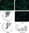
Primary, cultured pericyte-like lung cells. PDGFRβ+ cells were isolated from C57BL/6 mouse whole lung digest and expanded over two passages. Immunofluorescence for the pericyte-associated markers PDGFRβ (A), CD146 (B), and NG2 (D) is shown. C: representative flow cytometry data of magnetically sorted and cultured pericyte-like cells gated on single cells, viable cells, and immunolabeled with PE-anti-PDGFRβ and APC-anti-CD146. E: representative flow cytometry data of passage 2 cultured pericyte-like cells, which were originally isolated from FoxD1GC;R26TdT-R mice, gated on single cells, viable cells, and immunolabeled with PE-anti-PDGFRβ. PE gate was set to exclude 95% of cells labeled with a PE-isotype antibody and 99% of cells from a mouse lacking the R26TdT reporter construct. F: passage 2, primary pericyte-like cells were cultured with varying concentrations of PDGF-BB for 7 days, followed by assessment of angiopoietin-1 (Angpt1) expression normalized to Hprt1 expression by quantitative RT-PCR. Data are expressed relative to cells cultured in medium only. Values are means ± SE; n = 3.
Fig. 3.
Fig. 3.
Functional expression of Toll-like receptors (TLRs) by cultured pericyte-like cells. Expression is shown of Cxcl1 (A), Cxcl2 (B), Cxcl10 (C), Ccl2 (D), Tnf (E), and Icam1 (F) by cultured lung pericyte-like cells 24-h following exposure to IL-1β or agonists for different Toll-like receptors. L, low concentration; H, high concentration (please refer to Table 1 for concentrations). Expression of mRNA is normalized to Hprt1 and expressed relative to cells exposed to medium only. Values are means ± SE; n = 3. *P ≤ 0.05.
Fig. 4.
Fig. 4.
In vitro inflammatory activation of lung pericyte-like cells by bronchoalveolar lavage (BAL) fluid from normal mice and from mice with sterile lung injury. A: assessment of sterile lung injury caused by the combination of intratracheal rhFasL followed 18 h later by 5 h of mechanical ventilation. Lung injury is assessed by BALF polymorphonuclear cells and BALF total protein. Values are means ± SE; n = 4/group. *P < 0.05. B: expression of Cxcl1, Ccl2, and Il6 normalized to Hprt1 in cultured lung pericyte-like cells following 24 h of exposure to one of the following treatments: medium only, BALF from uninjured mice, or BALF from mice with sterile lung injury. Pericyte-like cells were isolated from normal mice [wild type (WT)] and mice lacking the TLR adapter protein, MyD88 (MyD88−/−). Values are means ± SE; n = 3. *P = 0.05. C: assessment of chemokine expression following treatment with BALF from mice without lung injury in alveolar epithelial cells (MLE12), primary lung endothelial cells, primary lung PDGFRβ-stromal cells, and primary bone marrow-derived macrophages. Values are means ± SE; n = 3. N.S., nonsignificant.
Fig. 5.
Fig. 5.
A protein component in uninjured BALF likely mediates the proinflammatory response in pericyte-like cells. Assessment of IL-6, CXCL1/KC, and CCL2/MCP-1 secretion by pericyte-like cells following exposure to trypsin-digested and untreated BALF. Cytokine levels in cell culture supernatant were measured by ELISA. Values are means ± SE; n = 3. *P < 0.05. n.s., Nonsignificant.
Fig. 6.
Fig. 6.
In vivo inflammatory activation of lung pericyte-like cells in response to LPS-induced lung inflammation. Top: single-cell preparations from lung digests were labeled with PE-conjugated anti-CD31, 45, and 326 antibodies, followed by fluorophore-conjugated anti-PDGFRβ antibody, and fluorophore-conjugated anti-CD54, anti-IL-6, anti-CXCL1, or anti-CCL2 antibodies. Lung stromal cells were identified by flow cytometry through gating of the CD31/CD45/CD326 cells (left), and the gated PDGFRβ+ stromal (pericyte-like) population (right) was evaluated for CD54, IL-6, CXCL1, or CCL2 expression by flow cytometry. Bottom right: representative flow plots of pericyte-like cells expressing CD54, IL-6, CXCL1, or CCL2 following PBS (control) or LPS administration. CD54+, IL-6+, CXCL1+, or CCL2+/PDGFRβ+ cells were identified by gates in the flow plots. Bottom left: pericyte-like cells from LPS-exposed mice showed significantly higher percentage of CD54+ cells (46.2 ± 1.8 vs. 22.5 ± 1.4%), IL-6+ cells (16.5 ± 2.2 vs. 3.2 ± 0.4%), CXCL1+ cells (40.3 ± 2.6 vs. 17 ± 2.2%), and CCL2+ cells (11.6 ± 0.2 vs. 2.7 ± 0.2%). Values are means ± SE; n = 3. *P ≤ 0.05.
Fig. 7.
Fig. 7.
Effect of lung pericyte ablation on response to sterile lung injury. A: scheme of pericyte ablation and acute lung injury model. Rs26-FoxD1GC;Rs26-iDTR (Cre+) mice and Cre littermate control mice (control) were given intratracheal diphtheria toxin, followed by administration of bleomycin (BLEO) or PBS. B: BALF was collected at the indicated time point and analyzed for white blood cell count (WBC), red blood cell count (RBC), and total protein content. Values are means ± SE; n ≥ 3 for the PBS group, n ≥ 13 for the bleomycin group. *P < 0.05. C: levels of IL-6, CXCL1, and CCL2 in the BALF collected from bleomycin-injured pericyte-ablated (Cre+) or nonablated (control) were measured by ELISA. Values are means ± SE; n ≥ 5. *P < 0.05.
Fig. 8.
Fig. 8.
Proposed model for the role of lung pericytes during acute lung inflammation. PDGFRβ+ perivascular stromal cells or pericytes recognize damage-associated molecular patterns and pathogen-associated molecular patterns and facilitate tissue leukocyte recruitment through interaction with endothelial cells and circulating leukocytes. AEC, alveolar epithelial cells.

References

    1. Altemeier WA, Zhu X, Berrington WR, Harlan JM, Liles WC. Fas (CD95) induces macrophage proinflammatory chemokine production via a MyD88-dependent, caspase-independent pathway. J Leukoc Biol 82: 721–728, 2007. doi: 10.1189/jlb.1006652. - DOI - PMC - PubMed
    1. Armulik A, Genové G, Betsholtz C. Pericytes: developmental, physiological, and pathological perspectives, problems, and promises. Dev Cell 21: 193–215, 2011. doi: 10.1016/j.devcel.2011.07.001. - DOI - PubMed
    1. Covas DT, Panepucci RA, Fontes AM, Silva WA Jr, Orellana MD, Freitas MCC, Neder L, Santos ARD, Peres LC, Jamur MC, Zago MA. Multipotent mesenchymal stromal cells obtained from diverse human tissues share functional properties and gene-expression profile with CD146+ perivascular cells and fibroblasts. Exp Hematol 36: 642–654, 2008. doi: 10.1016/j.exphem.2007.12.015. - DOI - PubMed
    1. Glavan BJ, Holden TD, Goss CH, Black RA, Neff MJ, Nathens AB, Martin TR, Wurfel MM; ARDSnet Investigators . Genetic variation in the FAS gene and associations with acute lung injury. Am J Respir Crit Care Med 183: 356–363, 2011. doi: 10.1164/rccm.201003-0351OC. - DOI - PMC - PubMed
    1. Guijarro-Muñoz I, Compte M, Álvarez-Cienfuegos A, Álvarez-Vallina L, Sanz L. Lipopolysaccharide activates Toll-like receptor 4 (TLR4)-mediated NF-κB signaling pathway and proinflammatory response in human pericytes. J Biol Chem 289: 2457–2468, 2014. doi: 10.1074/jbc.M113.521161. - DOI - PMC - PubMed

Substances