. 2017 Mar 24;292(12):4861-4872.
doi: 10.1074/jbc.M116.760322. Epub 2017 Feb 10.

Human Keratinocytes Respond to Extracellular UTP by Induction of Hyaluronan Synthase 2 Expression and Increased Hyaluronan Synthesis

Affiliations

Human Keratinocytes Respond to Extracellular UTP by Induction of Hyaluronan Synthase 2 Expression and Increased Hyaluronan Synthesis

Tiina Jokela et al. J Biol Chem. .

Abstract

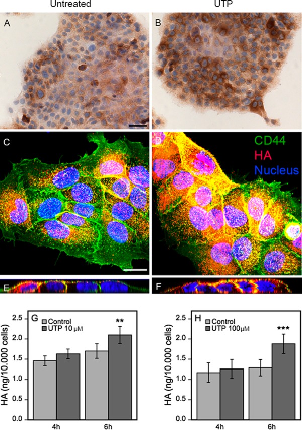
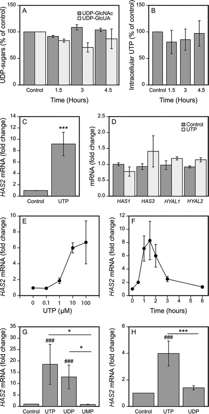
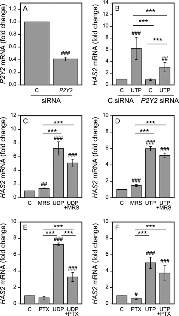
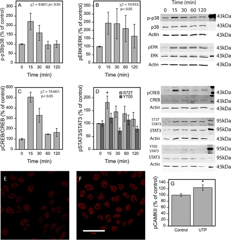
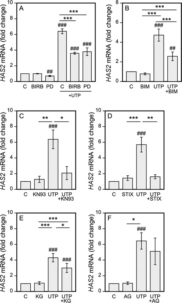
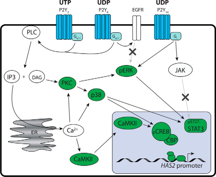
The release of nucleotides into extracellular space is triggered by insults like wounding and ultraviolet radiation, resulting in stimulatory or inhibitory signals via plasma membrane nucleotide receptors. As similar insults are known to activate hyaluronan synthesis we explored the possibility that extracellular UTP or its breakdown products UDP and UMP act as mediators for hyaluronan synthase (HAS) activation in human epidermal keratinocytes. UTP increased hyaluronan both in the pericellular matrix and in the culture medium of HaCaT cells. 10-100 μm UTP strongly up-regulated HAS2 expression, although the other hyaluronan synthases (HAS1, HAS3) and hyaluronidases (HYAL1, HYAL2) were not affected. The HAS2 response was rapid and transient, with the maximum stimulation at 1.5 h. UDP exerted a similar effect, but higher concentrations were required for the response, and UMP showed no stimulation at all. Specific siRNAs against the UTP receptor P2Y2, and inhibitors of UDP receptors P2Y6 and P2Y14, indicated that the response to UTP was mediated mainly through P2Y2 and to a lesser extent via UDP receptors. UTP increased the phosphorylation of p38, ERK, CREB, and Ser-727 of STAT3 and induced nuclear translocation of pCaMKII. Inhibitors of PKC, p38, ERK, CaMKII, STAT3, and CREB partially blocked the activation of HAS2 expression, confirming the involvement of these pathways in the UTP-induced HAS2 response. The present data reveal a selective up-regulation of HAS2 expression by extracellular UTP, which is likely to contribute to the previously reported rapid activation of hyaluronan metabolism in response to tissue trauma or ultraviolet radiation.

Keywords: P2Y; UTP; cell signaling; extracellular nucleotide; hyaluronan; hyaluronan synthesis; keratinocyte; nucleotide; purinergic receptor.


Conflict of interest statement

The authors declare that they have no conflicts of interest with the contents of this article

Figures

FIGURE 1.
FIGURE 1.
UTP addition in keratinocyte cultures rapidly increases pericellular hyaluronan, and induces a subsequent release of hyaluronan into the medium. HaCaT cultures were left untreated (A, C, and E) or treated with 100 μm UTP for 2 (B) and 4 h (D and F) and stained for hyaluronan using bHABC. In A and B, DAB was used as a chromogen (brown color). In C–F, the cultures were stained for hyaluronan using bHABC and TR-streptavidin (red), and for CD44 with a FITC-labeled secondary antibody (green). The nuclei were visualized with DAPI (blue). C and D are compressed stacks of the confocal images, E and F are side views cut through such stacks. The magnification bar for the bright field images is 50 μm, and for confocal images 20 μm. Culture media collected from HaCaT cells treated with 10 μm for 4 and 6 h (n = 3 for both) (G) and 100 μm (H) UTP for 4 and 6 h (n = 4 and n = 9, respectively) were analyzed for hyaluronan secretion. The data represent mean ± S.E. Mixed model ANOVA was used to calculate the significance of the difference to untreated cultures (**, p < 0.01; ***, p < 0.001).
FIGURE 2.
FIGURE 2.
UTP strongly up-regulates HAS2 expression. HaCaT cells were incubated for the indicated times with 100 μm UTP, the amounts of the intracellular UDP-sugar precursors of hyaluronan were measured (A) as well as the intracellular UTP (B). In panels C and D, HaCaT cells were incubated for 2 h with 100 μm UTP and the levels of HAS2 mRNA (C, n = 15) and other hyaluronan-related genes (D, HAS3, n = 4; others n = 3) were analyzed by qRT-PCR. E, HaCaT cells were treated with 0.1–100 μm UTP for 2 h (n = 3). F, 100 μm UTP was added to the cultures and the samples were collected after different incubation times for HAS2 mRNA assays (n = 3). G, HaCaT cells were treated for 2 h with 100 μm UTP, UDP, and UMP prior to qRT-PCR analysis (n = 3). H, HaCaT cells were treated for 2 h with 10 μm UTP and UDP prior to mRNA analysis (n = 3). Statistical significances of the differences between the groups were tested using (in C and D) one group t test (***, p < 0.001). In G and H, mixed model ANOVA was used for comparisons between the different treatments (indicated by *, p < 0.05) and comparisons of treatments to controls (set to 1) using pnorm (indicated by ###, p < 0.001). For the UDP-sugars (A) and concentration and time series (E and F) the non-parametric Friedman test was used due to unequal variances between the groups. Both the effects of the UTP concentration (E) and of the incubation time (F) were statistically significant (χ2 = 11.46, Friedman test p = 0.022 and χ2 = 16.7, Friedman test p = 0.01, respectively). No significance was found in UDP-GlcNAc and UDP-GlcUA (A) (χ2 = 4.667, p = 0.097, and χ2 = 5.0, p = 0.172, respectively) between the untreated and UTP-treated cultures.
FIGURE 3.
FIGURE 3.
The P2Y receptors and signaling pathways involved in the UTP-induced HAS2 up-regulation. HaCaT cells were transfected with control and P2Y2-specific siRNAs (A and B). A, 2 days after the transfection the samples were collected to test for the efficiency of the siRNAs (n = 4), or B, subjected to 100 μm UTP for 2 h prior to collecting the samples for HAS2 qRT-PCR (n = 5). C and D, cells were subjected to MRS2578 (a selective antagonist of P2Y6, 20 μm) for 30 min, and E and F, PTX (100 ng/ml) for 17 h prior to the addition of 100 μm UDP (C and E) and UTP (D and F) for 2 h before mRNA assays. Statistical significances of the differences between the groups were tested using one group t test in A (##, p < 0.01). Mixed model ANOVA was used for comparisons between the different treatments (indicated by **, p < 0.01; ***, p < 0.001) and comparisons of treatments to controls (set to 1) was tested by pnorm (indicated by #, p < 0.05; ##, p < 0.01; ###, p < 0.001) in B–F.
FIGURE 4.
FIGURE 4.
UTP induces rapid phosphorylations of p38, CREB, and Ser-727 of STAT3. HaCaT cells were subjected to 100 μm UTP for 15 min to 2 h before collecting the samples for Western blotting using antibodies against phosphorylated signaling proteins. For normalization the blots were reprobed for the total forms of signaling proteins and actin after a brief (10 min) stripping with 0.2 m NaOH. Representative blots out of 3 (p-p38, p38α (A); pERK, ERK (B); pCREB, CREB (C); pSTAT3-S727, pSTAT3-Y705, and total STAT3 (D)) experiments with the corresponding actins are shown, whereas mean ± S.E. of the quantifications of the phosphorylated forms per corresponding total proteins from all the experiments are presented as bar graphs. Statistical significances were tested using a non-parametric Friedman test because of unequal variances, indicating the overall significance values indicated in the figures. For STAT3-Y705 and -S727 the overall influence did not reach statistical significance. For Ser-727 of STAT3, allowing the use of a parametric test, pairwise comparisons of individual time points to the zero time point utilizing mixed model ANOVA and pnorm function were also performed, suggesting a significant influence at the 15-min time point (indicated by *, p < 0.05) (D). E and F, immunostainings for pCaMKII of the untreated (E) and the UTP-treated (100 μm) (F) cultures. The treatment time was 30 min. Mean ± S.E. of 5 experiments are shown in the chart (G). Statistical significance between the groups was tested using mixed model ANOVA (indicated by *, p < 0.05) (G).
FIGURE 5.
FIGURE 5.
Inhibition of p38, CaMKII, STAT3, and CREB reduce the UTP-induced HAS2 up-regulation. HaCaT cells were subjected to: A, the MEK kinase inhibitor PD98059 (PD, 0.5 μm) and the p38 inhibitor BIRB796 (BIRB, 2 μm); B, the PKC inhibitor (BIM, 10 μm); C, the CaMKII inhibitor KN93 (KN93, 25 μm); D, the STAT3 IX inhibitor (STIX, 50 μm); E, the CREB inhibitor KG501 (KG, 25 μm); and F, the JAK2/EGFR inhibitor AG490 (AG, 30 μm). Preincubations with the inhibitors were 0.5 h for ERK, p38, CREB, and PKC, and 2 h for CaMKII and STAT3 prior to the addition of 100 μm UTP for 2 h. Mean ± S.E. are shown. The number of experiments is 3 for ERK and p38, 6 for BIM, 4 for CREB, 6 for CaMKII, 5 for STAT3, and 4 for JAK2/EGFR. Statistical significances of the differences between the groups were tested using mixed model ANOVA (indicated by *, **, and ***) and comparisons of the treatments to the controls (set to 1, indicated by ## and ###) using pnorm. *, p < 0.05; ** and ##, p < 0.01; *** and ###, p < 0.001.
FIGURE 6.
FIGURE 6.
Schematic representation of the signaling pathways involved in the UTP-induced HAS2 up-regulation. Extracellular UTP and its breakdown product UDP activate the P2Y receptors that increase HAS2 expression via the indicated signaling steps. Those steps positively verified in the current study are marked green, whereas the pathways excluded are indicated by an “x.”

Comment in

  • Matrix Biology Highlights.
    [No authors listed] [No authors listed] Matrix Biol. 2017 May;59:1-2. doi: 10.1016/j.matbio.2017.03.001. Matrix Biol. 2017. PMID: 28335831 No abstract available.

References

    1. Inoue K., Hosoi J., and Denda M. (2007) Extracellular ATP has stimulatory effects on the expression and release of IL-6 via purinergic receptors in normal human epidermal keratinocytes. J. Invest. Dermatol. 127, 362–371 - PubMed
    1. Pastore S., Mascia F., Gulinelli S., Forchap S., Dattilo C., Adinolfi E., Girolomoni G., Di Virgilio F., and Ferrari D. (2007) Stimulation of purinergic receptors modulates chemokine expression in human keratinocytes. J. Invest. Dermatol. 127, 660–667 - PubMed
    1. Holzer A. M., and Granstein R. D. (2004) Role of extracellular adenosine triphosphate in human skin. J. Cutan. Med. Surg. 8, 90–96 - PubMed
    1. Ho C. L., Yang C. Y., Lin W. J., and Lin C. H. (2013) Ecto-nucleoside triphosphate diphosphohydrolase 2 modulates local ATP-induced calcium signaling in human HaCaT keratinocytes. PLoS ONE 8, e57666. - PMC - PubMed
    1. Azorin N., Raoux M., Rodat-Despoix L., Merrot T., Delmas P., and Crest M. (2011) ATP signalling is crucial for the response of human keratinocytes to mechanical stimulation by hypo-osmotic shock. Exp. Dermatol. 20, 401–407 - PubMed

Publication types