. 2017 Apr 14;292(15):6086-6093.
doi: 10.1074/jbc.M117.776047. Epub 2017 Feb 10.

Asymmetric configurations in a reengineered homodimer reveal multiple subunit communication pathways in protein allostery

Affiliations

Asymmetric configurations in a reengineered homodimer reveal multiple subunit communication pathways in protein allostery

Maria Fe Lanfranco et al. J Biol Chem. .

Abstract

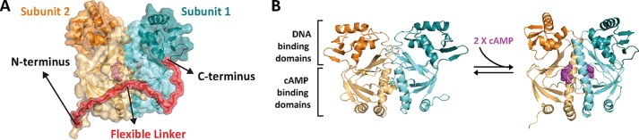
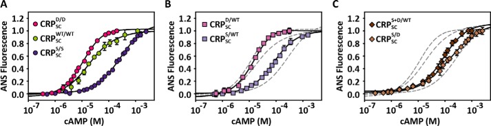
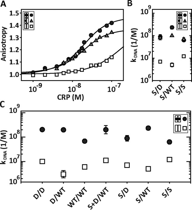
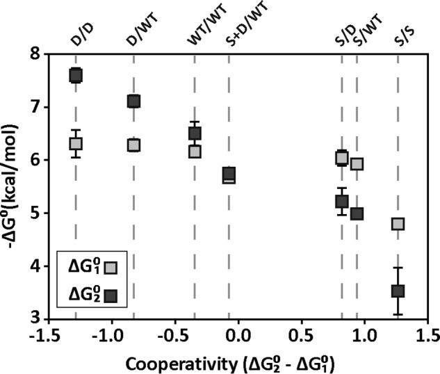
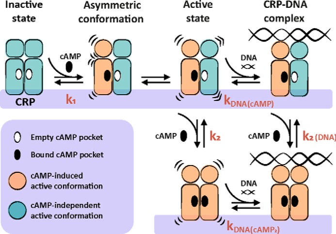
Many allosteric proteins form homo-oligomeric complexes to regulate a biological function. In homo-oligomers, subunits establish communication pathways that are modulated by external stimuli like ligand binding. A challenge for dissecting the communication mechanisms in homo-oligomers is identifying intermediate liganded states, which are typically transiently populated. However, their identities provide the most mechanistic information on how ligand-induced signals propagate from bound to empty subunits. Here, we dissected the directionality and magnitude of subunit communication in a reengineered single-chain version of the homodimeric transcription factor cAMP receptor protein. By combining wild-type and mutant subunits in various asymmetric configurations, we revealed a linear relationship between the magnitude of cooperative effects and the number of mutant subunits. We found that a single mutation is sufficient to change the global allosteric behavior of the dimer even when one subunit was wild type. Dimers harboring two mutations with opposite cooperative effects had different allosteric properties depending on the arrangement of the mutations. When the two mutations were placed in the same subunit, the resulting cooperativity was neutral. In contrast, when placed in different subunits, the observed cooperativity was dominated by the mutation with strongest effects over cAMP affinity relative to wild type. These results highlight the distinct roles of intrasubunit interactions and intersubunit communication in allostery. Finally, dimers bound to either one or two cAMP molecules had similar DNA affinities, indicating that both asymmetric and symmetric liganded states activate DNA interactions. These studies have revealed the multiple communication pathways that homo-oligomers employ to transduce signals.

Keywords: allosteric regulation; cAMP receptor protein (CRP); conformational change; cooperativity; cyclic AMP (cAMP); single-chain dimer; subunit communication; transcription factor.


Conflict of interest statement

The authors declare that they have no conflicts of interest with the contents of this article

Figures

Figure 1.
Figure 1.
Design and construction of a CRP single-chain dimer (CRPSC). A, model of a CRPSC that connects the two CRP subunits (cyan and orange) through a flexible polypeptide linker (red). The model was rendered in PyMOL based on the CRP structure bound to cyclic nucleotide (PDB 1G6N). B, structure of CRP in the unliganded (left, PDB 2WC2) and cAMP-bound conformations (right, PDB 1G6N). cAMP is shown in magenta.
Figure 2.
Figure 2.
Biophysical and functional characterization of CRPSC. A, SDS-PAGE showing the molecular mass (in kDa) of CRPSC versus wild-type CRP (labeled CRP in all figure panels). B, size-exclusion chromatogram of CRPSC and wild-type CRP. C, CD spectra of CRPSC and wild-type CRP. D, chemical denaturation of CRPSC and wild-type CRP monitored by changes in tryptophan fluorescence. The line corresponds to the fit of a two-state unfolding model as described under “Materials and Methods.” E, CRP-DNA interactions monitored by electrophoretic mobility shift assay using increasing lengths of the lac promoter in the absence and presence of 200 μm cAMP for CRPSC and wild-type CRP.
Figure 3.
Figure 3.
Quantification of the functional behavior of CRPSC. A, cAMP binding to CRPSC and wild-type CRP (labeled CRP in all figure panels) monitored by changes in ANS fluorescence. The solid lines represent the fit using a two-site binding model as described under “Materials and Methods.” B, binding of CRPSC or wild-type CRP to a 32-bp fluorescein-labeled lac promoter monitored by changes in fluorescence anisotropy. The solid lines represent the fit as described in Ref. . Residuals of the fit for both experiments are shown below the titrations.
Figure 4.
Figure 4.
Effect of symmetric and asymmetric mutations on cAMP binding affinity. A, cAMP titrations to CRPSCWT/WT (green circles) and the symmetric mutants CRPSCD/D (red circles) and CRPSCS/S (dark purple circles). B, cAMP titrations to the single asymmetric mutants CRPSCD/WT (light pink squares) and CRPSCS/WT (light purple squares). C, cAMP titrations to the double asymmetric mutants CRPSCS+D/WT (dark brown diamonds) and CRPSCS/D (beige diamonds). For comparison, the dashed lines corresponding to the fits of the parent symmetric proteins obtained from A were included in B and C. The solid lines in all three panels represent the fit using a two-site binding model as described under “Materials and Methods.” Error bars correspond to the standard deviation of at least three repeats.
Figure 5.
Figure 5.
DNA-CRPSC interactions using saturating and non-saturating cAMP concentrations. A, interaction of CRPSCS/D with the 32-bp lac promoter using 0, 30, and 1000 μm cAMP, which correspond to unbound (open squares), singly (gray triangles), and doubly (black circles) cAMP-bound states. The solid lines represent the fit as described in Ref. . B, DNA binding affinity constants of CRPSCS/D, CRPSCS/WT, and CRPSCS/S when the proteins are in the unbound (open squares), singly (gray triangles), and doubly (black circles) cAMP-bound states. C, DNA binding affinity constants obtained from fluorescence anisotropy experiments for symmetric and asymmetric CRPSC in the absence (open squares) and presence of saturating cAMP concentrations (black circles). For CRPSCWT/WT, CRPSCD/D, and CRPSCD/WT, [cAMP] = 200 μm. For CRPSCS/S, [cAMP] = 2000 μm. For CRPSCS/D, CRPSCS/WT, and CRPSCS+D/WT, [cAMP] = 1000 μm.
Figure 6.
Figure 6.
cAMP-binding energies and cooperativities of CRPSC variants. Light and dark squares correspond to the cAMP-binding energy for the first (ΔG01) and second (ΔG02) sites, respectively. The order of CRPSC mutants is sorted from most positive cooperativity (ΔG02 − ΔG01 < 0) to most negative (ΔG02 − ΔG01 > 0) as indicated by the dashed lines.
Figure 7.
Figure 7.
Mechanism of CRP activation and interaction with DNA. In the absence of cAMP, CRP is in an inactive state. Binding of cAMP to one CRP subunit triggers a conformational change in the DNA-binding domain of the bound subunit, generating an asymmetric conformation within the dimer. The cAMP-bound subunit induces a re-orientation in the unliganded neighboring subunit that is compatible with strong DNA interactions. Binding of a second cAMP molecule can occur to either the singly cAMP-bound CRP or to the ternary complex DNA-CRP-cAMP.

References

    1. Smock R. G., and Gierasch L. M. (2009) Sending signals dynamically. Science 324, 198–203 - PMC - PubMed
    1. Dayhoff J. E., Shoemaker B. A., Bryant S. H., and Panchenko A. R. (2010) Evolution of protein binding modes in homooligomers. J. Mol. Biol. 395, 860–870 - PMC - PubMed
    1. Goodsell D. S., and Olson A. J. (2000) Structural symmetry and protein function. Annu. Rev. Biophys. Biomol. Struct. 29, 105–153 - PubMed
    1. Changeux J.-P., and Edelstein S. J. (2005) Allosteric mechanisms of signal transduction. Science 308, 1424–1428 - PubMed
    1. Motlagh H. N., Wrabl J. O., Li J., and Hilser V. J. (2014) The ensemble nature of allostery. Nature 508, 331–339 - PMC - PubMed

MeSH terms