

A review of mallet finger and jersey finger injuries in the athlete
- PMID: 28188545
- PMCID: PMC5344866
- DOI: 10.1007/s12178-017-9395-6
A review of mallet finger and jersey finger injuries in the athlete
Abstract
Purpose of review: The purposes of this review are to discuss the diagnosis and management of mallet and jersey finger injuries in athletes and to highlight how treatment impacts return to play.
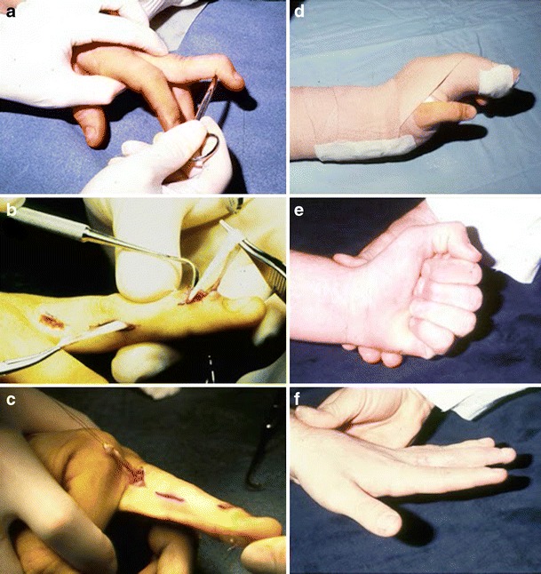
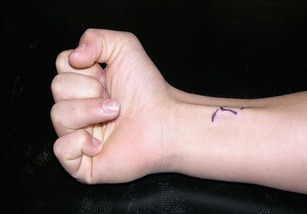
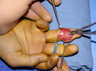
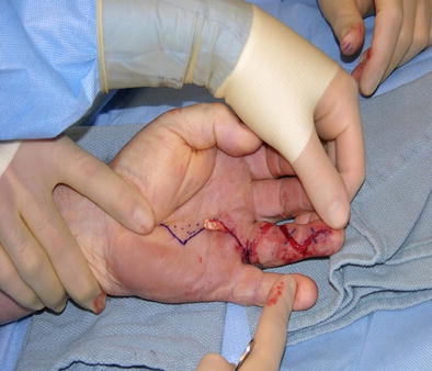
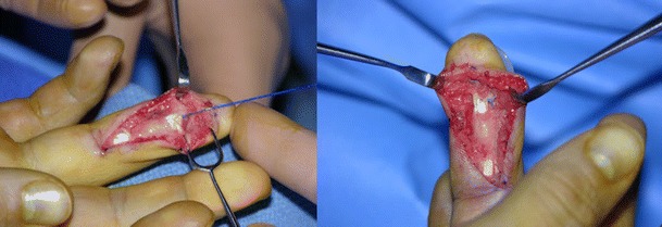
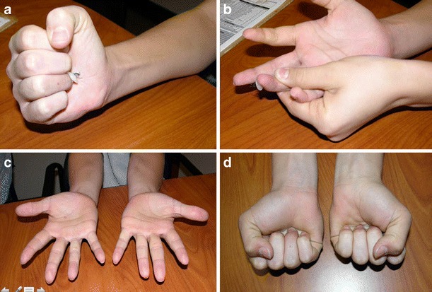
Recent findings: Mallet finger: although numerous non-operative and operative techniques have been described, there continues to be little consensus regarding the optimal procedure. Jersey finger: ultrasound appears to be a cost-effective imaging modality that may be useful for preoperative planning. Wide-awake surgery offers optimal intraoperative assessment of the tendon repair. Tendon repair with volar plate augmentation has been shown to improve the strength of the repair in the laboratory, and early clinical results are encouraging. Most mallet finger injuries will heal with non-operative treatment over a period of 8-12 weeks, even when treatment is delayed up to 3-4 months. An acute diagnosis of jersey finger requires surgical treatment and generally means 8-12 weeks of inability to compete in most contact sports.
Keywords: Athlete; Distal phalanx; Jersey finger; Mallet finger; Tendon avulsion.
Conflict of interest statement
Conflict of interest
The authors declare that they have no conflict of interest.
Human and animal rights and informed consent
This article does not contain any studies with human or animal subjects performed by any of the authors.
Figures
Similar articles
-
The Diagnosis and Management of Mallet Finger Injuries.Hand (N Y). 2017 May;12(3):223-228. doi: 10.1177/1558944716642763. Epub 2016 Mar 30. Hand (N Y). 2017. PMID: 28453357 Free PMC article. Review.
-
[Treatment of mallet finger with dorsal nail glued splint: retrospective analysis of 270 cases].Rev Chir Orthop Reparatrice Appar Mot. 2007 Nov;93(7):682-9. doi: 10.1016/s0035-1040(07)73253-1. Rev Chir Orthop Reparatrice Appar Mot. 2007. PMID: 18065879 French.
-
Review of Acute Traumatic Closed Mallet Finger Injuries in Adults.Arch Plast Surg. 2016 Mar;43(2):134-44. doi: 10.5999/aps.2016.43.2.134. Epub 2016 Mar 18. Arch Plast Surg. 2016. PMID: 27019806 Free PMC article. Review.
-
Mallet fingers with bone avulsion and DIP joint subluxation.J Hand Surg Eur Vol. 2015 Jan;40(1):8-15. doi: 10.1177/1753193414554772. Epub 2014 Oct 21. J Hand Surg Eur Vol. 2015. PMID: 25336471 Review.
-
Treatment of mallet finger deformity with a modified palmaris longus tendon graft through a bone tunnel.Int J Burns Trauma. 2018 Apr 5;8(2):34-39. eCollection 2018. Int J Burns Trauma. 2018. PMID: 29755840 Free PMC article.
Cited by
-
Causes of Procedural Failures of Closed Reductions using an Extension-Block Pin for Bony Mallet Finger.J Hand Microsurg. 2021 Apr;13(2):69-74. doi: 10.1055/s-0040-1701318. Epub 2020 Apr 7. J Hand Microsurg. 2021. PMID: 33867764 Free PMC article.
-
Secondary Repair of Jersey Finger: A Novel Method of Tendon Length Estimation via Measurement of Adjacent Landmarks.Cureus. 2024 Feb 10;16(2):e53963. doi: 10.7759/cureus.53963. eCollection 2024 Feb. Cureus. 2024. PMID: 38469003 Free PMC article.
-
Doigt en maillet chez un athlète âgé de 19 ans.CMAJ. 2025 Aug 24;197(28):E893-E894. doi: 10.1503/cmaj.250150-f. CMAJ. 2025. PMID: 40854607 Free PMC article. French. No abstract available.
-
Sonographic imaging of hand and wrist injuries: applications in the ER setting.Emerg Radiol. 2019 Apr;26(2):227-240. doi: 10.1007/s10140-018-1649-0. Epub 2018 Oct 16. Emerg Radiol. 2019. PMID: 30327891 Review.
-
Extensor tendon rupture and preoperative mri confirmations of suture anchor prolapse: a case report and literature review.BMC Musculoskelet Disord. 2024 May 4;25(1):355. doi: 10.1186/s12891-024-07476-0. BMC Musculoskelet Disord. 2024. PMID: 38704523 Free PMC article. Review.
References
-
- Schneider LH. Fractures of the distal interphalangeal joint. Hand Clin. 1994;10:277–85. - PubMed
Publication types
LinkOut - more resources
Full Text Sources
Other Literature Sources
Research Materials
