. 2017 Mar 28:250:36-47.
doi: 10.1016/j.jconrel.2017.02.001. Epub 2017 Feb 8.

Characterization of a novel intrinsically radiopaque Drug-eluting Bead for image-guided therapy: DC Bead LUMI™

Affiliations

Characterization of a novel intrinsically radiopaque Drug-eluting Bead for image-guided therapy: DC Bead LUMI™

Koorosh Ashrafi et al. J Control Release. .

Abstract

We have developed a straightforward and efficient method of introducing radiopacity into Polyvinyl alcohol (PVA)-2-Acrylamido-2-methylpropane sulfonic acid (AMPS) hydrogel beads (DC Bead™) that are currently used in the clinic to treat liver malignancies. Coupling of 2,3,5-triiodobenzaldehyde to the PVA backbone of pre-formed beads yields a uniformly distributed level of iodine attached throughout the bead structure (~150mg/mL) which is sufficient to be imaged under standard fluoroscopy and computed tomography (CT) imaging modalities used in treatment procedures (DC Bead LUMI™). Despite the chemical modification increasing the density of the beads to ~1.3g/cm3 and the compressive modulus by two orders of magnitude, they remain easily suspended, handled and administered through standard microcatheters. As the core chemistry of DC Bead LUMI™ is the same as DC Bead™, it interacts with drugs using ion-exchange between sulfonic acid groups on the polymer and the positively charged amine groups of the drugs. Both doxorubicin (Dox) and irinotecan (Iri) elution kinetics for all bead sizes evaluated were within the parameters already investigated within the clinic for DC Bead™. Drug loading did not affect the radiopacity and there was a direct relationship between bead attenuation and Dox concentration. The ability (Dox)-loaded DC Bead LUMI™ to be visualized in vivo was demonstrated by the administration of into hepatic arteries of a VX2 tumor-bearing rabbit under fluoroscopy, followed by subsequent CT imaging.

Keywords: Controlled drug release; Radiopaque Drug-eluting Beads; Transarterial chemoembolization; X-ray imaging.


Figures

Fig 1
Fig 1
(a) Embedded beads were sectioned using a microtome to yield 5μm thick sections from approximately the middle of the beads; (b) SEM of a bead cross-section and survey line profile for (c) EDAX elemental analysis mapping showing the uniform distribution of elements across the bead structure.
Fig 2
Fig 2
Optical micrographs of (top) unloaded, (middle) 37.5 mg/mL Dox-loaded and (bottom) 50 mg/mL Iri-loaded DC Bead LUMI of 40–90 μm, 70–150 μm and 100–300 μm size distributions.
Fig. 3
Fig. 3
(a) Box plots showing mean bead diameter (line), minimum and maximum range for three size ranges of DC Bead LUMI unloaded, loaded with 37.5 mg/mL Dox or loaded with 50 mg/mL Iri; inset (b) shows representation size distribution curves for unloaded and drug-loaded 70–150 μm DC Bead LUMI. ANOVA showed a statistically significant difference between beads sizes (p<0.0001) as anticipated, but also within each size group comparing unloaded, Dox and Iri (although the latter is not impactful on sedimented bead volume as the difference is driven by the large number of data points and the difference in the skewness of each of the distribution curves since they are not normally distributed populations).
Fig 4
Fig 4
Top row: μ-CT of homogenous bead phantoms, unloaded (left), beads loaded with 37.5 mg/mL Dox (middle) and beads loaded with 50 mg/mL Iri (right) showing consistency of intra-bead radiopacity; Middle row: representative cross-sectional images of this same phantom series; Bottom row: 3 representative cross-sections of individual beads demonstrating homogeneous iodine distribution.
Fig 5
Fig 5
Fluoroscopic evaluation under different imaging modes of various bead line phantoms (small and larger sizes, with and without loading with 37.5 mg/mL Dox) at two different BMI levels
Fig 6
Fig 6
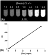
(A) Clinical CT images of various concentrations of DC Bead LUMI loaded with 37.5 mg/mL Dox in agar phantoms; (B) Correlation between DC Bead LUMI attenuation and Dox content (R2=0.9995).
Fig 7
Fig 7
In vivo CT image of Dox-loaded DC Bead LUMI (70–150 μm, 37.5 mg/mL) in a VX2 tumor (A): MDCT axial slice showing tumor position (hatched arrow pointing to an area of decreased enhancement) and beads in arteries at the periphery of the tumor and other non-tumor feeding vessels (yellow arrows); (B) axial Maximum Intensity Projections (MIPs) showing a 3D reconstruction of the distribution of beads around the area of the tumor (hatched arrow, area of decreased enhancement), within both tumor-feeding vessels and non-target arteries in the liver; (C) a comparative axial CT image of a human patient with hepatocellular carcinoma (hatched arrow) in which the beads can be clearly seen mainly filling the arteries in the interior of the tumor mass and some minor non-target arteries at the periphery (yellow arrows) [24].
Fig. 8
Fig. 8
(A) Effect of DC Bead LUMI bead size on Dox uptake rate (37.5 mg/mL); (B) Effect of DC Bead LUMI bead size and Dox target loading (25 or 37.5 mg/mL) on rate of Dox uptake from a 2 mg/mL loading solution. (C) Effect of bead size on Iri uptake rate (50 mg/mL). Constant agitation conditions.
Fig. 9
Fig. 9
(A) Dox raw data elution profile for DC Bead (DC) and for DC Bead LUMI (LUMI) (both 37.5 mg/mL Dox) showing a small decrease in the in vitro Cmax; (B) Cumulative elution data showing how Dox elution from DC Bead LUMI falls within the envelope of the clinically-studied sizes of DC Bead. Note that the entire drug is eventually eluted for both products but the study has been truncated to show the early time point data.
Fig. 10
Fig. 10
(A) Iri raw data elution profile for DC Bead and for DC Bead LUMI (50 mg/mL Iri) showing a marked reduction in the in vitro Cmax; (B) Cumulative elution data showing how Iri elution from DC Bead LUMI has a reduced burst effect compared to DC Bead.
Scheme 1
Scheme 1
Preparation of 2,3,5-Triiodobenzaldehyde
Scheme 2
Scheme 2
Preparation of PVA-AMPS hydrogel beads
Scheme 3
Scheme 3
Coupling of 2,3,5-triiodobenzaldehyde (TBA) to PVA-AMPS hydrogel beads

References

    1. Lewis AL. DC Bead(TM): a major development in the toolbox for the interventional oncologist. Expert Rev Med Devices. 2009;6:389–400. - PubMed
    1. Lewis AL, Gonzalez MV, Lloyd AW, Hall B, Tang Y, Willis SL, et al. DC bead: in vitro characterization of a drug-delivery device for transarterial chemoembolization. J Vasc Interv Radiol. 2006;17:335–42. - PubMed
    1. Nicolini A, Crespi S, Martinetti L. Drug delivery embolization systems: a physician’s perspective. Expert opinion on drug delivery. 2011;8:1071–84. - PubMed
    1. Liapi E, Geschwind JF. Transcatheter arterial chemoembolization for liver cancer: is it time to distinguish conventional from drug-eluting chemoembolization? Cardiovasc Intervent Radiol. 2011;34:37–49. - PMC - PubMed
    1. Lammer J, Malagari K, Vogl T, Pilleul F, Denys A, Watkinson A, et al. Prospective randomized study of doxorubicin-eluting-bead embolization in the treatment of hepatocellular carcinoma: results of the PRECISION V study. Cardiovasc Intervent Radiol. 2010;33:41–52. - PMC - PubMed

MeSH terms