. 2017 May;34(5):563-571.
doi: 10.1007/s10815-017-0881-y. Epub 2017 Feb 11.

Polar body transfer restores the developmental potential of oocytes to blastocyst stage in a case of repeated embryo fragmentation

Affiliations

Polar body transfer restores the developmental potential of oocytes to blastocyst stage in a case of repeated embryo fragmentation

Shuo-Ping Zhang et al. J Assist Reprod Genet. 2017 May.

Abstract

Purpose: We aimed to determine the developmental potential of human reconstructed oocytes after polar body genome transfer (PBT) and to report the case of a woman with multiple cycles of severe embryo fragmentation.

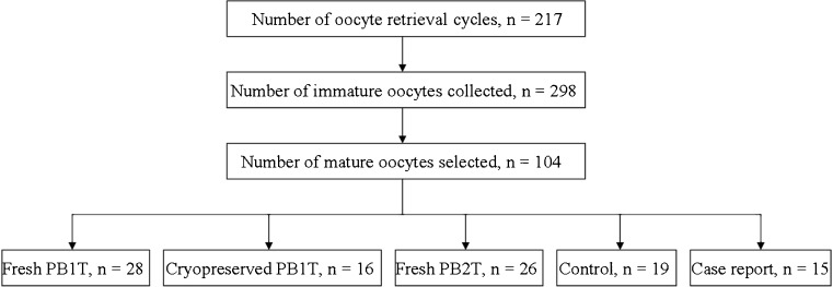
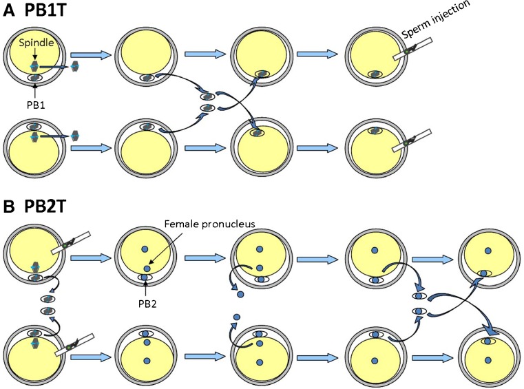
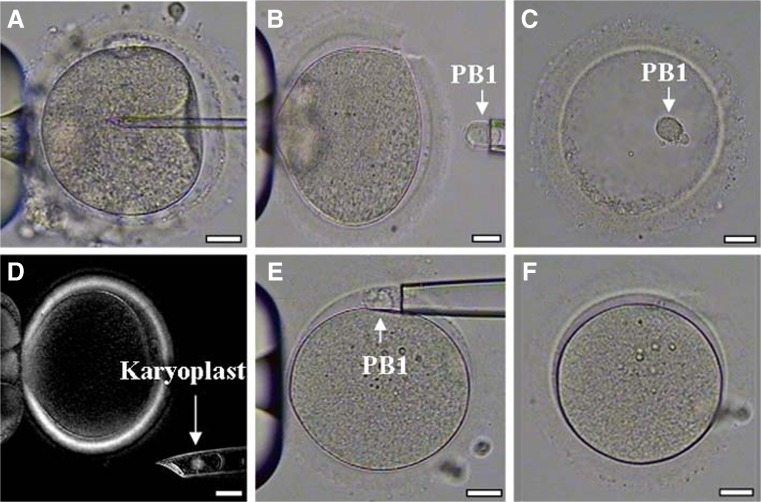
Methods: Fresh and cryopreserved first polar bodies (PB1s) were transferred to enucleated metaphase II oocytes (PB1T), while fresh PB2s were removed from fertilized oocytes and used instead of the female pronucleus in donor zygotes. Reconstructed oocytes underwent intracytoplasmic sperm injection (ICSI) and were cultured to blastocyst. Biopsied trophectoderm cells of PBT-derived blastocysts were screened for chromosomes by next-generation sequencing (NGS). Then, cryopreserved PB1T was carried out in one woman with a history of several cycles of extensive embryo fragmentation, and the blastocysts derived from PB1T were screened for aneuploidy but not transferred to the patient.

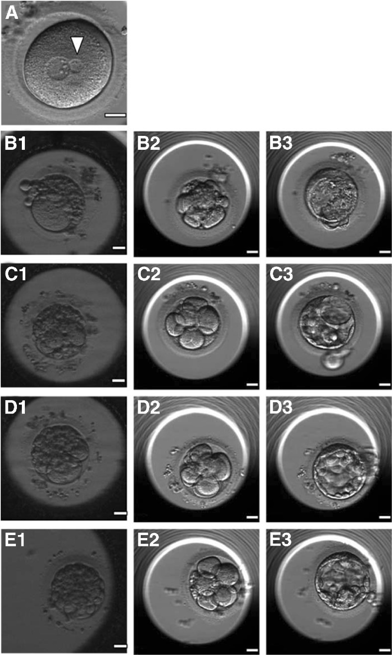
Results: There were no significant differences in the rates of normal fertilization and blastocyst formation between fresh and cryopreserved PB1T and control oocytes. Of the three fresh and three cryopreserved PB1T-derived blastocysts, two and one blastocysts exhibited normal diploidy respectively. In contrast, 17 PB2 transfers yielded 16 two pronuclei (2PN) zygotes with one normal and one small-sized pronucleus each and no blastocyst formation. In the female patient, 18 oocytes were inseminated by ICSI in the fourth cycle and the PB1s were biopsied. Although the embryos developed from the patient's own oocytes showed severe fragmentation, the oocytes reconstructed after PB1T produced three chromosomally normal blastocysts.

Conclusions: Normal blastocysts can develop from human reconstructed oocytes after PB1T. The application of the first PB transfers may be beneficial to patients with a history of poor embryo development and excessive fragmentation.

Keywords: Assisted reproductive technique; Blastocyst; Embryo fragmentation; Polar body transfer.


Conflict of interest statement

Competing interests

The authors declare that they have no competing interests.

Figures

Fig. 1
Fig. 1
Basic information of the experimental oocytes
Fig. 2
Fig. 2
Schematic drawing showing approaches of polar body (PB) transfer. a PB1T: oocytes are enucleated and the first PBs (PB1s) are biopsied. Then, the PB1s are interchanged and fused with the enucleated oocytes. The reconstituted oocytes are inseminated by ICSI. b PB2T: oocytes are inseminated by ICSI and the PB1s are removed simultaneously. After fertilization, the female pronuclei are removed, and then the PB2s are biopsied and fused with the enucleated zygotes
Fig. 3
Fig. 3
Polar body 1 transfer (PB1T) procedure. a Intracytoplasmic sperm injection (ICSI) of a patient’s oocyte. b The first polar body (PB1) was retrieved from the patient’s oocyte. c PB1 was placed into an empty zona pellucida for subsequent cryopreservation. d The spindle of a donor oocyte was visualized and removed under a polarized microscope. e Warmed PB1 derived from patient’s oocyte was transferred to the perivitelline space of the enucleated donor oocyte. f PB1 was fused with the cytoplast of the donor oocyte. Scale bars, 25 μm for all panels
Fig. 4
Fig. 4
a A 2PN zygote with a normal-sized pronucleus and a much smaller pronucleus (arrowhead) after polar body 2 transfer (PB2T). (b1, c1, d1, and e1) Day 3 embryos developing from the patient’s oocytes showed massive fragmentation, and the embryos reconstructed after PB1T of the four oocytes showed slight fragmentation on day 3 (b2, c2, d2, and e2) and developed to blastocyst stage (b3, c3, d3, and e3)

References

    1. Li R, Albertini DF. The road to maturation: somatic cell interaction and self-organization of the mammalian oocyte. Nat Rev Mol Cell Biol. 2013;14:141–52. doi: 10.1038/nrm3531. - DOI - PubMed
    1. Zamboni L. Ultrastructure of mammalian oocytes and ova. Biol Reprod Suppl. 1970;2:44–63. doi: 10.1095/biolreprod2.Supplement_2.44. - DOI - PubMed
    1. Verlinsky Y, Ginsberg N, Lifchez A, Valle J, Moise J, Strom CM. Analysis of the first polar body: preconception genetic diagnosis. Hum Reprod. 1990;5:826–9. doi: 10.1093/oxfordjournals.humrep.a137192. - DOI - PubMed
    1. Wakayama T, Hayashi Y, Ogura A. Participation of the female pronucleus derived from the second polar body in full embryonic development of mice. J Reprod Fertil. 1997;110:263–6. doi: 10.1530/jrf.0.1100263. - DOI - PubMed
    1. Wakayama T, Yanagimachi R. The first polar body can be used for the production of normal offspring in mice. Biol Reprod. 1998;59:100–4. doi: 10.1095/biolreprod59.1.100. - DOI - PubMed

LinkOut - more resources