Review
. 2017 Mar;40(3):151-166.
doi: 10.1016/j.tins.2017.01.002. Epub 2017 Feb 9.

Mitophagy and Alzheimer's Disease: Cellular and Molecular Mechanisms

Affiliations
Review

Mitophagy and Alzheimer's Disease: Cellular and Molecular Mechanisms

Jesse S Kerr et al. Trends Neurosci. 2017 Mar.

Abstract

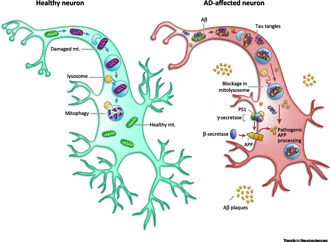
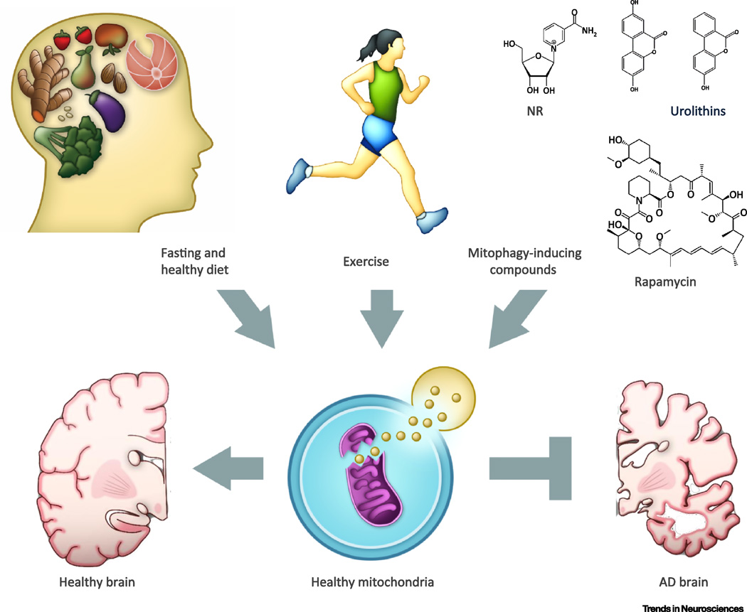
Neurons affected in Alzheimer's disease (AD) experience mitochondrial dysfunction and a bioenergetic deficit that occurs early and promotes the disease-defining amyloid beta peptide (Aβ) and Tau pathologies. Emerging findings suggest that the autophagy/lysosome pathway that removes damaged mitochondria (mitophagy) is also compromised in AD, resulting in the accumulation of dysfunctional mitochondria. Results in animal and cellular models of AD and in patients with sporadic late-onset AD suggest that impaired mitophagy contributes to synaptic dysfunction and cognitive deficits by triggering Aβ and Tau accumulation through increases in oxidative damage and cellular energy deficits; these, in turn, impair mitophagy. Interventions that bolster mitochondrial health and/or stimulate mitophagy may therefore forestall the neurodegenerative process in AD.


Figures

Figure 1
Figure 1. Mechanisms of mitophagy
A. A simplified overview of the mitophagy pathway. A damaged mitochondrion is marked and recognized by the autophagic machinery, forming an autophagosome. This then fuses to a lysosome to be degraded. B. Major autophagy/mitophagy proteins in the process of mitophagosome elongation and maturation across species. Major homology has been found in the higher eukaryotes; however, mammalian orthologues of many autophagy related proteins (ATGs) in yeast have not yet been identified. See Supplementary Table 1 for more details of the functions of each of the proteins. ‘-’, unknown. *In yeast, ATG7 activates ATG12, passes to ATG10, then to ATG5-ATG12 complex. Yeast cells add ATG16L to form complex at membrane. This complex facilitates ATG8-phosphatidylethanolamine (PE) formation through stimulating ATG3 [122, 123]. C. Proteins involved in mitophagy in mammalian cells. PINK1 translocation into mitochondria is stopped upon loss of mitochondrial membrane potential, causing PINK1 to accumulate on mitochondria. Together with Parkin, these proteins form phospho-ubiquitin chains on mitochondrial outer membrane proteins such as VDAC1, which recruit autophagy receptors such as optineurin (OPTN) and NDP52, which bind to both ubiquitin and LC3 (an autophagosomal protein), inducing the formation of the autophagosome. Abbreviations: FIS1- fission, mitochondrial 1; MGM1- mitochondrial dynamin-like GTPase; DNM1- dynamin 1; OPA1- Optic atrophy 1; DRP1- Dynamin-Related Protein; MARF- Mitochondrial assembly regulatory factor; PINK1- PTEN induced putative kinase 1; FZO1- mitofusin; VDAC1- voltage dependent anion channel 1; TBK1- TANK binding kinase 1; TAX1BP1- Tax1 binding protein 1; OPTN- Optineurin; NDP52(CALCOCO2)-calcium binding and coiled-coil domain 2; SQST1- SeQueSTosome related; REF2P–refractory to sigma P; NBR1- neighbor of Brca1 gene 1; DFCP1(ZFYVE1)- zinc finger FYVE-type containing 1; ATG- autophagy related gene; WIPI1- WD repeat domain, phosphoinositide interacting 1; FUNDC1- FUN14 domain containing 1; MAP1LC3-microtubule associated protein 1 light chain 3; BNIP3- BCL2 interacting protein 3; UNC51-Serine/threonine-protein kinase unc-51; ULKB- unc-51 like autophagy activating kinase 1; BEC1- Beclin homolog; BCL2L13- BCL2 like 13; PE- phosphatidylethanolamine; AMBRA1- autophagy and beclin 1 regulator 1; USP30- ubiquitin specific peptidase 30; LRPPRC- leucine rich pentatricopeptide repeat containing; P - Phosphate; Ub – Ubiquitin.
Figure 2
Figure 2. Mitochondrial dysfunction and compromised mitophagy in AD
In unaffected neurons, healthy mitochondria are distributed through the neuron. When they become dysfunctional (shown here in purple), they are packaged into autophagosomes and trafficked to lysosomes to be degraded. In AD affected neurons, comprised mitophagy causes decreased energy production and increased oxidative stress. This leads to increased amyloidogenic processing of APP by β-secretase and γ-secretase/PS1 and, in parallel, the accumulation of pTau aggregates. Pathogenic Aβ and pTau can impair mitophagy, leading to a subsequent increase in damaged mitochondria, and initiation of a self-propagating vicious cycle. Mechanisms of compromised mitophagy in AD are still elusive.
Figure 3
Figure 3. Physiological and pharmacological stimulation of mitophagy as potential therapeutic approaches for AD
Maintenance of a healthy mitochondrial pool in neurons may be paramount in healthy aging and avoidance of AD development. We highlight several lifestyle factors that can contribute to mitochondrial health, such as decreased energy consumption and suitable exercise, as well as certain compounds that pharmacologically induce mitophagy and mitochondrial health, such as rapamycin, spermidine, and the urolithins, as possible methods of AD prevention. Research into candidate drugs that mimic these lifestyle changes and mitophagy-inducing compounds, holds promise in AD treatment.

References

    1. Citron M. Alzheimer’s disease: strategies for disease modification. Nat Rev Drug Discov. 2010;9(5):387–398. - PubMed
    1. Hampel H, et al. The future of Alzheimer’s disease: the next 10 years. Prog Neurobiol. 2011;95(4):718–728. - PubMed
    1. Perry RJ, Watson P, Hodges JR. The nature and staging of attention dysfunction in early (minimal and mild) Alzheimer’s disease: relationship to episodic and semantic memory impairment. Neuropsychologia. 2000;38(3):252–271. - PubMed
    1. Braak H, Braak E. Staging of Alzheimer’s disease-related neurofibrillary changes. Neurobiol Aging. 1995;16(3):271–278. discussion 278–84. - PubMed
    1. Price JL, et al. The distribution of tangles, plaques and related immunohistochemical markers in healthy aging and Alzheimer’s disease. Neurobiol Aging. 1991;12(4):295–312. - PubMed