Review
. 2017 Feb 7:14:6.
doi: 10.1186/s12983-017-0192-2. eCollection 2017.

From egg to "no-body": an overview and revision of developmental pathways in the ancient arthropod lineage Pycnogonida

Affiliations
Review

From egg to "no-body": an overview and revision of developmental pathways in the ancient arthropod lineage Pycnogonida

Georg Brenneis et al. Front Zool. .

Abstract

Background: Arthropod diversity is unparalleled in the animal kingdom. The study of ontogeny is pivotal to understand which developmental processes underlie the incredible morphological disparity of arthropods and thus to eventually unravel evolutionary transformations leading to their success. Work on laboratory model organisms has yielded in-depth data on numerous developmental mechanisms in arthropods. Yet, although the range of studied taxa has increased noticeably since the advent of comparative evolutionary developmental biology (evo-devo), several smaller groups remain understudied. This includes the bizarre Pycnogonida (sea spiders) or "no-bodies", a taxon occupying a crucial phylogenetic position for the interpretation of arthropod development and evolution.

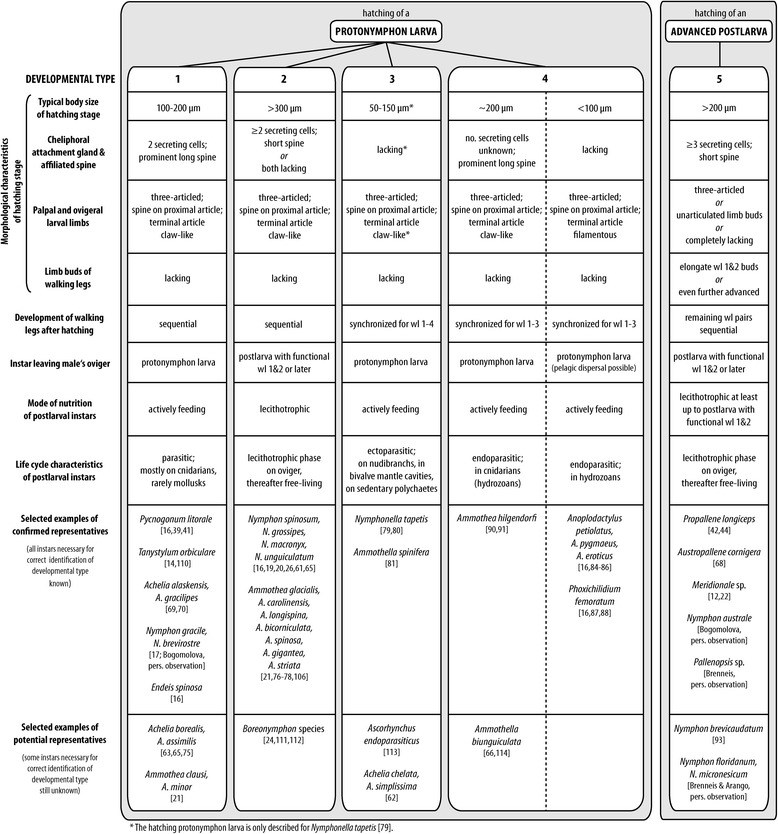
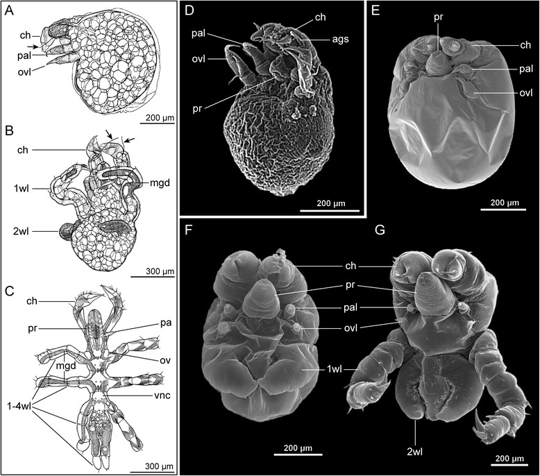
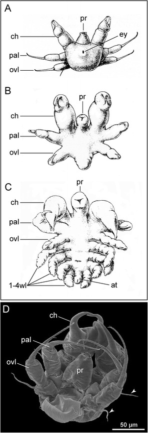
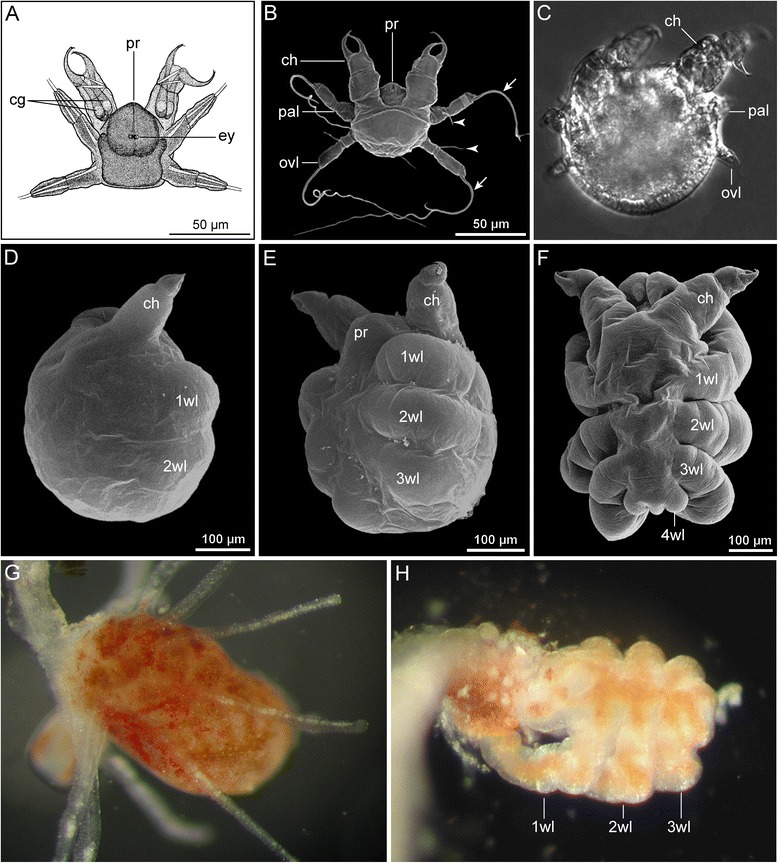
Results: Pycnogonid development is variable at familial and generic levels and sometimes even congeneric species exhibit different developmental modes. Here, we summarize the available data since the late 19th century. We clarify and resolve terminological issues persisting in the pycnogonid literature and distinguish five developmental pathways, based on (1) type of the hatching stage, (2) developmental-morphological features during postembryonic development and (3) selected life history characteristics. Based on phylogenetic analyses and the fossil record, we discuss plausible plesiomorphic features of pycnogonid development that allow comparison to other arthropods. These features include (1) a holoblastic, irregular cleavage with equal-sized blastomeres, (2) initiation of gastrulation by a single bottle-shaped cell, (3) the lack of a morphologically distinct germ band during embryogenesis, (4) a parasitic free-living protonymphon larva as hatching stage and (5) a hemianamorphic development during the postlarval and juvenile phases. Further, we propose evolutionary developmental trajectories within crown-group Pycnogonida.

Conclusions: A resurgence of studies on pycnogonid postembryonic development has provided various new insights in the last decades. However, the scarcity of modern-day embryonic data - including the virtual lack of gene expression and functional studies - needs to be addressed in future investigations to strengthen comparisons to other arthropods and arthropod outgroups in the framework of evo-devo. Our review may serve as a basis for an informed choice of target species for such studies, which will not only shed light on chelicerate development and evolution but furthermore hold the potential to contribute important insights into the anamorphic development of the arthropod ancestor.

Keywords: Anamorphic development; Arthropoda; Embryology; Evo-devo; Evolution; Gastrulation; Postembryonic development; Protonymphon larva; Sea spider.


Figures

Fig. 1
Fig. 1
Adult morphology of Pycnogonida and male paternal brood care. a Colossendeis australis, dorsal view. Note small body and prominent proboscis and long walking legs. b Nymphon australe, lateral view of anterior body region of an egg-carrying male, autofluorescence image. For better view of proboscis, cheliphores, palps and ovigers, the walking legs have been removed. c Nymphon molleri, ventral view of live male carrying egg packages (arrowheads) of different matings on each oviger. Note color change of egg packages from proximal (orange) to distal (light yellow) along the oviger, being indicative of different developmental stages of the embryos. d Ascorhynchus ramipes, ventral view of male carrying four egg packages (arrowheads). Note that both ovigers insert into each of the midline-spanning packages. e Nymphon micronesicum, ventral view of male carrying far advanced postlarval instars, autofluorescence image. In some pycnogonid species, the offspring leaves the male’s ovigers only at far advanced developmental stages
Fig. 2
Fig. 2
Embryonic development of Pycnogonida. a-c Pycnogonum litorale (Pycnogonidae), representing ‘small egg’ pycnogonids. a Four cell stage (Sytox nucleic acid staining). The blastomeres are of equal size. Asterisks mark cell nuclei, arrows indicate two brightly stained granules. b Initiation of gastrulation (Sytox nucleic acid staining). Note the immigration of the large bottle-shaped cell that is still attached to the embryo’s surface (arrowhead). c Embryonic morphogenesis (SEM). In the shown developmental stage, the proboscis, cheliphores and palpal and ovigeral larval limbs of the prospective protonymphon larva are recognizable. a&b modified from [10] and reproduced with permission of Springer; c modified from [11] and reproduced with permission of John Wiley and Sons. d-f Meridionale sp. (Callipallenidae), representing ‘large egg’ pycnogonids. d Early germ band stage (SEM). One embryonic hemisphere is covered by the densely packed small germ band cells, whereas the other hemisphere features few large yolk-rich cells (arrowheads). Asterisk indicates a damaged region. e Slightly later germ band stage (Sytox nucleic acid staining). Note stomodeum (arrow) in a far anterior position, being posteriorly followed by the cheliphore limb buds. Scattered nuclei around the germ band illustrate successive overgrowing of the large yolk-rich cells of the other embryonic hemisphere. f Late embryonic morphogenesis (SEM). Note that Meridionale sp. hatches as an advanced postlarva and develops walking leg pairs 1 and 2 before hatching. d&f modified from [12] and reproduced with permission of Springer
Fig. 3
Fig. 3
The protonymphon larva of Pycnogonida. a Ventral view of egg-carrying male of Tanystylum sp., SEM (modified from [73], therein published as “Tanystylum bealensis”, reproduced with permission of John Wiley and Sons). Arrowheads mark newly hatched protonymphon larvae. b Anterolateral view of protonymphon larva of Achelia assimilis, SEM (modified from [63], reproduced with permission of Cambridge University Press). Arrowheads mark gland processes of the palpal and ovigeral larval limbs. c, d Internal anatomy of the protonymphon larva of Nymphon brevirostre (modified from [61], reproduced with permission of Springer). Arrowheads mark gland processes of palpal and ovigeral larval limbs. The arrow highlights thread-like secretion of the cheliphoral attachment gland. c Ventral view. d Dorsal view
Fig. 4
Fig. 4
Type 1 postembryonic development of Pycnogonida. a-f Sequence of postembryonic instars of Achelia alaskensis up to the first juvenile instar (modified from [70], reproduced with permission of Hokkaido University). Dorsal view always on the left side, ventral view on the right side. Note strictly sequential development of the walking legs. The late protonymphon larva in (a) shows slight elevations of walking leg pair 1 posterior to the ovigeral larval limb (potentially the second larval instar of postembryonic development, the actual hatching having not been observed)
Fig. 5
Fig. 5
Overview of the different modes of postembryonic development in Pycnogonida. The general structure of the diagram is adopted from [19] but was extended and modified to accommodate additional details and terminological changes [–114]
Fig. 6
Fig. 6
Type 2 postembryonic development of Pycnogonida. a-c Three attaching postembryonic instars of Nymphon grossipes (modified from [65]). Arrows mark thread-like secretions of the cheliphoral attachment gland. a Lecithotrophic protonymphon larva, lateral view. b Postlarval instar with articulated walking leg 1 and limb bud of walking leg 2, ventral view. c Oldest attaching instar, a late postlarva with articulated walking legs 1–3 and elongate limb bud of walking leg 4 (the latter considered as three-articled in [65]), ventral view. d Lecithotrophic protonymphon larva of Nymphon unguiculatum, ventral view, SEM. e Lecithotrophic protonymphon larva of Ammothea carolinensis, ventral view, SEM. f, g Postlarval instars 1 and 2 of Ammothea bicorniculata, ventral views, SEM. Note increasing reduction of palpal and especially ovigeral larval limbs. d-g modified from [20, 21] and reproduced with permission of Springer
Fig. 7
Fig. 7
Type 3 postembryonic development of Pycnogonida. a-c Three postembryonic instars of Nymphonella tapetis, parasitizing in the mantle cavity of the lamellibranch bivalve Paphia philippinarum (modified from [79]). a Newly hatched protonymphon larva 1, dorsal view. b Presumable postembryonic instar 2 (modified protonymphon larva 2), ventral view. c Older postlarval instar, ventral view. Note incompletely differentiated walking leg pairs 1–4. d Protonymphon larva of Achelia chelata, its further developmental having been suggested to follow type 3, ventral view, SEM (modified from [62] and reproduced with permission of Cambridge University Press). Arrowheads mark gland processes of palpal and ovigeral larval limbs
Fig. 8
Fig. 8
Type 4 postembryonic development of Pycnogonida. a Newly hatched protonymphon larva 1 of Phoxichilidium femoratum, ventral view (modified from [65]). b-f Sequence of larval and postlarval instars of Anoplodactylus eroticus, endoparasitic in the hydrozoan Pennaria disticha. SEM (b, d-f) and brightfield (c) micrographs (modified from [86]). Reproduced with permission of Amy Maxmen. b Newly hatched protonymphon larva 1, dorsal view, note modified filamentous terminal articles of palpal and ovigeral larval limbs (arrows). Arrowheads mark gland processes of palpal and ovigeral larval limbs. c Postembryonic instar 2 (=modified protonymphon larva 2), dorsal view. Note significant reduction of palpal and ovigeral larval limbs. c Instar with primordia of walking leg pairs 1 and 2, lateral view. d Slightly later than (c), ventrolateral view. Note distinct limb buds of walking leg pairs 1–3 and lack of walking leg 4 primordia. e Old postlarval instar, shortly before molt and emergence from the hydranth, dorsal view. Note elongate anlagen of walking leg pairs 1–3 and tiny limb bud of walking leg 4. g Hydranth of live Pennaria disticha, infested by A. eroticus (Original: Amy Maxmen). g A. eroticus old postlarval instar (compare to (f)) protruding from ruptured hydrant of P. disticha. (Original: Amy Maxmen). Note orange color of the midgut diverticula extending into the walking legs
Fig. 9
Fig. 9
Type 5 postembryonic development of Pycnogonida. a Newly hatched postlarva of Pseudopallene spinipes, lateral view (modified from [65]). b, c Postlarval instars of Meridionale sp., SEM (modified from [22], reproduced with permission of Springer). b Newly hatched postlarva, lateral view. Arrow head marks short cheliphoral attachment gland spine with protruding thread-like secretions. c Postlarval instar 2, ventral view. This instar leaves the oviger and commences active feeding. d Hatching postlarva of Propallene kempi, ventral view. Left: surface of the postlarval cuticle through which anlagen of walking leg pairs 1 and 2 can be discerned. Right: combination of autofluorescence (white) and fluorescent marker FM1-43FX (glow). Walking leg pairs 1 and 2 underlie the cuticle, being extremely compressed and curved (black dashed line for walking leg 1). Glowing regions represent ventral nerve cord ganglia. e Pallenopsis hodgsoni. Left: autofluorescence image of egg package containing postlarvae about to hatch. Center: ventral view of late embryo (propidium iodide staining) showing anlagen of three walking leg pairs. Right: lateral view of hatched postlarva (propidium iodide staining). Note the presence of palpal and ovigeral larval limbs and the elongate walking leg pairs 1 and 2 still folded at the ventral side
Fig. 10
Fig. 10
Distribution and evolution of different developmental pathways in Pycnogonida. The shown cladograms have been simplified from [96] and [97]. On the right, the different developmental types are indicated by schematic drawings of their hatching stages and a color code. The gray area in each drawing indicates the post-ovigeral body region from which the walking leg segments develop. The developmental pathways have been mapped on the cladograms according to their color code. Note that in the case of Ascorhynchidae and Eurycydidae developmental type 1 has been inferred based on hatching protonymphon larva only, since no descriptions of subsequent postembryonic development exist. Taxa names with white background indicate that no developmental data are available. In both shown scenarios, developmental type 1 (green) has been given preference during the reconstruction of the single nodes whenever it is found in one of the two sister groups in question (therefore also the reconstruction of type 1 as an ancestral feature in scenario two). Accordingly, only the controversial grouping of paraphyletic callipallenids with respect to nymphonids results in developmental type 5 as their ancestral developmental pathway

References

    1. Giribet G, Edgecombe GD. The Arthropoda: A Phylogenetic Framework. In: Minelli A, Boxshall G, Fusco G, editors. Arthropod Biology and Evolution Molecules, Development, Morphology. Heidelberg: Springer Verlag; 2013. pp. 17–40.
    1. Richter S, Wirkner C. A research program for Evolutionary Morphology. J Zool Syst Evol Res. 2014;52:338–350. doi: 10.1111/jzs.12061. - DOI
    1. Maxmen A. The sea spider’s contribution to T.H. Morgan’s (1866–1945) development. J Exp Zool (Mol Dev Evol) 2008;310B:203–215. doi: 10.1002/jez.b.21202. - DOI - PubMed
    1. Dunlop JA. Geological history and phylogeny of Chelicerata. Arthropod Struct Dev. 2010;39:124–142. doi: 10.1016/j.asd.2010.01.003. - DOI - PubMed
    1. Dunlop JA, Arango CP. Pycnogonid affinities: a review. J Zool Syst Evol Res. 2005;43:8–21. doi: 10.1111/j.1439-0469.2004.00284.x. - DOI

LinkOut - more resources