

doi: 10.1002/j.2205-0140.2011.tb00131.x.
Epub 2015 Dec 31.
Ultrasonography of sternal fractures
Affiliations
- PMID: 28191123
- PMCID: PMC5024903
- DOI: 10.1002/j.2205-0140.2011.tb00131.x
Item in Clipboard
Ultrasonography of sternal fractures
Australas J Ultrasound Med.
2011 Nov.
Abstract
This paper describes the use of clinician-performed ultrasound to detect sternal fractures in trauma patients. It is a pictorial essay that describes the ultrasound technique, the normal anatomy and ultrasound findings, variants, potential pitfalls and the appearance of fractures when they occur in both children and adults.
Keywords: sternal fracture; sternum; ultrasonography; ultrasound.
Figures
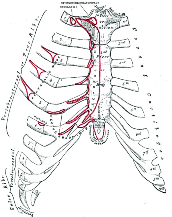
The anterior surface of the sternum and costal cartilages
18
.
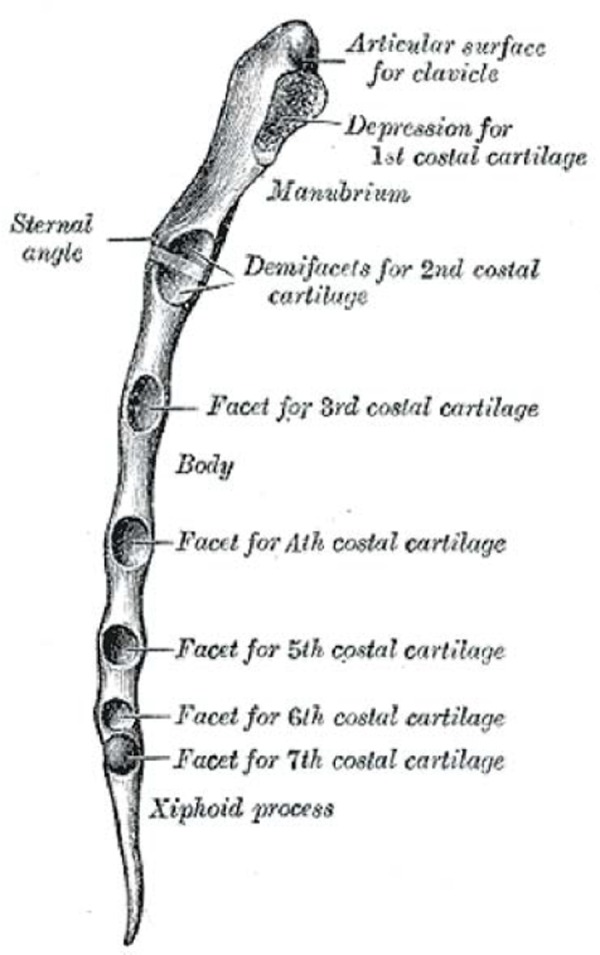
Lateral view of the sternum
18
.
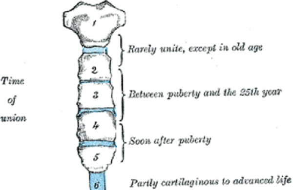
Development of the sternum; the sternebrae
18
.
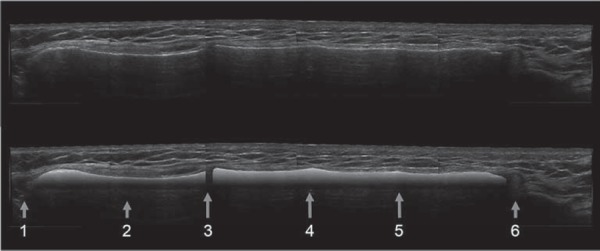
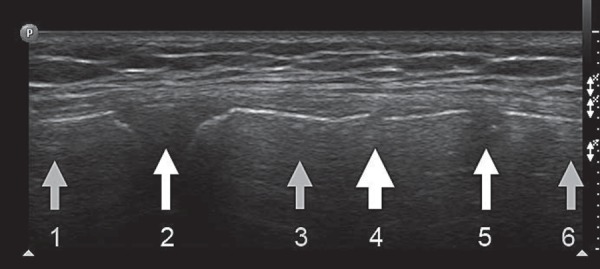
Ultrasound of normal adult sternum (longitudinal composite view). The anterior cortex has been highlighted in the lower image. (1) Jugular notch; (2) manubrium; (3) sternomanubrial junction (4) ridge at level of 3rd costal cartilage (5) ridge at level of 4th costal cartilage (6) xiphisternum.
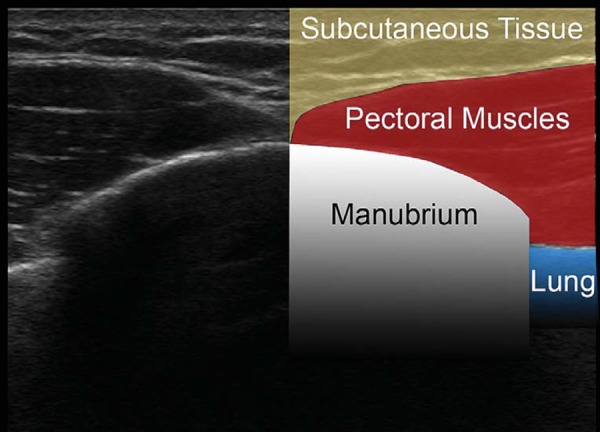
Transverse ultrasound image of the manubrium.
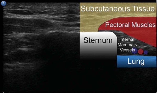
Transverse ultrasound image of the sternum.
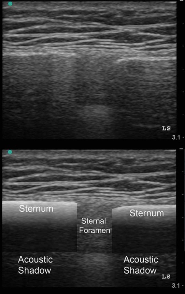
Sternal foramen.
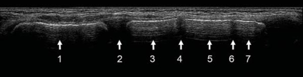
Normal paediatric sternum. This is a longitudinal panoramic ultrasound view of the sternum of a 6 year‐old. (1) Manubrium; (2) sternomanubrial junction; (3) 1st sternebra; (4) intersternebral cartilaginous junction; (5) 2nd sternebra, (6) intersternebral cartilaginous junction; (7) 3rd sternebra.
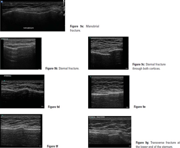
Manubrial and sternal fractures in adults. A step in the cortex at the site of maximal tenderness, running transversely across the sternum is likely to be a fracture. If the fracture traverses both cortices the proximal and distal portions may move independently through the respiratory cycle.
Paediatric sternal fracture. Here a step is seen in the anterior cortex of the first sternebrae. This 9 year‐old child had been in a car accident, was wearing a seatbelt and complained of pain and tenderness at exactly the site of the cortical step. (1) Manubrium; (2) sternomanubrial junction; (3) 1st sternebra; (4) fracture; (5) cartilaginous junction; (6) 2nd sternebra.
References
-
- Rose JS. Ultrasound in abdominal trauma. Emerg Med Clin North Am 2004; 22(3): 581–99, vii. - PubMed
-
- McGahan JP, Richards J, Fogata ML. Emergency ultrasound in trauma patients. Radiol Clin North Am 2004; 42 (2): 417–25. - PubMed
-
- Brookes JG, Dunn RJ, Rogers IR. Sternal fractures: a retrospective analysis of 272 cases. J Trauma 1993; 35 (1): 46–54. - PubMed
-
- Recinos G, Inaba K, Dubose J, Barmparas G, Teixeira PG, Talving P, et al. Epidemiology of sternal fractures. Am Surg 2009; 75 (5): 401–4. - PubMed
-
- Mazzocca AD, Garretson R, Romeo AA. Section I: Sternum and Rib Fractures in Adults and Children. In: DeLee JC, Drez D, Miller MD, editors. DeLee and Drez's Orthopaedic Sports Medicine. 2nd ed. Philadelphia: Elsevier Science; 2003.
LinkOut - more resources
Full Text Sources