Review
. 2013 Nov;16(4):158-167.
doi: 10.1002/j.2205-0140.2013.tb00242.x. Epub 2015 Dec 31.

Ultrasound assessment of fetal cardiac function

Affiliations
Review

Ultrasound assessment of fetal cardiac function

Fàtima Crispi et al. Australas J Ultrasound Med. 2013 Nov.

Abstract

Introduction: Fetal heart evaluation with US is feasible and reproducible, although challenging due to the smallness of the heart, the high heart rate and limited access to the fetus. However, some cardiac parameters have already shown a strong correlation with outcomes and may soon be incorporated into clinical practice. Materials and Methods: Cardiac function assessment has proven utility in the differential diagnosis of cardiomyopathies or prediction of perinatal mortality in congenital heart disease. In addition, some cardiac parameters with high sensitivity such as MPI or annular peak velocities have shown promising results in monitoring and predicting outcome in intrauterine growth restriction or congenital diaphragmatic hernia. Conclusion: Cardiac function can be adequately evaluated in most fetuses when appropriate expertise, equipment and time are available. Fetal cardiac function assessment is a promising tool that may soon be incorporated into clinical practice to diagnose, monitor or predict outcome in some fetal conditions. Thus, more research is warranted to further define specific protocols for each fetal condition that may affect cardiac function.

Keywords: 4D STIC; echocardiography; fetal cardiac function; myocardial imaging; tissue Doppler.


Figures

Figure 1
Figure 1
Graphic representation of the three‐directional myocardial motility involving longitudinal, radial and circumferential contraction. The motion is shown as a single point motility determined by displacement and systolic (s') and early diastolic (E') annular peak velocities; and deformation by the change in length or thickness between two points represented as strain or strain rate.
Figure 2
Figure 2
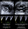
Illustration of the right and left ventricular outflow tracts for measuring stroke volume (SV) and cardiac output (CO). The valve diameter (D) is measured in a 2D image. Velocity time integral (VTI) of the blood flow and heart rate (HR) are evaluated in the spectral Doppler waveform. Combined cardiac output (CCO) is calculated by the sum of both CO, and cardiac index (CI) represents the normalization by estimated fetal weight (EFW).
Figure 3
Figure 3
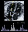
Image of the E/A ratio evaluation. The sample gate is placed just below the atrio‐ventricular valves in a four‐chamber view in order to display biphasic inflow (including the E (early diastole) and A (atrial contraction).
Figure 4
Figure 4
Illustration of myocardial performance index (MPI) assessment by spectral Doppler. Placing the Doppler sample volume in a four‐chamber view on the medial wall of the ascending aorta, the mitral biphasic inflow (grey arrow, early (E) and atrial (A) waveforms) and the aortic outflow (blue arrow and waveform (Ao)) are displayed in the same spectral image. The MPI is calculated by measuring time intervals including: isovolumic contraction time (ICT) from the closure of the mitral valve to the opening of the aortic valve; ejection time (ET) from the opening to closure of the aorta; and isovolumic relxaxation time (IRT) from the closure of the aortic valve to the opening of the mitral valve.
Figure 5
Figure 5
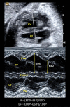
Illustration of a transverse four‐chamber view in order to measure shortening (SF) and ejection fractions (EF) of the right (RV) and left ventricles (LV) by M‐mode. The arrows between the septal and right free walls show the measurement of end‐diastolic (EDD) and end systolic (ESD) diameters required for the SF and EF calculation. RVW= right ventricle wall; LVW= left ventricle wall.
Figure 6
Figure 6
Illustration of the tricuspid annular plane systolic excursion (TAPSE) measurement by applying M‐mode at the tricuspid valve annulus in an apical four‐chamber view.
Figure 7
Figure 7
Example of early (E') and late (A') diastolic and systolic (s') peak annular velocities obtained by spectral tissue Doppler at the right annulus.
Figure 8
Figure 8
Offline analysis of strain (above) and strain rate (below) waveforms at the right basal free wall using color tissue Doppler.
Figure 9
Figure 9
Offline analysis of regional and global strain waveforms of the left ventricle wall using 2D speckle tracking.
Figure 10
Figure 10
Post‐processing analysis of the left ventricular volume through virtual organ computerized analysis using 4D‐spatio temporal correlation.
Figure 11
Figure 11
Graphic representation of cardiac function parameters at the different stages of the fetal adaptation to disease, illustrating longitudinal and diastolic function becoming abnormal at earlier stages than radial and systolic. Ductus venosus, E/A ratios, myocardial performance index (MPI) and cardiac output are measured by conventional Doppler; ejection fraction and longitudinal displacement by M‐mode; systolic (s') and early diastolic (E') annular pea k velocities by spectral tissue Doppler; and strain and strain‐rate by offline tissue Doppler or 2D speckle tracking.

References

    1. Carvalho JS, Chaoui R, Copel JA, DeVore GR, Hecher K, Lee W, et al. ISUOG practice guidelines (updated): sonographic screening examination of the fetal heart. Ultrasound Obstet Gynecol 2013; (41): 348–59. - PubMed
    1. Lee W, Allan L, Carvalho JS, Chaoui R, Copel J, Devore G, et al. ISUOG consensus statement: what constitutes a fetal echocardiogram? Ultrasound Obstet Gynecol 2008; 32 (2): 239–42. - PubMed
    1. Rychik J, Tian Z, Bebbington M, Xu F, McCann M, Mann S, et al. The twin‐twin transfusion syndrome: spectrum of cardiovascular abnormality and development of a cardiovascular score to assess severity of disease. Am J Obstet Gynecol 2007; 197 (4): 392. e1–8. - PubMed
    1. Crispi F, Hernandez‐Andrade E, Pelsers MM, Plasencia W, Benavides‐Serralde JA, Eixarch E, et al. Cardiac dysfunction and cell damage across clinical stages of severity in growth‐restricted fetuses. Am J Obstet Gynecol 2008; 199 (3): 254. e1–8. - PubMed
    1. Van Mieghem T, Gucciardo L, Done E, Van Schoubroeck D, Graatsma EM, Visser GH, et al. Left ventricular cardiac function in fetuses with congenital diaphragmatic hernia and the effect of fetal endoscopic tracheal occlusion. Ultrasound Obstet Gynecol 2009; 34 (4): 424–29. - PubMed

LinkOut - more resources