Review
. 2017 Jun 15;595(12):3907-3917.
doi: 10.1113/JP273049. Epub 2017 Mar 13.

The role of epicardial adipose tissue in cardiac biology: classic concepts and emerging roles

Affiliations
Review

The role of epicardial adipose tissue in cardiac biology: classic concepts and emerging roles

Alexios S Antonopoulos et al. J Physiol. .

Abstract

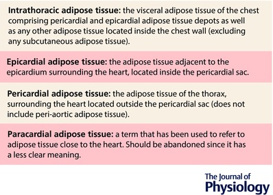
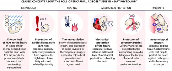
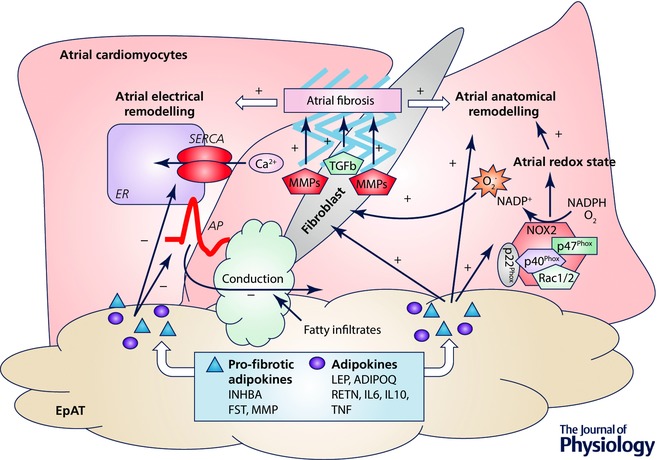
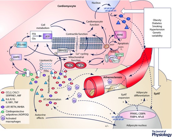
Classic concepts about the role of epicardial adipose tissue (EpAT) in heart physiology include its role in cardiac metabolism, mechanical protection of coronaries, innervation and possibly cryoprotection of the heart too. Nevertheless, recent evidence has revealed that epicardial adipose tissue regulates multiple aspects of cardiac biology including myocardial redox state, intracellular Ca2+ cycling, the electrophysiological and contractile properties of cardiomyocytes, cardiac fibrosis as well as coronary atherosclerosis progression. Moreover, it is now understood that the communication between EpAT and the heart is regulated by complex bidirectional pathways, since not only do adipokines regulate cardiac function, but also the heart affects EpAT biology via paracrine 'reverse' signalling. Such complex interactions as well as epicardial fat accumulation as a consequence of cardiac disease and epicardium to adipocyte differentiation should be taken into account by the clinical studies investigating EpAT as a risk marker and its potential as a therapeutic target against cardiovascular disease. Further in-depth exploration of the molecular mechanisms regulating the cross-talk between the heart and EpAT is expected to enhance our understanding regarding the role of the latter in cardiac physiology and relevant disease mechanisms.

Keywords: adipokines; cardiac biology; epicardial adipose tissue; myocardium; redox state.


Figures

Figure 1
Figure 1. Definitions of epicardial, pericardial and paracardial adipose tissue
Figure 2
Figure 2. The classic concepts about the role of epicardial adipose tissue in heart physiology
Figure 3
Figure 3. Adipokines and atrial fibrillation development
Adipokines have an impact on atrial electrophysiological properties, action potential (AP) duration and sarco/endoplasmic reticulum Ca2+‐ATPase (SERCA) activity of atrial cardiomyocytes, affecting thus arrhythmiogeneicity. Fibrofatty infiltrates into subepicardium also affect per se the electrical conduction properties of atrium. Adipokines can modulate NADPH oxidase activity (mainly NOX2) and myocardial redox state in human atria, which is causally involved in atrial fibrillation development. Through the direct effects of adipokines on extracellular matrix (e.g. matrix metalloproteinases) or via their indirect effects on activation of fibroblasts and modulation of myocardial redox state, and promote of atrial fibrosis. The latter is centrally involved in atrial anatomical and electrical remodelling, which disrupts the electrical conduction properties of atrial tissue and favours atrial fibrillation development. ADIPOQ, adiponectin; FST, follistatin; IL, interleukin; INHBA, activin A; LEP, leptin; MMP, matrix metallopeptidases; RETN, resistin; TGFb, transforming growth factor β; TNF, tumour necrosis factor α; list of adipokines is indicative; +, stimulate/induce; –, decrease/impair.
Figure 4
Figure 4. Communication between the cardiomyocytes and epicardial adipose tissue
Epicardial adipose tissue (EpAT) and the cardiomyocyte transcriptomic profile are altered in the presence of cardiovascular risk factors or by genetic variability. Nevertheless, further to any systemic effects, a local cross‐talk takes place between cardiomyocytes and EpAT which determines aspects of myocardial biology, cardiac function and coronary atherosclerosis progression. Secreted adipokines (e.g. adiponectin or leptin) differentially affect AMP‐activated kinase (AMPK) and phosphoinositide 3‐kinase (PI3K)/Akt signalling in cardiomyocytes, which are centrally involved in cardiomyocyte metabolism and substrate utilization. Free fatty acid (FFA)‐related lipotoxicity results in mitochondrial dysfunction, impaired oxidative metabolism and increased oxidative stress. NADPH oxidase activity is also enhanced by mitochondrial dysfunction, and alterations in AMPK signalling induced by EpAT‐secreted adipokines. Increased phsopshorylation of SMAD2 (e.g. by activin A) and/or reduced PI3K/Akt signalling by pro‐inflammatory adipokines negatively affect Ca2+ cycling and cardiomyocyte contractility. Increased cardiomyocyte oxidative stress has also direct effects on redox‐sensitive proteins of the contractile apparatus and cell apoptosis. Cardiomyocyte stress due to impaired substrate utilization, contractile dysfunction and increased oxidative burden leads to respective changes in cardiomyocyte transcriptome. Products of increased myocardial oxidative stress, such as 4‐hydroxynonenal (4HNE, an end product of lipid oxidation) and possibly others among the cardiomyocyte secretome may signal back to EpAT and affect key aspects of its biology, such as the differentiation of adipocytes, adipose tissue expansion and its infiltration by inflammatory cells as well as the regulation of transcriptional factors and relevant gene expression profile, which is shifted towards a pro‐inflammatory phenotype. The concept of a bidirectional signalling between the heart and EpAT is represented with continuous (‘outside‐to‐inside’ signalling) and dashed arrows (‘inside‐to‐outside’ signalling) respectively. ADIPOQ, adiponectin; CEBPA, CCAAT/enhancer‐binding protein α; CXCL1, C‐X‐C motif chemokine ligand 1; FABP4, fatty acid binding protein‐4; IL, interleukin; CCL2, C‐C motif chemokine ligand; INHBA, activin A; LEP, leptin; MIF, macrophage migration inhibitory factor; NFκB, nuclear factor κB; PPARG, peroxisome proliferator activator receptor γ; RETN, resistin; SERPINE1, serpin family E member 1; SMAD2, mothers against decapentaplegic homolog 2; TNF, tumour necrosis factor α; list of adipokines is indicative. +, stimulate/induce; –, decrease/impair; short white arrows represent diffusion of adipokines.

References

    1. Anan M, Uchihashi K, Aoki S, Matsunobu A, Ootani A, Node K & Toda S (2011). A promising culture model for analyzing the interaction between adipose tissue and cardiomyocytes. Endocrinology 152, 1599–1605. - PubMed
    1. Antoniades C, Demosthenous M, Reilly S, Margaritis M, Zhang MH, Antonopoulos A, Marinou K, Nahar K, Jayaram R, Tousoulis D, Bakogiannis C, Sayeed R, Triantafyllou C, Koumallos N, Psarros C, Miliou A, Stefanadis C, Channon KM & Casadei B (2012). Myocardial redox state predicts in‐hospital clinical outcome after cardiac surgery effects of short‐term pre‐operative statin treatment. J Am Coll Cardiol 59, 60–70. - PubMed
    1. Antonopoulos AS, Margaritis M, Coutinho P, Digby J, Patel R, Psarros C, Ntusi N, Karamitsos TD, Lee R, De Silva R, Petrou M, Sayeed R, Demosthenous M, Bakogiannis C, Wordsworth PB, Tousoulis D, Neubauer S, Channon KM & Antoniades C (2014). Reciprocal effects of systemic inflammation and brain natriuretic peptide on adiponectin biosynthesis in adipose tissue of patients with ischemic heart disease. Arterioscler Thromb Vasc Biol 34, 2151–2159. - PubMed
    1. Antonopoulos AS, Margaritis M, Coutinho P, Shirodaria C, Psarros C, Herdman L, Sanna F, De Silva R, Petrou M, Sayeed R, Krasopoulos G, Lee R, Digby J, Reilly S, Bakogiannis C, Tousoulis D, Kessler B, Casadei B, Channon KM & Antoniades C (2015). Adiponectin as a link between type 2 diabetes and vascular NADPH oxidase activity in the human arterial wall: the regulatory role of perivascular adipose tissue. Diabetes 64, 2207–2219. - PubMed
    1. Antonopoulos AS, Margaritis M, Verheule S, Recalde A, Sanna F, Herdman L, Psarros C, Nasrallah H, Coutinho P, Akoumianakis I, Brewer AC, Sayeed R, Krasopoulos G, Petrou M, Tarun A, Tousoulis D, Shah AM, Casadei B, Channon KM & Antoniades C (2016a). Mutual regulation of epicardial adipose tissue and myocardial redox state by PPAR‐γ/adiponectin signalling. Circ Res 118, 842–855. - PMC - PubMed

Publication types