

The role of neuroinflammation and amyloid in cognitive impairment in an APP/PS1 transgenic mouse model of Alzheimer's disease
- PMID: 28191738
- PMCID: PMC6492725
- DOI: 10.1111/cns.12677
The role of neuroinflammation and amyloid in cognitive impairment in an APP/PS1 transgenic mouse model of Alzheimer's disease
Abstract
Aims: Both amyloid deposition and neuroinflammation appear in the early course of Alzheimer's disease (AD). However, the progression of neuroinflammation and its relationship with amyloid deposition and behavioral changes have not been fully elucidated. A better understanding the role of neuroinflammation in AD might extend our current knowledge to therapeutic intervention possibilities.
Methods: This study systematically characterized changes in behavioral abnormalities in APP/PS1 transgenic mice. Brain pathology measures were performed in post-mortem brain tissues of mice from 2 to 22 months.
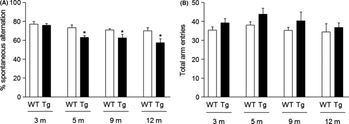
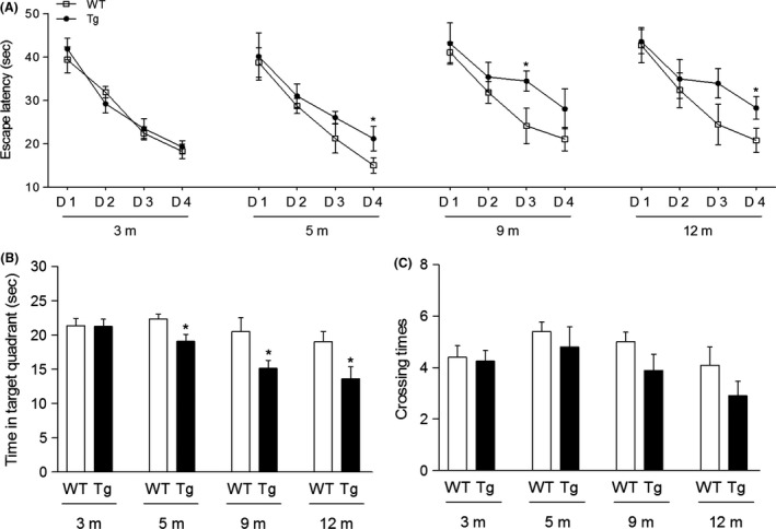
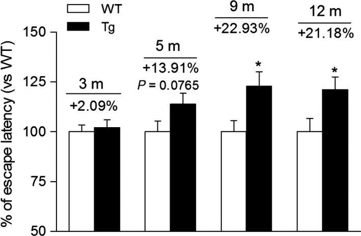
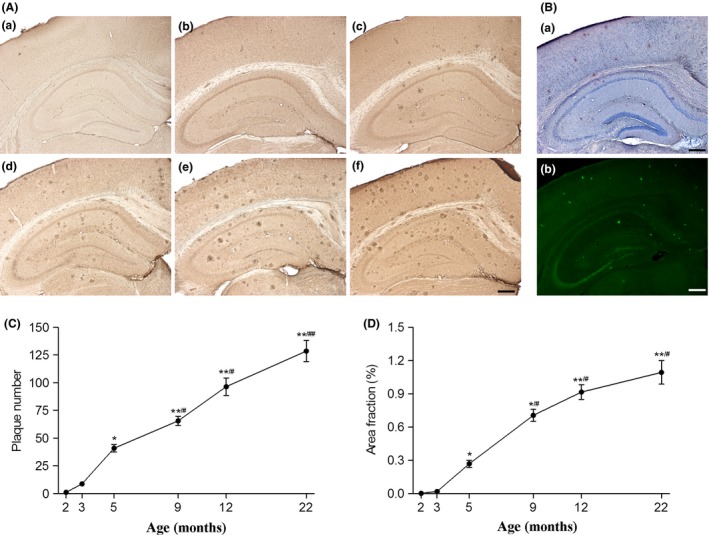
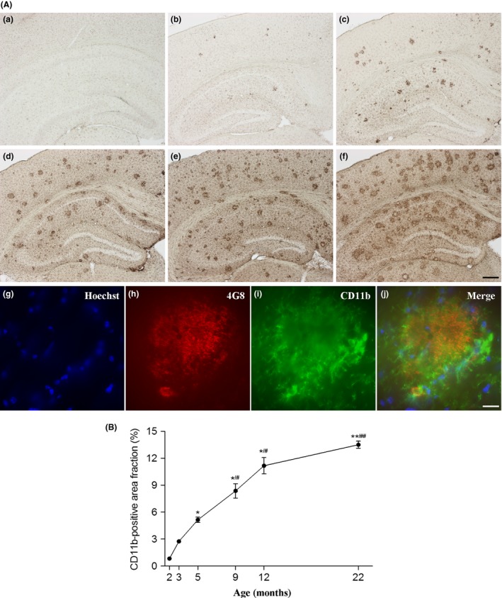
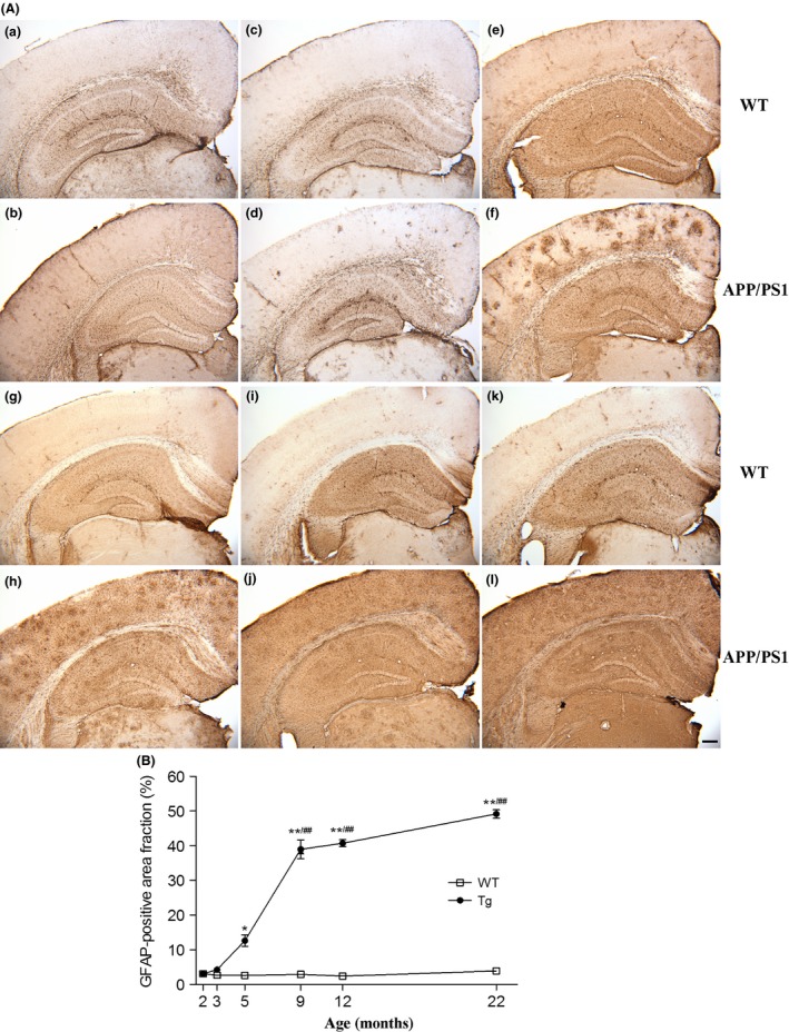
Results: APP/PS1 mice exhibited significant memory deficits from 5 months old, which were aggravated at the later stage of life. However, the degree of memory impairments reached a plateau at 12 months. An early appearance of amyloid plaques was at 3 months with a linear increase throughout the disease course. CD11b-positive microglia and glial fibrillary acidic protein-(GFAP) positive astrocytes were first detected at 3 months with a close association with amyloid plaques. Yet, the rate of changes in glial activation slowed down from 12 months despite the steady increase in Aβ.
Conclusion: These findings provided evidence that neuroinflammation might be involved in the development and progression of cognitive deficits in APP/PS1 mice, suggesting novel intervention and prevention strategies for AD.
Keywords: Alzheimer's disease; astrocyte; microglia; neuroinflammation.
© 2017 John Wiley & Sons Ltd.
Conflict of interest statement
The authors declare no conflict of interest.
Figures
Similar articles
-
Dihydromyricetin inhibits microglial activation and neuroinflammation by suppressing NLRP3 inflammasome activation in APP/PS1 transgenic mice.CNS Neurosci Ther. 2018 Dec;24(12):1207-1218. doi: 10.1111/cns.12983. Epub 2018 Jun 4. CNS Neurosci Ther. 2018. PMID: 29869390 Free PMC article.
-
Fruitless Wolfberry-Sprout Extract Rescued Cognitive Deficits and Attenuated Neuropathology in Alzheimer's Disease Transgenic Mice.Curr Alzheimer Res. 2018;15(9):856-868. doi: 10.2174/1567205015666180404160625. Curr Alzheimer Res. 2018. PMID: 29623840
-
Effects of accelerated senescence on learning and memory, locomotion and anxiety-like behavior in APP/PS1 mouse model of Alzheimer's disease.J Neurol Sci. 2013 Dec 15;335(1-2):145-54. doi: 10.1016/j.jns.2013.09.018. Epub 2013 Sep 21. J Neurol Sci. 2013. PMID: 24095271
-
Alzheimer's disease in the retina: imaging retinal aβ plaques for early diagnosis and therapy assessment.Neurodegener Dis. 2012;10(1-4):285-93. doi: 10.1159/000335154. Epub 2012 Feb 10. Neurodegener Dis. 2012. PMID: 22343730 Review.
-
Microglia and Astrocytes in Alzheimer's Disease: Implications for Therapy.Curr Neuropharmacol. 2018;16(5):508-518. doi: 10.2174/1570159X15666170720095240. Curr Neuropharmacol. 2018. PMID: 28730967 Free PMC article. Review.
Cited by
-
Long-Term Treatment with Fluoroethylnormemantine (FENM) Alleviated Memory Deficits, Amyloid Pathology, and Microglial Reaction in APP/PS1 Mice.ACS Pharmacol Transl Sci. 2024 Dec 2;7(12):4069-4082. doi: 10.1021/acsptsci.4c00522. eCollection 2024 Dec 13. ACS Pharmacol Transl Sci. 2024. PMID: 39698294
-
Disease Stage-Associated Alterations in Learning and Memory through the Electroacupuncture Modulation of the Cortical Microglial M1/M2 Polarization in Mice with Alzheimer's Disease.Neural Plast. 2020 Aug 28;2020:8836173. doi: 10.1155/2020/8836173. eCollection 2020. Neural Plast. 2020. PMID: 32908486 Free PMC article.
-
ΔFosB is part of a homeostatic mechanism that protects the epileptic brain from further deterioration.Front Mol Neurosci. 2024 Jan 12;16:1324922. doi: 10.3389/fnmol.2023.1324922. eCollection 2023. Front Mol Neurosci. 2024. PMID: 38283700 Free PMC article.
-
Effects of Qi-Fu-Yin on aging of APP/PS1 transgenic mice by regulating the intestinal microbiome.Front Cell Infect Microbiol. 2023 Jan 13;12:1048513. doi: 10.3389/fcimb.2022.1048513. eCollection 2022. Front Cell Infect Microbiol. 2023. PMID: 36710967 Free PMC article.
-
Curcumin inhibits APOE4-induced injury by activating peroxisome proliferator-activated receptor-γ (PPARγ) in SH-SY5Y cells.Iran J Basic Med Sci. 2020 Dec;23(12):1576-1583. doi: 10.22038/ijbms.2020.47184.10858. Iran J Basic Med Sci. 2020. PMID: 33489032 Free PMC article.
References
-
- Hardy J, Selkoe DJ. The amyloid hypothesis of Alzheimer's disease: progress and problems on the road to therapeutics. Science. 2002;297:353–356. - PubMed
-
- Itagaki S, McGeer PL, Akiyama H, Zhu S, Selkoe D. Relationship of microglia and astrocytes to amyloid deposits of Alzheimer disease. J Neuroimmunol. 1989;24:173–182. - PubMed
-
- Ho GJ, Drego R, Hakimian E, Masliah E. Mechanisms of cell signaling and inflammation in Alzheimer's disease. Curr Drug Targets Inflamm Allergy. 2005;4:247–256. - PubMed
MeSH terms
Substances
LinkOut - more resources
Full Text Sources
Other Literature Sources
Medical
Research Materials
Miscellaneous
