. 2017 Feb;6(2):527-538.
doi: 10.5966/sctm.2015-0428. Epub 2016 Sep 2.

Heparin Promotes Cardiac Differentiation of Human Pluripotent Stem Cells in Chemically Defined Albumin-Free Medium, Enabling Consistent Manufacture of Cardiomyocytes

Affiliations

Heparin Promotes Cardiac Differentiation of Human Pluripotent Stem Cells in Chemically Defined Albumin-Free Medium, Enabling Consistent Manufacture of Cardiomyocytes

Yongshun Lin et al. Stem Cells Transl Med. 2017 Feb.

Abstract

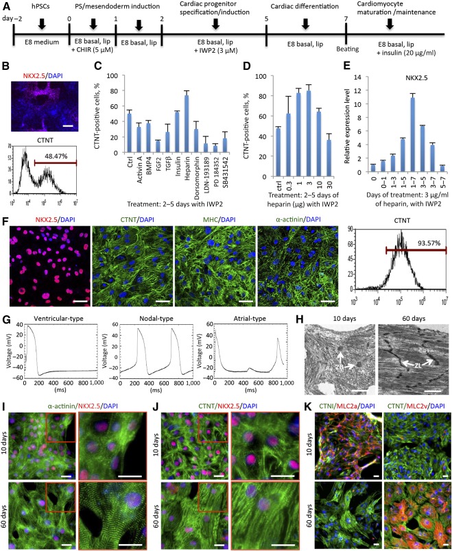
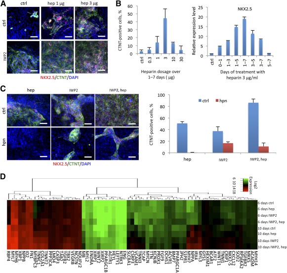
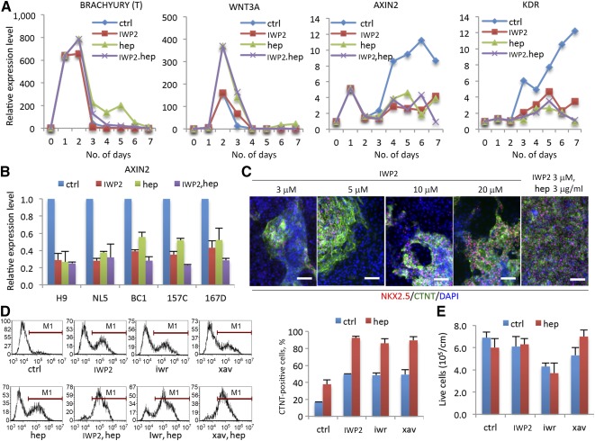
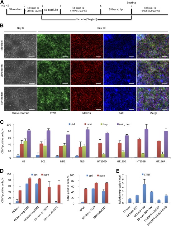
Cardiomyocytes can be differentiated from human pluripotent stem cells (hPSCs) in defined conditions, but efficient and consistent cardiomyocyte differentiation often requires expensive reagents such as B27 supplement or recombinant albumin. Using a chemically defined albumin-free (E8 basal) medium, we identified heparin as a novel factor that significantly promotes cardiomyocyte differentiation efficiency, and developed an efficient method to differentiate hPSCs into cardiomyocytes. The treatment with heparin helped cardiomyocyte differentiation consistently reach at least 80% purity (up to 95%) from more than 10 different hPSC lines in chemically defined Dulbecco's modified Eagle's medium/F-12-based medium on either Matrigel or defined matrices like vitronectin and Synthemax. One of heparin's main functions was to act as a Wnt modulator that helped promote robust and consistent cardiomyocyte production. Our study provides an efficient, reliable, and cost-effective method for cardiomyocyte derivation from hPSCs that can be used for potential large-scale drug screening, disease modeling, and future cellular therapies. Stem Cells Translational Medicine 2017;6:527-538.

Keywords: Cardiac; Cell culture; Embryonic stem cells; Heparin; Induced pluripotent stem cells.


Figures

Figure 1
Figure 1
Heparin enhances hPSC cardiomyocyte differentiation along with Wnt modulation in E8 medium. (A): Schematic of hPSC differentiation protocol with singular modulation of Wnt signaling in the absence of B27. The experiments were carried out in chemically defined E8 basal medium (Dulbecco's modified Eagle's medium/F‐12 plus l‐ascorbic acid, selenium, and transferrin) with chemically defined lipid concentrate. (B): Cardiac differentiation efficiency without B27 in an E8‐based platform displayed by immunofluorescence of NKX2.5 (top) and flow cytometry of CTNT (bottom). Scale bar = 100 μm. (C): Screen of signaling pathway regulation during cardiac specification (treated at days 2–5). Data are presented as the mean ± SD of three or more independent experiments. (D): Heparin dosage screen for CTNT‐positive cell percentage by flow cytometry at day 10 when treated at days 2–5 in the presence of Wnt inhibitor IWP2. Data are presented as the mean ± SD of three or more independent experiments. (E): Real‐time polymerase chain reaction of NKX2.5 indicates the effect of various heparin time‐course (3 μg/ml) treatments in the presence of the Wnt inhibitor IWP2. Data are presented as the mean ± SD of three or more independent experiments. (F): Confocal immunofluorescence and flow cytometry of cardiac markers indicate that the derived cardiomyocyte population was up to 95% pure with heparin and IWP2 treatment. (G): Electrophysiological characterization by representative action potential activities of 3 cardiomyocyte subtypes at day 30 after differentiation (n = 3 cells for atrial and ventricular types and n = 2 cells for nodal type). (H–K): Maturation of cardiomyocytes on culture plates. Transmission electron microscopy showed the more highly organized and clearly aligned myofibrils of day‐60 cardiomyocytes compared with that of day‐10 cells, as well as the Z‐lines of myofibril bundles at day 60 rather than the Z‐bodies of premyofibrils at day 10 (H). Immunofluorescence of α‐actinin (I) and CTNT (J) showed the increased sarcomere alignment in cardiomyocytes from 10 days to 60 days of culture. (K): Costaining of MLC2a and MLC2v with CTNI or CTNT showed that the majority of cells at early stage (10 days) expressed MLC2a, whereas the majority of cells at late stage (60 days) expressed MLC2v. (H): Scale bar = 1 μm. (F, I–K): Scale bar = 25 μm. Abbreviations: CTNI, cardiac troponin I; CTNT, cardiac troponin T; ctrl, control; hep, heparin; DAPI, 4′,6‐diamidino‐2‐phenylindole; hPSC, human pluripotent stem cell; lip, chemically defined lipid; MHC, myosin heavy chain; MLC2a, myosin light chain 2a; MLC2v, myosin light chain 2v; PS, primitive streak; ZB, Z‐body; ZL, Z‐line.
Figure 2
Figure 2
Heparin alone promoted cardiac differentiation from human pluripotent stem cells without treatment of Wnt inhibitors. (A): Immunofluorescence for NKX2.5 (red) and CTNT‐positive cells (green) at day 10 after treatment with 1 μg/ml or 3 μg/ml heparin in the absence or presence of IWP2. Scale bar = 50 μm. (B): Flow cytometry for CTNT (left) and real‐time polymerase chain reaction of NKX2.5 (right) comparing the dosage (treatment from day 1 to day 7) and time course (3 μg/ml treatment) of heparin treatment to promote cardiac differentiation in the absence of IWP2 treatment. Data are presented as the mean ± SD of three or more independent experiments. (C): Immunofluorescence (left) of CTNT (green) and NKX2.5 (red) as well as flow cytometry (right) with CTNT antibody, demonstrating that heparinase I blocked the effects of heparin and inhibited cardiomyocyte derivation. Data are presented as the mean ± SD of three or more independent experiments. Scale bar = 50 μm. (D): Heat map illustrating the promotion of cardiogenesis by heparin during differentiation at day 6 and day 10 in either absence or presence of IWP2. Abbreviations: CTNT, cardiac troponin T; ctrl, control; DAPI, 4′,6‐diamidino‐2‐phenylindole; hep, heparin; hpn, heparinase I.
Figure 3
Figure 3
Heparin modulated canonical Wnt signaling during human pluripotent stem cell (hPSC) cardiac differentiation. (A): Heparin upregulated WNT3A expression at mesoderm induction stage (days 2–3) but inhibited canonical Wnt signaling downstream target AXIN2 and downregulated cardiovascular progenitor cell marker KDR during cardiac specification stage (days 3–6) as indicated by reverse transcription‐quantitative polymerase chain reaction (RT‐qPCR). Data are presented as the mean ± SD of three or more independent experiments. (B): RT‐qPCR for AXIN2 at day 5 with IWP2, heparin, and combination treatments in 5 hPSC lines. Data are presented as the mean ± SD of three or more independent experiments. (C): Immunofluorescence of NKX2.5 (red) and CTNT (green) at day 10 shows the differentiation efficiency among different dosages of IWP2 alone and in combination with heparin. Data are presented as the mean ± SD of three or more independent experiments. Scale bar = 50 μm. (D): Comparison of different Wnt inhibitors on cardiac differentiation with or without heparin treatment. Flow cytometry of CTNT‐positive cell population after 10 days of differentiation (left); percentage of CTNT positive cells is quantified (right). Data are presented as the mean ± SD of three or more independent experiments. (E): The yield of live cells with different Wnt inhibitor treatments after 10 days of differentiation in the absence (ctrl) or presence (hep) of heparin. Abbreviations: CTNT, cardiac troponin T; ctrl, control; DAPI, 4′,6‐diamidino‐2‐phenylindole; hep, heparin; iwr, IWR1; xav, XAV939.
Figure 4
Figure 4
Heparin promoted cardiac differentiation from multiple human pluripotent stem cell (hPSC) lines in DMEM/F‐12‐based media. (A): Schematic of the hPSC differentiation protocol with modulation of Wnt signaling and treatment of heparin in chemically defined E8 medium. (B): Heparin promotes cardiomyocyte differentiation on Matrigel, vitronectin, and Synthemax matrices. Phase contrast images show the day 0 colony morphology before inducing differentiation. Immunofluorescence of CTNT‐positive (green) and NKX2.5‐positive (red) cardiomyocytes at day 10 of differentiation. Scale bar = 50 μm. (C): The cardiac derivation efficiency at day 10 as determined by flow cytometry with CTNT antibody. Data are presented as the mean ± SD of three or more independent experiments. (D): Flow cytometry using CTNT antibody comparing the effects of different batches of heparin and recombinant human albumin on cardiac differentiation with E8 basal medium (left) or RPMI medium (right) in either absence or presence of IWP2 treatment. The batch numbers of the reagents are listed. Data are presented as the mean ± SD of three or more independent experiments. (E): Quantitative reverse transcription‐polymerase chain reaction for CTNT comparing the effects of heparin and B27 (without insulin) on cardiomyocyte differentiation in E8 basal medium and DMEM/F‐12 medium. Data are presented as the mean ± SD of three or more independent experiments. Abbreviations: alb, albumin; B27, B27 without insulin; CTNI, cardiac troponin I; CTNT, cardiac troponin T; ctrl, control; hep, heparin; DAPI, 4′,6‐diamidino‐2‐phenylindole; DMEM, Dulbecco's modified Eagle's medium; lip, chemically defined lipid; RPMI, Roswell Park Memorial Institute medium.
Figure 5
Figure 5
In vivo transplantation of human pluripotent stem cell (hPSC)‐derived cardiomyocytes in NSG mice. (A): GFP‐labeled human induced PSC (hiPSC)‐derived cardiomyocytes (green: GFP labeled hiPSCs; red: CTNT; blue: DAPI stain). Scale bar = 50 μm. (B): Light microscopy (left) and GFP fluorescence (right) of a NSG mouse heart 2 weeks after infarction and hiPSC‐derived cardiomyocyte injection. Scale bar = 0.4 mm. (C): Immunohistochemistry for CTNT (antibody recognizes both human and mouse antigens; top) and desmin (antibody recognizes only human, and not mouse, antigen; bottom) on cross sections from the infarcted area of a NSG mouse heart performed at 7 weeks after injection of enhanced GFP‐labeled induced pluripotent stem cells‐derived cardiomyocytes. Abbreviations: CTNT, cardiac troponin T; DAPI, 4′,6‐diamidino‐2‐phenylindole; GFP, green fluorescent protein.

References

    1. Thomson JA, Itskovitz‐Eldor J, Shapiro SS et al. Embryonic stem cell lines derived from human blastocysts. Science 1998;282:1145–1147. - PubMed
    1. Takahashi K, Tanabe K, Ohnuki M et al. Induction of pluripotent stem cells from adult human fibroblasts by defined factors. Cell 2007;131:861–872. - PubMed
    1. Yu J, Vodyanik MA, Smuga‐Otto K et al. Induced pluripotent stem cell lines derived from human somatic cells. Science 2007;318:1917–1920. - PubMed
    1. Cherry AB, Daley GQ. Reprogrammed cells for disease modeling and regenerative medicine. Annu Rev Med 2013;64:277–290. - PMC - PubMed
    1. Burridge PW, Keller G, Gold JD et al. Production of de novo cardiomyocytes: human pluripotent stem cell differentiation and direct reprogramming. Cell Stem Cell 2012;10:16–28. - PMC - PubMed

Publication types

MeSH terms