. 2017 Feb;6(2):490-501.
doi: 10.5966/sctm.2016-0088. Epub 2016 Aug 24.

Directing Differentiation of Pluripotent Stem Cells Toward Retinal Pigment Epithelium Lineage

Affiliations

Directing Differentiation of Pluripotent Stem Cells Toward Retinal Pigment Epithelium Lineage

Parul Choudhary et al. Stem Cells Transl Med. 2017 Feb.

Abstract

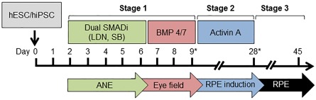
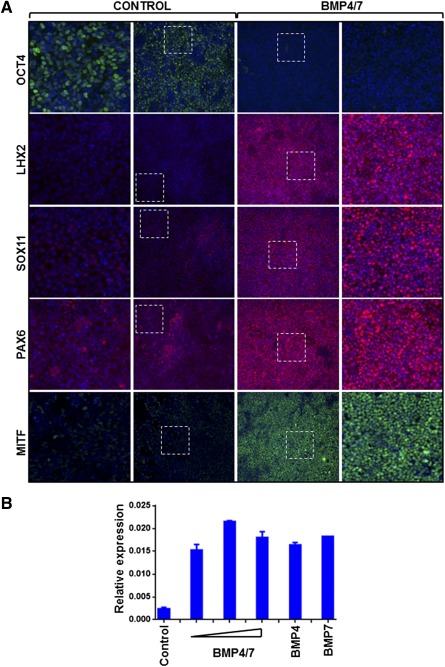
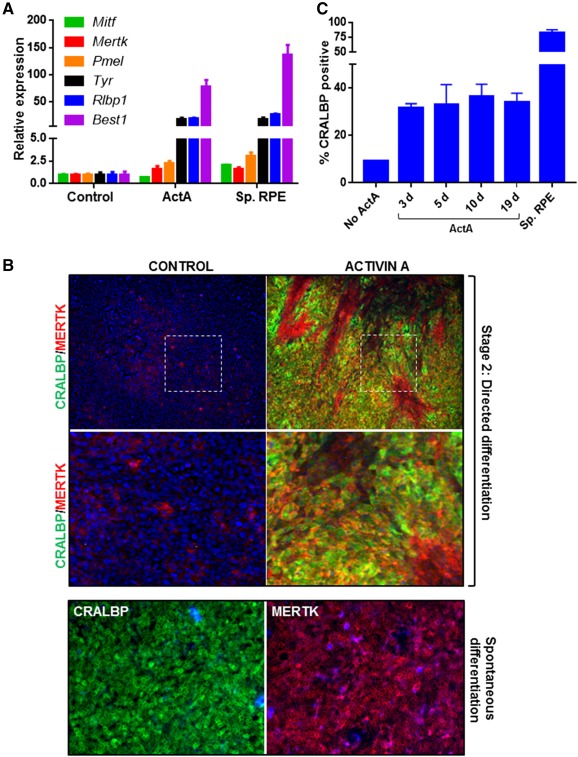
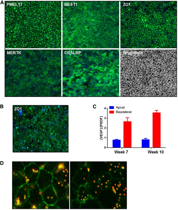
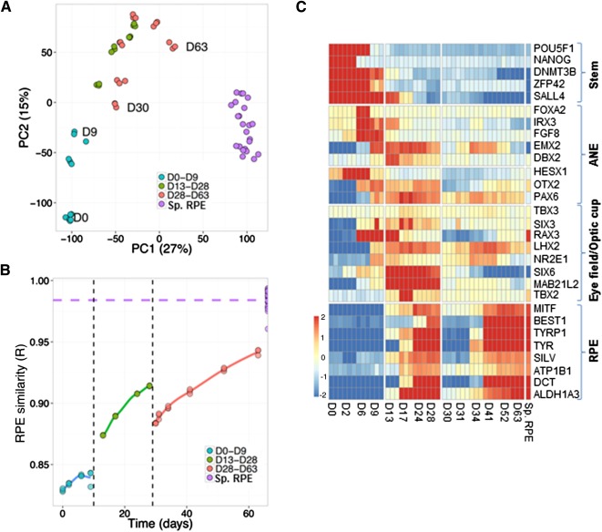
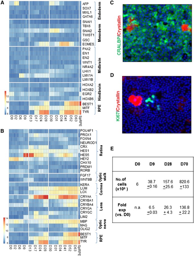
Development of efficient and reproducible conditions for directed differentiation of pluripotent stem cells into specific cell types is important not only to understand early human development but also to enable more practical applications, such as in vitro disease modeling, drug discovery, and cell therapies. The differentiation of stem cells to retinal pigment epithelium (RPE) in particular holds promise as a source of cells for therapeutic replacement in age-related macular degeneration. Here we show development of an efficient method for deriving homogeneous RPE populations in a period of 45 days using an adherent, monolayer system and defined xeno-free media and matrices. The method utilizes sequential inhibition and activation of the Activin and bone morphogenetic protein signaling pathways and can be applied to both human embryonic stem cells and induced pluripotent stem cells as the starting population. In addition, we use whole genome transcript analysis to characterize cells at different stages of differentiation that provides further understanding of the developmental dynamics and fate specification of RPE. We show that with the described method, RPE develop through stages consistent with their formation during embryonic development. This characterization- together with the absence of steps involving embryoid bodies, three-dimensional culture, or manual dissections, which are common features of other protocols-makes this process very attractive for use in research as well as for clinical applications. Stem Cells Translational Medicine 2017;6:490-501.

Keywords: Activin; Bone morphogenetic protein; Directed differentiation; Retinal pigment epithelium; Stem cells.


Figures

Figure 1
Figure 1
Directed differentiation of stem cells to retinal pigment epithelium. Schematic of the directed differentiation protocol described in this study. ∗, replating steps. Abbreviations: ANE, anterior neuroectoderm; BMP, bone morphogenetic protein; RPE, retinal pigment epithelium; hESC, human embryonic stem cells; hiPSC, human induced pluripotent stem cells; RPE, retinal pigment epithelium; SMADi, inhibitor of SMAD pathway.
Figure 2
Figure 2
Stage 1: induction of eye field from human embryonic stem cells (hESC). (A): Two days’ postseeding, SHEF1 hESC were treated with Dulbecco's modified Eagle's medium KSR‐XF alone (Control) or Induction Medium 1 for 4 days, followed by Induction Medium 2 for 3 days (BMP4/7). Representative images showing immunocytochemistry for the pluripotency marker OCT4 and eye field markers LHX2, SOX11, PAX6, and MITF are shown. Dotted squares indicate the region that is magnified in the adjacent panels. Images are captured at ×10 magnification. (B): Quantitative polymerase chain reaction was carried out to measure expression of Mitf transcript in SHEF1 hESC treated with Induction Medium 1 alone (Control) or Induction Medium 1 supplemented with BMP4/7 (50 ng/ml, 100 ng/ml, 200 ng/ml; shown by triangle in order of ascending concentration); BMP4 (200 ng/ml); or BMP7 (200 ng/ml) (n = 3, ±SD). Abbreviation: BMP, bone morphogenetic protein.
Figure 3
Figure 3
Stage 2: Generation of a mixed RPE population. (A): Two days’ postseeding, SHEF1 human embryonic stem cells were treated with Induction Medium 1 for 4 days, followed by Induction Medium 2 for 3 days. At day 9, cells were replated in Dulbecco's modified Eagle's medium (DMEM) KSR‐XF alone (Control) or Induction Medium 3 (ActA) for 19 days. Quantitative polymerase chain reaction was used to measure expression of a panel of RPE markers. RPE generated by using spontaneous differentiation (Sp. RPE) were used for comparison (n = 3, ±SD). (B): Representative images showing immunocytochemistry for CRALBP and MERTK in cells derived by directed differentiation and cultured as in panel A. Dotted squares indicate the region that is magnified in the panels below. RPE generated by spontaneous differentiation were used for comparison. Images are captured at ×10 magnification. (C): Quantification of CRALBP immunocytochemistry at day 28 in which cells during stage 2 were treated with DMEM KSR‐XF (Control) or Induction Medium 3 (ActA) for 3 days, 5 days, 10 days, or 19 days. For fewer than 19‐day ActA treatments, DMEM KSR‐XF was used for the remainder of the 19‐day period. RPE derived by using spontaneous differentiation (Sp. RPE) were used for comparison (n = 3, ±SD). Abbreviations: ActA, Activin A; d, days; RPE, retinal pigment epithelium; Sp. RPE, spontaneous differentiation retinal pigment epithelium.
Figure 4
Figure 4
Stage 3: Generation of a homogenous and functional retinal pigment epithelium (RPE) population. (A): Two days’ postseeding, SHEF1 human embryonic stem cells were treated with Induction Medium 1 for 4 days, followed by Induction Medium 2 for 3 days. At day 9, cells were replated in Induction Medium 3 for a period of 19 days. At day 28, cells were replated in Dulbecco's modified Eagle's medium KSR‐XF and cultured for a period of 14 days. Representative images showing immunocytochemistry for indicated RPE markers are shown. Images are captured at ×10 magnification. (B): Cells cultured as in panel A were further dissociated and replated on transwells and cultured for a period of 10 weeks. A representative image showing en face immunocytochemistry for ZO‐1 is shown. Images are captured at ×20 magnification. (C): Spent medium from transwells described in panel B was collected from the top and bottom chambers and quantified for vascular endothelial growth factor (VEGF) and pigment epithelium‐derived factor (PEDF) concentration. The ratio of [VEGF]:[PEDF] was quantified in media from the two compartments (n = 3, ±SD). (D): Representative confocal images showing phagocytosis of fluorescent bead (red) by RPE. ZO‐1 immunocytochemistry (green) shows the cell edge, and the presence of bead within the cell boundary indicates internalization by phagocytosis. Images are captured at ×63 magnification. Abbreviations: PEDF, pigment epithelium‐derived factor; VEGF, vascular endothelial growth factor.
Figure 5
Figure 5
Transcriptome analysis of the directed differentiation protocol. (A, B): Human embryonic stem cells were cultured as described in Figure 4A, and samples were collected at different time points of the differentiation protocol. Similarity between samples at different points of the protocol is shown by principal components analysis (A) and Pearson's correlation coefficient (B). (C): Microarray heatmap showing expression of markers representative of different stages of retinal pigment epithelium differentiation. The x‐axis refers to samples at different time points of directed differentiation. Abbreviations: ANE, anterior neuroectoderm; D, day; PC1, first principal component; PC2, second principal component; RPE, retinal pigment epithelium; Sp. RPE, RPE generated by spontaneous differentiation.
Figure 6
Figure 6
Specificity and efficiency of retinal pigment epithelium (RPE) differentiation. (A, B): Microarray heatmap showing expression of markers representative of different lineages and brain regions (A) and regions of the eye (B). The x‐axis refers to samples at different time points of directed differentiation and the Sp. RPE samples refer to RPE generated by spontaneous differentiation. (C, D): Representative images for immunocytochemistry of CRALBP (green) and β‐crystallin (red) (C) and Ki67 (green) and β‐crystallin (red) (D) are shown. Images are captured at ×10 magnification. (E): Table summarizing yield and fold expansion of cells at various stages of the directed differentiation protocol. Abbreviations: D, day; exp, expansion; n.a., not applicable; RPE, retinal pigment epithelium; SpRPE, RPE generated by spontaneous differentiation.

References

    1. Friedman DS, O'Colmain BJ, Muñoz B et al. Prevalence of age‐related macular degeneration in the United States. Arch Ophthalmol 2004;122:564–572. - PubMed
    1. Strauss O. The retinal pigment epithelium in visual function. Physiol Rev 2005;85:845–881. - PubMed
    1. Gehrs KM, Anderson DH, Johnson LV et al. Age‐related macular degeneration—Emerging pathogenetic and therapeutic concepts. Ann Med 2006;38:450–471. - PMC - PubMed
    1. Schwartz SD, Hubschman JP, Heilwell G et al. Embryonic stem cell trials for macular degeneration: A preliminary report. Lancet 2012;379:713–720. - PubMed
    1. Schwartz SD, Regillo CD, Lam BL et al. Human embryonic stem cell‐derived retinal pigment epithelium in patients with age‐related macular degeneration and Stargardt's macular dystrophy: Follow‐up of two open‐label phase 1/2 studies. Lancet 2015;385:509–516. - PubMed

MeSH terms