Comparative Study
. 2017 Feb;6(2):601-612.
doi: 10.5966/sctm.2016-0050. Epub 2016 Sep 14.

Angiogenic Potential of Human Bone Marrow-Derived Mesenchymal Stem Cells in Chondrocyte Brick-Enriched Constructs Promoted Stable Regeneration of Craniofacial Cartilage

Affiliations
Comparative Study

Angiogenic Potential of Human Bone Marrow-Derived Mesenchymal Stem Cells in Chondrocyte Brick-Enriched Constructs Promoted Stable Regeneration of Craniofacial Cartilage

Zhiye Li et al. Stem Cells Transl Med. 2017 Feb.

Abstract

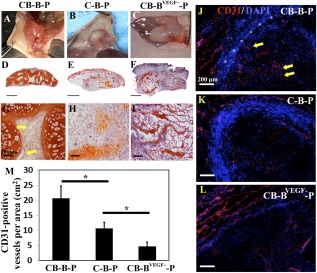
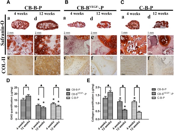
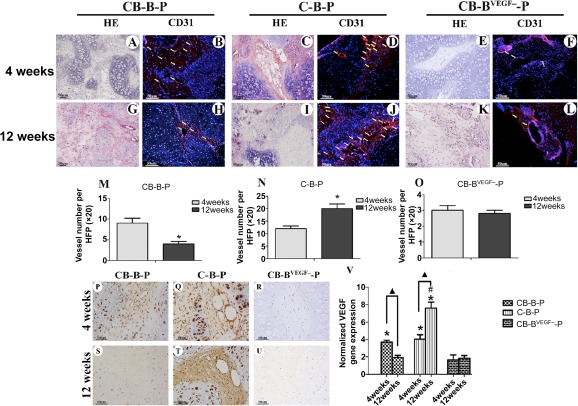
Craniofacial deformities caused by congenital defects or trauma remain challenges for clinicians, whereas current surgical interventions present limited therapeutic outcomes. Injection of bone marrow-derived mesenchymal stem cells (BMSCs) into the defect is highly desirable because such a procedure is microinvasive and grafts are more flexible to fill the lesions. However, preventing hypertrophic transition and morphological contraction remain significant challenges. We have developed an "all host derived" cell transplantation system composed of chondrocyte brick (CB)-enriched platelet-rich plasma (P) gel and BMSCs (B). Without exogenous biomaterials or growth factors, such grafts regenerate cartilage efficiently and present great clinical promise. In immunodeficient mice, we compared performance of BMSCs and BMSCs lacking angiogenic potential in CB-B-P constructs and followed the cartilage maturation process by histology, immunostaining, micro-computed tomography, and protein analysis. We determined that angiogenesis occurred quickly inside rudimentary cartilage derived from CB-B-P constructs after implantation, which improved tissue survival, tissue growth, and production of chondrogenic signals from chondrocytes. In contrast, silencing angiogenic potential of BMSCs led to poor chondrogenesis accompanied by necrosis. Chondrocyte bricks merged rapidly with angiogenesis, which constituted an enclosed chondrogenic niche and effectively inhibited runt-related transcription factor-2-dependent hypertrophic transition of BMSCs as well as endochondral ossification; progressive chondrogenic differentiation of BMSCs resulted in vascularization regression, thus favoring persistent chondrogenesis and effectively augmenting nasal cartilage. In conclusion, these findings provided a novel, efficient approach to regenerating cartilage tissues in vivo. Chondrocyte bricks mixed with P provide transient vascularization and a persistently chondrogenic microenvironment for BMSCs; this provides a mini-invasive approach for craniofacial cartilage reconstruction. Stem Cells Translational Medicine 2017;6:601-612.

Keywords: Adult human bone marrow; Angiogenesis; Bone marrow stromal cells; Cell transplantation; Chondrogenesis.


Figures

Figure 1
Figure 1
Characterization of CB‐based injectable cell aggregate. (A): Schematic depiction of the strategy for CB‐based cell transplantation and in vivo implantation. (B): A chondrocyte sheet was cultured, harvested, and embedded for fragmenting in a net cutting system (by multiple blades). (C): The CBs or chondrocytes and cultured BMSCs were mixed together or BMSCs alone were suspended in PRP, so that the injectable constructs were formed and injected subcutaneously into nude mice. (Da, Db): Scanning electron microscopy images show that CBs mixed with PRP gel formed small cavities and connected with each other. (Dc): BMSCs filled in cavities among chondrocytes bricks. (Dd): BMSCs aggregated among CBs and reconstituted into macroaggregate along with CBs (arrowheads). ([Da, Dc]: low magnification, scale bars = 1 mm; [Db, Dd]: high magnification, scale bars = 100 μm). Abbreviations: BMSC, bone marrow‐derived mesenchymal stem cells; CB, chondrocyte brick; P and PRP, platelet‐rich plasma.
Figure 2
Figure 2
Proangiogenic role of BMSCs in coculturing system in vitro. (A): Representative images showed cell migration of HUVECs after coculturing with conditioned medium derived from BMSCs, C, CBs, BMSCVEGF−, C + BMSC, CB + BMSC, and CB + BMSCVEGF−, respectively. Scale bars = 50 μm. (B): Bar graph shows quantitative analysis of cell migration of HUVECs in each group; n = 4, ∗, p < .05; #, p > .05. (C): Protein expression of VEGF and actin by Western blot in BMSC, CB + BMSC, CB + BMSCVEGF−., and C + BMSC. (D): Detection of VEGF concentration by using enzyme‐linked immunosorbent assay in conditioned medium of BMSC, C + BMSC, CB + BMSC, CB + BMSCVEGF−; n = 4 for each group. Abbreviations: BMSC, bone marrow‐derived mesenchymal stem cells; BMSCVEGF−, vascular endothelial growth factor‐silencing bone marrow‐derived mesenchymal stem cells; C, chondrocyte; CB, chondrocyte brick; DMEM, Dulbecco's modified Eagle's medium; HUVEC, human umbilical vein endothelial cell; VEGF, vascular endothelial growth factor.
Figure 3
Figure 3
Sprouting of new blood vessels from the host to the engineered tissue grafts 2 weeks after implantation. (A–C): Macroscopic appearances of constructs after 2 weeks in vivo. (D–F): Safranin‐O staining of grafts 2 weeks after implantation (scale bars = 2 mm). (G): Orange staining depicts the CBs; spindle cells among CBs are biomaterial seeded with mesenchymal stem cells. Yellow arrows, host vessel sprouting to the constructs. (G–I): High magnification of images showing details; yellow arrows, new capillaries (original magnification, ×20; scale bars = 100 μm). (J–L): Representative images of CD31‐stained blood vessels (red) in grafts at 2 weeks after implantation. (J): CB‐B‐P grafts. (K): C‐B‐P grafts. (L): CB‐BVEGF−‐P grafts. For J–L, the nuclei are stained blue (scale bar = 200 μm). (M): Vascularization quantification of CB‐B‐P versus C‐B‐P or CB‐BVEGF−‐P constructs. The density of CD31‐positive vessels was measured at 2 weeks after implantation. All values are normalized to the graft area (cm2). ∗, p < .05. For all determinations, the sample size was n = 3, and all values are represented as mean ± SEM. Abbreviations: B, bone marrow‐derived mesenchymal stem cells; BVEGF−, vascular endothelial growth factor‐silencing bone marrow‐derived mesenchymal stem cells; C, chondrocyte; CB, chondrocyte brick; DAPI, 4′,6‐diamidino‐2‐phenylindole; P, platelet‐rich plasma.
Figure 4
Figure 4
Evaluation of cartilage formation for CB‐B‐P, C‐B‐P, and CB‐BVEGF‐‐P constructs. (A–C): Histological analysis of chondrogenesis of grafts in vivo after 4 weeks (left lane of [A–C]) and 12 weeks (right lane of [A–C]). Merged images (Safranin‐O staining) showed that the CB‐B‐P and C‐B‐P groups acquired tissue survival throughout, whereas CB‐BVEGF‐‐P group was characterized by central necrosis and poor tissue merging. The CB‐B‐P group presented good chondrogenic differentiation in the biomaterial seeded with mesenchymal stem cell regions, as was confirmed by Safranin‐O and COL‐II immunostaining (Aa–Af). In contrast, progressive osteogenesis and calcification occurred in the C‐B‐P group (Ca–Cf). Scale bars = 2 mm (Aa Ad, Ba, Bd), 1 mm (Ca, Cd), and 50 μm (Ab, Ac, Ae, Af, Bb, Bc, Be, Bf, Cb, Cc, Ce, Cf). (D, E): Quantitative evaluation of cartilaginous extracellular matrix, including glycosaminoglycans and collagen. ∗, p < .05 compared with the same time points of CB‐B‐P constructs; ▲, p < .05 for comparision of the same group between 4 and 12 weeks (n = 4 in each group). Abbreviations: B, bone marrow‐derived mesenchymal stem cells; BVEGF−, vascular endothelial growth factor‐silencing bone marrow‐derived mesenchymal stem cells; C, chondrocyte; CB, chondrocyte brick; P, platelet‐rich plasma.
Figure 5
Figure 5
Evaluation of angiogenesis at weeks 4 and 12. Representative images of CB‐B‐P, C‐B‐P and C‐BVEGF‐‐P constructs revealed infiltration of capillaries at week 4 (A, C, E) and 12 (G, I, K), which was predominantly distributed in bone marrow‐derived mesenchymal stem cells (BMSC) regions, as confirmed by CD31 immunofluorescent staining (4 weeks: [B, D, F]; 12 weeks: [H, J, K]); scale bars = 50 μm. (M–O): Comparison of the number of blood vessels between weeks 4 and 12; the value represents the average number of all fields examined (n = 6). (P–U): Immunostaining of VEGF reveals decreasing expression of VEGF in BMSC regions through 4 and 12 weeks in the CB‐B‐P group, whereas increasing expression in the C‐B‐P group, in addition, C‐BVEGF‐‐P constructs showed weak VEGF expression; scale bars = 50 μm. (V): Real‐time polymerase chain reaction quantitatively revealed the expression profile of VEGF in all groups. ∗, p < .05 compared with CB‐BVEGF−‐P at the same time point, #, p < .05 compared with CB‐B‐P at week 12; triangles, p < .05 for comparision of the same group between 4 and 12 weeks (n = 4 in each group). Abbreviations: B, bone marrow‐derived mesenchymal stem cells; BVEGF−, vascular endothelial growth factor‐silencing bone marrow‐derived mesenchymal stem cells; C, chondrocyte; CB, chondrocyte brick; HE, hematoxylin and eosin; HPF, high‐power field; P, platelet‐rich plasma; VEGF, vascular endothelial growth factor.
Figure 6
Figure 6
Osteogenesis of grafts in vivo at 4 weeks (A–H) and 12 weeks (I–P). The CB‐B‐P group did not present ossification of BMSCs through 12 weeks (A–D; I–L), as was confirmed by Von Kossa, Masson trichrome, COL‐I, and COL‐X immunostaining. In contrast, BMSCs in C‐B‐P presented early calcification, and progressive ossification occurred through 12 weeks (E–H, M–P); scale bars = 50 μm. Quantitative analysis of osteogenic markers including SOX‐9 (Q), RUNX‐2 (R), and COL‐X (S) by using real‐time polymerase chain reaction and Western blotting (T, U). ∗, p < .05 compared with native cartilage at the same time point; #, p < .05 compared with CB‐B‐P at the same time point; ▲, p < .05 for comparision of the same group between 4 and 12 weeks (n = 4 in each group). Abbreviations: B, bone marrow‐derived mesenchymal stem cells; C, chondrocyte; CB, chondrocyte brick; P, platelet‐rich plasma; RUN‐X, runt‐related transcription factor‐2.
Figure 7
Figure 7
Nasal augmentation by injecting chondrocyte bricks enriched constructs. (A, E): Macroscopic appearance of nasal dorsum at week 12 after injection of CB‐B‐P and CB‐BVEGF−‐P. (B, F): Examination of injected constructs by micro‐computed tomography. (C, D): Safranin‐O staining of the cross‐sections presented cartilage‐like tissue without ossification and higher magnification for above staining. (G, H): Safranin‐O staining for samples from CB‐BVEGF−‐P constructs. Scale bars = 1 mm (C, G), 50 μm (D, H). (I): Quantitative evaluation of peak thickness between CB‐B‐P and CB‐BVEGF−‐P constructs. ∗, p < .05 (n = 3 in each group). Abbreviations: B, bone marrow‐derived mesenchymal stem cells; BVEGF−, vascular endothelial growth factor‐silencing bone marrow‐derived mesenchymal stem cells; C, chondrocyte; CB, chondrocyte brick; CT, computed tomography; P, platelet‐rich plasma.

References

    1. Fulco I, Miot S, Haug MD et al. Engineered autologous cartilage tissue for nasal reconstruction after tumour resection: An observational first‐in‐human trial. Lancet 2014;384:337–346. - PubMed
    1. Hollander AP, Dickinson SC, Kafienah W. Stem cells and cartilage development: Complexities of a simple tissue. Stem Cells 2010;28:1992–1996. - PMC - PubMed
    1. Sasai Y. Next‐generation regenerative medicine: Organogenesis from stem cells in 3D culture. Cell Stem Cell 2013;12:520–530. - PubMed
    1. Scotti C, Tonnarelli B, Papadimitropoulos A et al. Recapitulation of endochondral bone formation using human adult mesenchymal stem cells as a paradigm for developmental engineering. Proc Natl Acad Sci USA 2010;107:7251–7256. - PMC - PubMed
    1. Ravindran S, Roam JL, Nguyen PK et al. Changes of chondrocyte expression profiles in human MSC aggregates in the presence of PEG microspheres and TGF‐β3. Biomaterials 2011;32:8436–8445. - PMC - PubMed

Publication types

MeSH terms

Substances