Comparative Study
. 2017 Feb;6(2):512-526.
doi: 10.5966/sctm.2015-0226. Epub 2016 Sep 7.

Generation of Osteosarcomas from a Combination of Rb Silencing and c-Myc Overexpression in Human Mesenchymal Stem Cells

Affiliations
Comparative Study

Generation of Osteosarcomas from a Combination of Rb Silencing and c-Myc Overexpression in Human Mesenchymal Stem Cells

Jir-You Wang et al. Stem Cells Transl Med. 2017 Feb.

Abstract

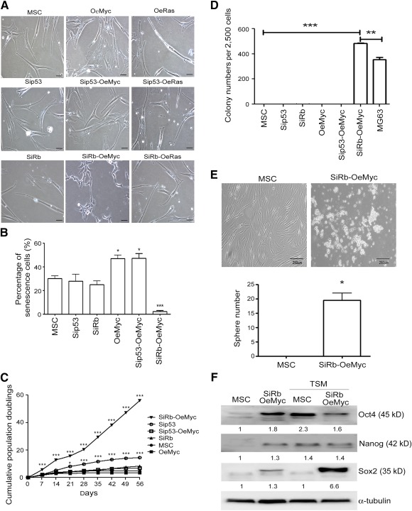
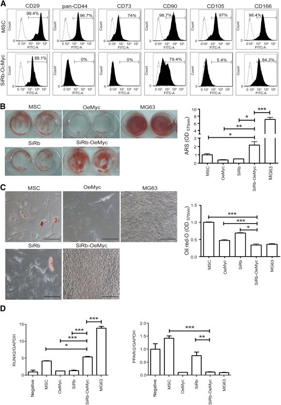
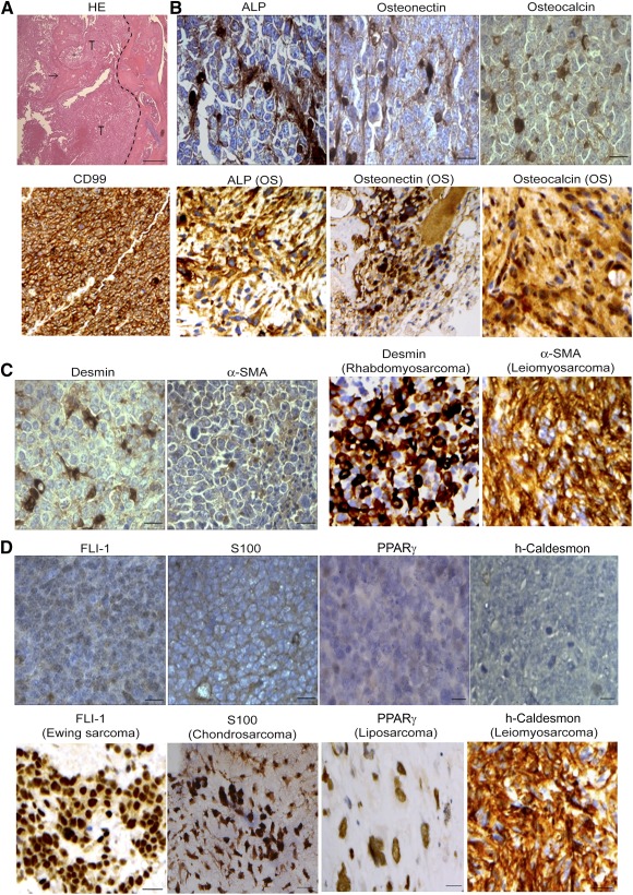
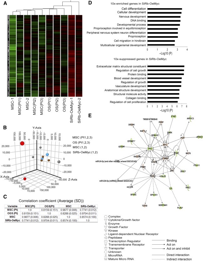
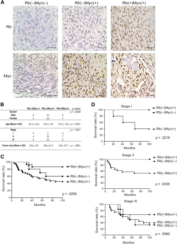
Osteosarcoma (OS) was a malignant tumor occurring with unknown etiology that made prevention and early diagnosis difficult. Mesenchymal stem cells (MSCs), which were found in bone marrow, were claimed to be a possible origin of OS but with little direct evidence. We aimed to characterize OS cells transformed from human MSCs (hMSCs) and identify their association with human primary OS cells and patient survival. Genetic modification with p53 or retinoblastoma (Rb) knockdown and c-Myc or Ras overexpression was applied for hMSC transformation. Transformed cells were assayed for proliferation, differentiation, tumorigenecity, and gene expression profile. Only the combination of Rb knockdown and c-Myc overexpression successfully transformed hMSCs derived from four individual donors, with increasing cell proliferation, decreasing cell senescence rate, and increasing ability to form colonies and spheres in serum-free medium. These transformed cells lost the expression of certain surface markers, increased in osteogenic potential, and decreased in adipogenic potential. After injection in immunodeficient mice, these cells formed OS-like tumors, as evidenced by radiographic analyses and immunohistochemistry of various OS markers. Microarray with cluster analysis revealed that these transformed cells have gene profiles more similar to patient-derived primary OS cells than their normal MSC counterparts. Most importantly, comparison of OS patient tumor samples revealed that a combination of Rb loss and c-Myc overexpression correlated with a decrease in patient survival. This study successfully transformed human MSCs to OS-like cells by Rb knockdown and c-Myc overexpression that may be a useful platform for further investigation of preventive and target therapy for human OS. Stem Cells Translational Medicine 2017;6:512-526.

Keywords: Mesenchymal stem cell; Osteosarcoma; Retinoblastoma (Rb) knockdown; Transformation; c-Myc.


Figures

Figure 1
Figure 1
Combination of Rb knockdown and c‐Myc overexpression transforms MSCs. (A): Parental MSCs or MSCs with genetic modification were grown in complete medium. Morphological changes were observed after genetic modification. (B): β‐galactosidase staining for analyzing senescence showed abundant senescent cells in OeMyc and Sip53‐OeMyc. Percentage of senescent cells in MSCs with different genetic modification. ∗, p < .05; ∗∗∗, p < .005 compared with MSC. (C): Cells were continuously subcultured in complete medium, and cumulative population doublings were calculated. (D): Cells were assayed for soft agar colony formation ability. (E): Cells were assayed for sphere formation in TSM. (F): Cells in TSM were subjected to Western blotting analysis for pluripotency gene expression. ∗, p < .05; ∗∗, p < .01; ∗∗∗, p < .005 compared with MSC. Bar = 500 μm. Abbreviations: MSC, mesenchymal stem cell; OeMyc, overexpression of c‐Myc; Rb, retinoblastoma; Sip53, silence of p53; SiRb, silence of retinoblastoma; TSM, tumor sphere medium.
Figure 2
Figure 2
Combination of retinoblastoma (Rb) knockdown and c‐Myc overexpression caused phenotypic changes. (A): Flow cytometric analysis for cell surface markers in parental MSCs and MSCs with SiRb and c‐Myc overexpression (SiRb‐OeMyc). (B left): Cells were induced for osteogenesis in osteogenic induction medium for 2 weeks, followed by ARS staining. (B right): Stained dye was extracted, and OD values were measured. (C left): Cells were induced for adipogenesis in adipogenic induction medium for 2 weeks, followed by Oil Red O staining. (C right): Stained dye was extracted, and OD values were measured. Bars = 100 μm. The gene expression levels of RUNX2 at 7 days of osteogenic induction and PPARγ2 at 7 days of adipogenic induction were analyzed by quantitative reverse‐transcriptase polymerase chain reaction assay. (D): Compared with parental MSCs, SiRb‐OeMyc cells significantly increased in RUNX2 mRNA level (left), whereas they decreased in PPARγ2 mRNA level (right). ∗, p < .05; ∗∗, p < .01; ∗∗∗, p < .005 compared with MSC. Abbreviations: ARS, Alizarin Red S; GAPDH, glyceraldehyde‐3‐phosphate dehydrogenase; MSC, mesenchymal stem cell; OD, optical density; OeMyc, overexpression of c‐Myc; PPARγ2, peroxisome proliferator‐activated receptor γ2; Rb, retinoblastoma; RUNX2, runt‐related gene 2; SiRb, silence of Rb.
Figure 3
Figure 3
Intraosseous tumor formation by SiRb‐OeMyc cells in nude mice. (A): After intraosseous injection, only SiRb‐OeMyc and MG63 cells formed tumor (arrows) at 4 weeks. (B): The ratio of tumor formation for each injection at the indicated time period after injection is shown. (C): Tumor growth curve after intraosseous injection. ∗, p < .05; ∗∗∗, p < .005. (D): Representative x‐ray plain films show tumor formation in SiRb‐OeMyc and MG63. The magnified rectangle shows sunburst features (arrows). (E): Representative magnetic resonance imaging images of intraosseous tumor formed by SiRb‐OeMyc. Dotted lines in coronal and sagittal views are tumor ranges and arrows in axial view show the location of tibia and fibula. (F): Representative micro‐positron emission tomography/computed tomography images for tumor location outlined by dotted lines. Abbreviations: MSC, mesenchymal stem cell; OeMyc, overexpression of c‐Myc; SiRb, silence of retinoblastoma; W, week.
Figure 4
Figure 4
Tumors formed by retinoblastoma SiRb‐OeMyc express OS features. (A): HE staining of intraosseous tumor formed by SiRb‐OeMyc showed new bone formation (arrow) within tumor. Dotted line indicates the tumor margin. (B–D): Immunohistochemistry shows tumors formed by SiRb‐OeMyc are positive for OS markers such as CD99, ALP, osteonectin, and osteocalcin (B, upper panels; brown); and slightly positive for sarcoma markers such as desmin and α‐SMA (C, left panels; brown); but negative for markers of Ewing sarcoma (FLI‐1), chondrosarcoma (S100), liposarcoma (PPARγ), and leiomyosarcoma (h‐caldesmon) (D). Positive controls of immunohistochemistry are from human pathological sections of OS (B, lower panels), rhabdomyosarcoma, leiomyosarcoma (C, right panels), Ewing sarcoma, chondrosarcoma, liposarcoma, and leiomyosarcoma (D, upper panels). Bars = 100 μm. Abbreviations: ALP, alkaline phosphatase; HE, hematoxylin and eosin; OS, osteosarcoma; PPARγ, peroxisome proliferator‐activated receptor γ; α‐SMA, α‐smooth muscle actin; T, tumor.
Figure 5
Figure 5
The SiRb‐OeMyc cells have similar gene profile as human primary OS cells. (A): Analyzing microarray data using cluster analysis. Heat map of genes with significant induction (red) or repression (green) showed the location of SiRb‐OeMyc behind three primary OS cells isolated from OS patients, whereas parental MSCs were close to primary MSCs from OS patients. (B): The principal component analysis shows both SiRb‐OeMyc and primary OS cells located far away from parental and patients’ derived MSCs, and scattered at different locations reflecting the diversity of tumor cells. (C): The correlation coefficient between SiRb‐OeMyc cells and primary OS cells is nearly 1, indicating the similarity of SiRb‐OeMyc cells and OS cells. (D): Gene Ontology analysis for 10‐fold upregulated or downregulated genes compared between SiRb‐OeMyc and parental MSCs. (E): Ingenuity pathway analysis for 10‐fold difference genes. The upregulated genes are marked as red and downregulated genes are marked as green, and no‐difference genes are shown as white. Abbreviations: MSC, mesenchymal stem cell; MSC‐1, parental MSC 1; OeMyc, overexpression of c‐Myc; OGS, osteogenic sarcoma; OS, osteosarcoma; Pt, OS patient; SiRb, silence of retinoblastoma.
Figure 6
Figure 6
The expression level of Rb and c‐Myc correlated of human osteosarcoma (OS) survival rate. (A): The immunohistochemistry of Rb and c‐Myc in 72 human OS patients’ tissue array showed three expression patterns, Rb(−)Myc(−), Rb(−)Myc(+), and Rb(+)Myc(+). (B): The patients’ information showed no significant difference in gender, age, and stage. The tumor size in Rb(+)Myc(+) seemed smaller than the other two groups, but showed no significant difference. (C): The survival rate in Rb(−)Myc(+) group was significant lower than the Rb(−)Myc(−) and Rb(+)Myc(+) groups. (D): The survival rate was significantly different in stage II, but not in stages I and III. Bars = 100 μm. Abbreviation: Rb, retinoblastoma.

References

    1. Ottaviani G, Jaffe N. The epidemiology of osteosarcoma. Cancer Treat Res 2009;152:3–13. - PubMed
    1. Briccoli A, Rocca M, Salone M et al. High grade osteosarcoma of the extremities metastatic to the lung: Long‐term results in 323 patients treated combining surgery and chemotherapy, 1985‐2005. Surg Oncol 2010;19:193–199. - PubMed
    1. Yamamoto N, Tsuchiya H. Chemotherapy for osteosarcoma—where does it come from? What is it? Where is it going?. Expert Opin Pharmacother 2013;14:2183–2193. - PubMed
    1. Friedenstein AJ, Chailakhyan RK, Latsinik NV et al. Stromal cells responsible for transferring the microenvironment of the hemopoietic tissues. Cloning in vitro and retransplantation in vivo. Transplantation 1974;17:331–340. - PubMed
    1. Pittenger MF, Mackay AM, Beck SC et al. Multilineage potential of adult human mesenchymal stem cells. Science 1999;284:143–147. - PubMed

Publication types

MeSH terms

Substances