Comparative Study
. 2017 Feb;6(2):382-393.
doi: 10.5966/sctm.2016-0201. Epub 2016 Sep 7.

Preventing Pluripotent Cell Teratoma in Regenerative Medicine Applied to Hematology Disorders

Affiliations
Comparative Study

Preventing Pluripotent Cell Teratoma in Regenerative Medicine Applied to Hematology Disorders

Aurelie Bedel et al. Stem Cells Transl Med. 2017 Feb.

Abstract

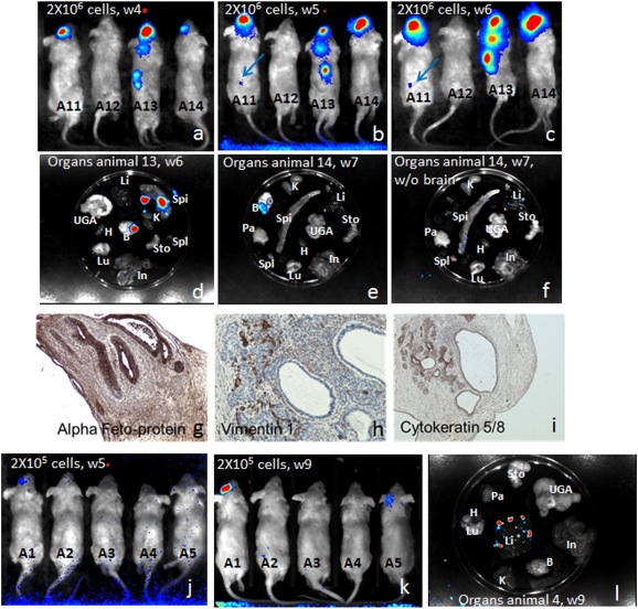
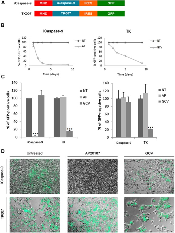
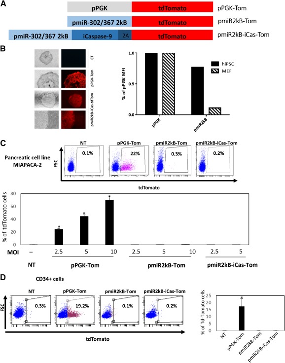
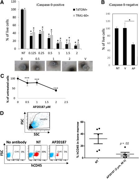
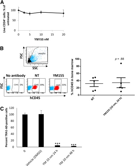
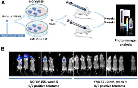
Iatrogenic tumorigenesis is a major limitation for the use of human induced pluripotent stem cells (hiPSCs) in hematology. The teratoma risk comes from the persistence of hiPSCs in differentiated cell populations. Our goal was to evaluate the best system to purge residual hiPSCs before graft without compromising hematopoietic repopulation capability. Teratoma risk after systemic injection of hiPSCs expressing the reporter gene luciferase was assessed for the first time. Teratoma formation in immune-deficient mice was tracked by in vivo bioimaging. We observed that systemic injection of hiPSCs produced multisite teratoma as soon as 5 weeks after injection. To eliminate hiPSCs before grafting, we tested the embryonic-specific expression of suicide genes under the control of the pmiR-302/367 promoter. This promoter was highly active in hiPSCs but not in differentiated cells. The gene/prodrug inducible Caspase-9 (iCaspase-9)/AP20187 was more efficient and rapid than thymidine kinase/ganciclovir, fully specific, and without bystander effect. We observed that iCaspase-9-expressing hiPSCs died in a dose-dependent manner with AP20187, without reaching full eradication in vitro. Unexpectedly, nonspecific toxicity of AP20187 on iCaspase-9-negative hiPSCs and on CD34+ cells was evidenced in vitro. This toxic effect strongly impaired CD34+ -derived human hematopoiesis in adoptive transfers. Survivin inhibition is an alternative to the suicide gene approach because hiPSCs fully rely on survivin for survival. Survivin inhibitor YM155 was more efficient than AP20187/iCaspase-9 for killing hiPSCs, without toxicity on CD34+ cells, in vitro and in adoptive transfers. hiPSC purge by survivin inhibitor fully eradicated teratoma formation in immune-deficient mice. This will be useful to improve the safety management for hiPSC-based medicine. Stem Cells Translational Medicine 2017;6:382-393.

Keywords: Hematology; Hematopoietic stem cell; Induced pluripotent stem cells; Regenerative medicine; Safety; Suicide gene; Survivin inhibitor; Teratoma; Thymidine kinase; iCaspase-9.


Figures

Figure 1
Figure 1
Monocellular suspensions of human induced pluripotent stem cells (hiPSCs) are highly teratogenic when injected in the systemic circulation. Luciferase‐expressing hiPSCs were injected i.v. in the retro‐orbital sinus of recipient NSG mice (A, B, C: 2 × 10⁶ cells or J, K, L: 2 × 10⁵ cells) and imaging was performed on live animals at several time points after injection (A: 4 weeks; B, J: 5 weeks; C: 6 weeks; and K: 9 weeks). For some animals, organs were collected and imaged directly after 5–10 minutes in luciferin‐containing phosphate‐buffered saline incubation (D, E, F, and L). Brain teratoma from animal 13 was fixed and sectioned and immunohistochemistry showing the presence of several tissues was performed with anti‐α‐fetoprotein (G: endoderm; original magnification ×200), antivimentin‐1 (H: connective tissues but epithelium, original magnification ×200), and anticytokeratin 5/8 (I: epithelial structures; original magnification ×100). Abbreviations: B, brain; H, heart; In, intestine; K: kidneys; Li, liver; Lu, lungs; Pa: pancreas; Spi: spine; Spl: spleen; Sto: stomach; UGA: urogenital tract, w, week.
Figure 2
Figure 2
The ganciclovir/thymidine kinase couple presents a bystander effect, whereas AP20187/iCaspase‐9 does not in HEK293T cells. (A): Either the iCaspase‐9 suicide gene coding sequence or the codon‐optimized TK007 coding sequence were placed under the control of the strong ubiquitous pMND (myeloproliferative sarcoma virus enhancer, negative control region deleted) promoter. IRES allowed the production of bicistronic mRNAs for GFP expression together with the suicide genes. (B): 293T HEK cells were transduced and treated for several days with prodrugs AP20187 (1 nM) or GCV (1 µM) or were treated with vehicle. Mean values ± SD of the percentages of GFP‐positive remaining cells are plotted against time in days. (C): Prodrug specificity was tested by treating for 9 days cells transduced with either suicide genes with AP20187 or GCV. Mean values ± SD of the percentages of GFP‐positive remaining cells (left) or GFP‐negative cells are represented as histogram bars. ∗∗∗, p < .01 with Student's t tests against the NT condition. (D): Representative photos of cells transduced with iCaspase‐9 (top) or with TK007 (bottom) lentivectors, after 9 days of treatment with the indicated prodrugs. Original magnification ×200. Transduced cells are green. Abbreviations: AP, AP20187; GCV, ganciclovir; GFP, green fluorescent protein; iCaspase‐9, inducible Caspase‐9; NT, treated with vehicle; TK, thymidine kinase.
Figure 3
Figure 3
The promoter region of pmiR‐302‐367 is specifically active in embryonic stem cells, but not in hematopoietic stem cells or pancreatic tumor cells. (A): iPSCs were transduced with lentivectors carrying the tdTomato reporter gene under the control of the pPGK ubiquitous gene, or the pmiR302‐367 embryonic‐specific promoter. The third construct carries the iCaspase‐9 suicide gene and the 2A sequence upstream the reporter gene. (B): Left panel: iPSCs were transduced with pmiR2kB‐iCas‐Tom or the pPGK‐Tom lentivectors. Transduced cells were sorted by flow cytometry, and fluorescence was observed by microscopy using the same gain (CT: untransduced cells). Right panel: MFIs were measured by flow cytometry and expressed as a ratio of the pPGK MFI. (C): pmiR2kB‐Tom activity was tested in pancreatic cells at increasing multiplicities of infection (MOIs), compared with pPGK. Top: Flow cytometry dot plots with % tdTomato‐positive cells. Bottom: Bars represent mean ± SD % of tdTomato‐positive cells with increasing MOIs. (D): pmiR2kB‐Tom activity was tested in hCD34+ cells, compared with pPGK. Left: Flow cytometry dot plots with % tdTomato‐positive cells. Right: Bars represent mean ± SD % of tdTomato‐positive cells. ∗, p < .05. Abbreviations: hiPSC, human induced pluripotent stem cells; iCaspase‐9, inducible Caspase‐9; iPSCs, induced pluripotent stem cells; MFIs, mean fluorescent intensities; NT, nontransduced cells; pPGK, phosphoglycerate kinase.
Figure 4
Figure 4
iCaspase‐9 dimerization drug AP20187 presents an unspecific toxic effect on iCaspase‐negative iPSCs or CD34+ cells. (A): Transduced iPSCs were treated with increasing doses of AP20187 (µM) for 48 hours. Toxicity was assessed by counting by flow cytometry tdTomato‐positive or Tra1‐60‐positive cells. (B): Percentage of live cells was determined in the iCaspase‐9‐negative cell population (expressed as mean ± SD % of nontransduced cells). (C): hCD34+ cells were treated with increasing doses of AP20187. Results are expressed as mean ± SD percentages of untreated cells. ***p < .01 as compared by a Student's t test with the untreated condition. (D): hCD34+ cells were treated with a single dose of AP20187 (2µM, 48 hours) toxic for iPSCs and grafted by i.v. injections in NSG mice. Eight weeks after the injections, bone marrow cells obtained from the femurs were analyzed by flow cytometry with anti‐hCD45 pan hematologic marker. Left: Representative dot plots are presented with % of hCD45‐positive cells. Right: Mean ± SD of hCD45‐positive cells for each condition. ∗, p < .05, n = 5 mice in each group. Abbreviations: iCaspase‐9, inducible Caspase‐9; iPSCs, induced pluripotent stem cells; NT, treated with vehicle; V, vehicle.
Figure 5
Figure 5
The survivin inhibitor YM155 is very efficient in killing iPSCs and spares hematopoietic stem cells. (A): hCD34+ cells from cord blood were cultured in the presence of increasing doses of YM155 (24 hours). Cell viability was evaluated by direct counting with Turque Blue vital stain. Results are expressed as mean ± SD percentages of untreated cells. (B): hCD34+ cells were treated with a single dose of YM155 (10 nM) toxic for iPSCs and grafted by i.v. injections in NSG mice. Eight weeks after the injections, bone marrow cells obtained from the femurs were analyzed by flow cytometry with anti‐hCD45 pan hematologic marker. Left: Representative dot plots are presented with % of hCD45‐positive cells. Right: Mean ± SD of hCD45‐positive cells for each condition, n = 5 mice in each group. (C): hiPSCs were treated with a single dose (10 nM) of YM155 for 24 hours or 48 hours. Cells were stained with anti‐TRA1‐60 and % of positive cells were determined by flow cytometry. ∗, p < .05; ∗∗∗, p < .005. Abbreviations: iPSCs, induced pluripotent stem cells; NT, treated with vehicle.
Figure 6
Figure 6
Purge of iPSCs in vitro, before systemic transplantation completely prevents teratoma formation. (A): Schematic representation of the experimental schedule for testing YM155 in preventing teratoma formation in vivo. Human induced pluripotent stem cells expressing luciferase were plated in equal numbers in sixteen 10‐mm dishes and cultured for 10 days, until colonies reached approximately 1 mm width, containing approximately 2 × 10⁶ cells. Nine plates were treated with YM155 (10 nM) for 24 hours, and 7 plates were not. Mice were injected i.v. with the whole content of monocellular suspensions from each plate, one plate per mouse. (B): Luciferase signal presence was assessed by bioimaging the mice 5 or 7 weeks after the injections. Abbreviation: iPSCs, induced pluripotent stem cells.

References

    1. Okita K, Ichisaka T, Yamanaka S. Generation of germline‐competent induced pluripotent stem cells. Nature 2007;448:313–317. - PubMed
    1. Yu J, Vodyanik MA, Smuga‐Otto K et al. Induced pluripotent stem cell lines derived from human somatic cells. Science 2007;318:1917–1920. - PubMed
    1. Bedel A, Taillepierre M, Guyonnet‐Duperat V et al. Metabolic correction of congenital erythropoietic porphyria with iPSCs free of reprogramming factors. Am J Hum Genet 2012;91:109–121. - PMC - PubMed
    1. Focosi D, Amabile G, Di Ruscio A et al. Induced pluripotent stem cells in hematology: Current and future applications. Blood Cancer J 2014;4:e211. - PMC - PubMed
    1. Robinton DA, Daley GQ. The promise of induced pluripotent stem cells in research and therapy. Nature 2012;481:295–305. - PMC - PubMed

Publication types

MeSH terms