Review
. 2017 Feb 10:79:335-356.
doi: 10.1146/annurev-physiol-022516-034202.

Tongue and Taste Organ Biology and Function: Homeostasis Maintained by Hedgehog Signaling

Affiliations
Review

Tongue and Taste Organ Biology and Function: Homeostasis Maintained by Hedgehog Signaling

Charlotte M Mistretta et al. Annu Rev Physiol. .

Abstract

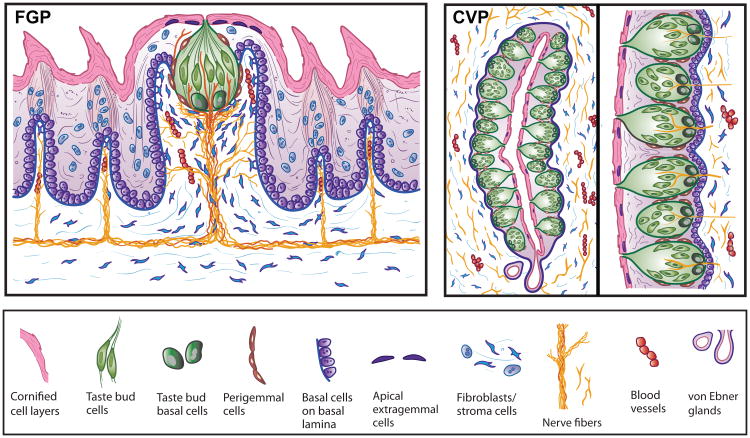
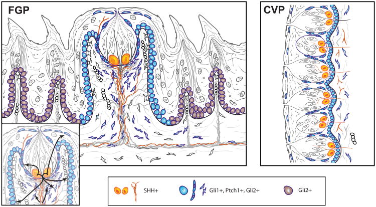
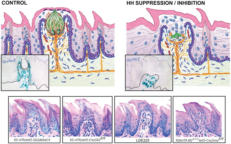
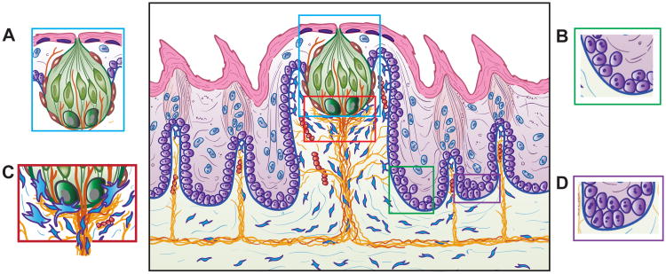
The tongue is an elaborate complex of heterogeneous tissues with taste organs of diverse embryonic origins. The lingual taste organs are papillae, composed of an epithelium that includes specialized taste buds, the basal lamina, and a lamina propria core with matrix molecules, fibroblasts, nerves, and vessels. Because taste organs are dynamic in cell biology and sensory function, homeostasis requires tight regulation in specific compartments or niches. Recently, the Hedgehog (Hh) pathway has emerged as an essential regulator that maintains lingual taste papillae, taste bud and progenitor cell proliferation and differentiation, and neurophysiological function. Activating or suppressing Hh signaling, with genetic models or pharmacological agents used in cancer treatments, disrupts taste papilla and taste bud integrity and can eliminate responses from taste nerves to chemical stimuli but not to touch or temperature. Understanding Hh regulation of taste organ homeostasis contributes knowledge about the basic biology underlying taste disruptions in patients treated with Hh pathway inhibitors.

Keywords: Hh pathway disruption; fungiform and circumvallate papillae; taste and cancer treatments; taste cell progenitors; taste organ niches; tongue innervation.


Figures

Figure 1
Figure 1
Diagram of tissue architecture for the (a) fungiform (FGP) and (b) circumvallate (CVP) papillae. The dorsal tongue and luminal surfaces of the taste organs are covered with layers of cornified, desquamating cells. Within the tongue, FGP and CVP are complex organs that are home to specialized collections of taste cells, the taste buds, and are composed of an epithelium covering stromal cell compartments, with fibroblasts, nerves, and blood vessels in the lamina propria. The taste buds and general papilla tissues share several cell characteristics; however, the FGP and CVP embryonic derivations are ectodermal and endodermal, respectively, and the comparative tissue structure is very different. The FGP (illustrated in sagittal section) in mouse and rat accommodates one apical taste bud and is innervated by lingual and taste bud-specific, chorda tympani nerves. Spinous filiform, nontaste papillae surround the FGP, innervated by the lingual nerve. The CVP is illustrated in an orientation horizontal to the dorsal tongue surface. (i) One half or one wall of the CVP is included to the left, with an inset on the right (ii) to emphasize taste bud distribution along the epithelium. The CVP contains a few hundred taste buds in mouse and rat and is innervated by the glossopharyngeal nerve. The luminal aspects of taste bud cells in the CVP open onto a papilla trench, and contents from lingual von Ebner's glands empty into the base of the trenches.
Figure 2
Figure 2
Hh signaling components in the (a) FGP and (b) CVP. Building on Figure 1, Hh signaling components are located in FGP and CVP. Within the epithelium of taste organs, Shh is principally restricted in taste bud cells, but Shh expression is also in nerve fibers of the tongue and FGP or CVP core, identified with mouse reporter lines (B.L. Allen, A.A. Dlugosz, A. Kumari, C.M. Mistretta, unpublished data). Gli1+, Ptch1+, and Gli2+ cells are within the basal layer of the epithelium of the lateral FGP walls, in perigemmal and apical extragemmal cells, and in stromal cells of the papilla core. Gli2+ cells also extend in basal epithelial cells beyond the FGP. The inset illustrates paracrine signaling flow from Shh+ taste bud cells to Hh-responding cells in the lateral papilla walls, perigemmal and apical extragemmal cells, to stromal cells, and from Shh+ nerves to fibroblasts/stromal cells. Within the CVP, Gli1+, Ptch1+, and Gli2+ cells are within the basal layer of the papilla epithelium, in perigemmal and apical extragemmal cells, and in stromal cells of the papilla core. Paracrine signaling from Shh+ cells within the taste bud to Hh-responding cells is proposed similar to the FGP. Abbreviations: CVP, circumvallate papillae; FGP, fungiform papillae; Hh, Hedgehog; Shh, Sonic Hedgehog.
Figure 3
Figure 3
Effects of Hh signaling suppression or inhibition on FGP morphology and Hh-responding cells. FGP diagrams illustrate Gli1lacZ+, Hh-responding cells (insets), for (a) control mouse tongue and (b) after Hh signaling suppression or inhibition. An H&E stained FGP image is included as an inset in a, the control diagram. This provides a comparison for images in cf. (cf) Four examples of papilla morphology after Hh signaling repression with taste bud cell loss are presented in H&E staining. Examples represent four different models for Hh/Gli suppression, or Smo inhibition or Smo deletion: (c) epithelial expression of a dominant negative-repressor form of Gli2C4); (d) genetic conditional deletion of Gli2 from epithelium; (e) pharmacological blockade of Smo with the Hh pathway inhibitor LDE225; and (f) global conditional deletion of Smo. After Hh signaling suppression in all models, Hh-responding cells are eliminated in the epithelium but retained in papilla stroma (as shown in the inset in panel b from the Gli1lacz Hh signaling reporter mouse). With Hh signaling disruption, the FGP loses an intact apical taste bud, and across models, the papilla acquires a pointed, heavily keratinized spinous apex. Abbreviations: FGP, fungiform papillae; Hh, Hedgehog; Shh, Sonic Hedgehog; Smo, smoothened receptor.
Figure 4
Figure 4
FGP with signaling niches or compartments. Building on Figures 1 and 2, four niches are proposed for Hh signaling in specific FGP cell compartments. (a) Niche 1 is within and around the taste bud, including taste bud and perigemmal cells. Signaling from Shh+ taste bud cells to HH-responding, perigemmal proliferative cells, extragemmal apical cells, and extragemmal apical nerves is proposed. (b) Niche 2 represents the highly proliferative, basal epithelial cells at the base or bottom of the FGP walls, including Gli1+ Hh-responding cells that are the progenitors of taste bud cells, perigemmal cells, and stratified keratinocytes of the FGP. These cells are positioned over stromal cells and lingual innervation. (c) In the apical FGP core and base of the taste bud, Niche 3 includes Shh+ basal taste bud cells and the basal lamina, taste cell bipolar extensions to the basal lamina, Hh-responding cells of the stroma, nerve fibers and Shh+ nerve fibers, and fibroblasts with filopodia in close association with the basal lamina. Multiple Shh signaling exchanges are likely, including those to the stromal compartment, based on taste bud-derived Shh secretion and sequestration of Shh ligand by basal lamina components. This also provides a signaling domain for stromal cells to regulate epithelial progenitors, and importantly, a particular perineural environment around lingual and taste nerve fibers to regulate FGP and taste bud progenitor and stem cells. (d) Niche 4 is in basal epithelial cells at the base or near bottom of the FILIFP, including proliferative cells seated on the basal lamina positioned over vimentin+ stromal cells and nerve fibers. Bmi1+, Tcf3+, and Gli2+ cells include progenitors for keratinocytes of the interpapilla epithelium and FILIFP. Abbreviations: FGP, fungiform papillae; FILIFP, spinous filiform papillae; Hh, Hedgehog; Shh, Sonic Hedgehog

Similar articles

Cited by

References

    1. Barker N, Bartfeld S, Clevers H. Tissue-resident adult stem cell populations of rapidly self-renewing organs. Cell Stem Cell. 2010;7:656–70. - PubMed
    1. Potten CS, Saffhill R, Maibach HI. Measurement of the transit time for cells through the epidermis and stratum corneum of the mouse and guinea-pig. Cell Tissue Kinet. 1987;20:461–72. - PubMed
    1. Cameron IL. Cell proliferation migration and specialization in epithelium of mouse tongue. J Exp Zool. 1966;163:271–83.
    1. Toto PD, Ojha G. Generation cycle of oral epithelium in mice. J Dent Res. 1962;41:388–91. - PubMed
    1. Beidler LM, Smallman RL. Renewal of cells within taste buds. J Cell Biol. 1965;27:263–72. - PMC - PubMed

Publication types

Substances

LinkOut - more resources