. 2017 Aug;14(4):343-356.
doi: 10.1089/zeb.2016.1366. Epub 2017 Feb 13.

Vascular Endothelial Growth Factor A and Leptin Expression Associated with Ectopic Proliferation and Retinal Dysplasia in Zebrafish Optic Pathway Tumors

Affiliations

Vascular Endothelial Growth Factor A and Leptin Expression Associated with Ectopic Proliferation and Retinal Dysplasia in Zebrafish Optic Pathway Tumors

Laura E Schultz et al. Zebrafish. 2017 Aug.

Abstract

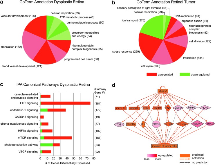
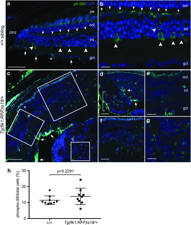
In the central nervous system injury induces cellular reprogramming and progenitor proliferation, but the molecular mechanisms that limit regeneration and prevent tumorigenesis are not completely understood. We previously described a zebrafish optic pathway tumor model in which transgenic Tg(flk1:RFP)is18/+ adults develop nonmalignant retinal tumors. Key pathways driving injury-induced glial reprogramming and regeneration contributed to tumor formation. In this study, we examine a time course of proliferation and present new analyses of the Tg(flk1:RFP)is18/+ dysplastic retina and tumor transcriptomes. Retinal dysplasia was first detected in 3-month-old adults, but was not limited to a specific stem cell or progenitor niche. Pathway analyses suggested a decrease in cellular respiration and increased expression of components of Hif1-α, VEGF, mTOR, NFκβ, and multiple interleukin pathways are associated with early retinal dysplasia. Hif-α targets VEGFA (vegfab) and Leptin (lepb) were both highly upregulated in dysplastic retina; however, each showed distinct expression patterns in neurons and glia, respectively. Phospho-S6 immunolabeling indicated that mTOR signaling is activated in multiple cell populations in wild-type retina and in the dysplastic retina and advanced tumor. Our results suggest that multiple pathways may contribute to the continuous proliferation of retinal progenitors and tumor growth in this optic pathway tumor model. Further investigation of these signaling pathways may yield insight into potential mechanisms to control the proliferative response during regeneration in the nervous system.

Keywords: VEGFA; dysplasia; leptin; progenitor; proliferation; retina.


Conflict of interest statement

No competing financial interests exist.

Figures

<b>FIG. 1.</b>
FIG. 1.
Detection of aberrant cell migration and ectopic proliferation in young adult Tg(flk1:RFP)is18 dysplastic retina. (a, e) Histological staining of retina section from 3-month-old adult wild-type zebrafish showing nuclear layers and organized laminar structure. (b, f) Representative image of section through retina of 3-month-old Tg(flk1:RFP)is18 adult reveals aberrant migration of cells across the inner nuclear layer (arrows, f) (n = 9). (c–h) Representative images of retinal sections from 4-month-old Tg(flk1:RFP)is18 adults (n = 15). (c, g) Retina from 4-month-old Tg(flk1:RFP)is18 adult with dysplasia reveals disruption of inner, outer, and ganglion cell layers with numerous mitotic figures (arrows, g). (d, h) Four-month-old Tg(flk1:RFP)is18 adult with advanced retinal tumor filling the vitreous space. Tumor tissue is composed of fibrous material interspersed with numerous mitotic figures, cells showing heterogeneous nuclear morphology and forming occasional rosettes (arrows, h), and blood vessels (arrowheads, h). pe, pigmented epithelium; onl, outer nuclear layer; inl, inner nuclear layer; gcl, ganglion cell layer. Scale bars (a, b) 200 μm; (c, d) 500 μm; (e–h) 50 μm.
<b>FIG. 2.</b>
FIG. 2.
Ectopic proliferation and Sox2 expressing cells in young adult Tg(flk1:RFP)is18 dysplastic retina. (a–e) Wild-type 4-month-old adult (n = 3) shows that PCNA positive cells are restricted to stem/progenitor cells at the ciliary marginal zone (cmz, arrow, c) and rod precursor cells in the photoreceptor outer nuclear layer (arrowheads, e). Sox2-expressing cells overlap with PCNA positive stem cells at the ciliary marginal zone (bracket, c) and are present in amacrine/displaced amacrine cells in the inner nuclear and ganglion cell layers (c, e). (f–j) Retina from 4-month-old Tg(flk1:RFP)is18 adult (n = 3) shows numerous regions of proliferation distributed throughout the neural retina (f). The ciliary marginal zone (g, h) and ventral ciliary circumferential artery (asterisk, g) are expanded. Small masses of PCNA and Sox2 positive cells (arrowheads, g, h) are present in the normally single-cell layered nonpigmented epithelium extending from the ciliary marginal zone (small arrow, g, h–compare to small arrow in wild type b). Ectopic regions of proliferation in the neural retina contain disorganized cells expressing PCNA and Sox2 (arrowheads, j). cmz, ciliary marginal zone; gcl, ganglion cell layer; inl, inner nuclear layer; onl, outer nuclear layer; pe, pigmented epithelium. Scale bars (a, f) 200 μm; all other panels 50 μm.
<b>FIG. 3.</b>
FIG. 3.
The ciliary marginal zone and dysplastic regions of Tg(flk1:RFP)is18/+ retina in 4-month-old adults contain increased numbers of PCNA and Sox2 positive cells. A total number of PCNA and Sox2 labeled cells were counted at the ciliary marginal zone and in a 300 μm long section of the internal retina in three wild-type and three age-matched 4-month-old Tg(flk1:RFP)is18/+ siblings containing a region of dysplasia identified by Hematoxylin and Eosin staining. (a) Ciliary marginal zone PCNA positive cells 19 ± 4 versus 7 ± 1, p = 0.0082. (b) Ciliary marginal zone Sox2 positive cells 19 ± 4 versus 7 ± 1, p = 0.0168. (c) Internal retina PCNA positive cells: 11 ± 2 versus <1, p = 0.0001. (d) Internal retina Sox2 positive cells: 47 ± 5 versus 28 ± 3, p = 0.007. n = 9 retinal sections, three each from three individuals of each genotype. Error bars represent standard error of the mean. cmz, ciliary marginal zone; ir, internal retina.
<b>FIG. 4.</b>
FIG. 4.
Tg(flk1:RFP)is18/+ dysplastic retina associated GoTerm and IPA canonical pathways. (a) GoTerm analysis of DGE of dysplastic retinal tissue suggests decreased oxidative metabolism and increased vasculogenesis pathways. (b) GoTerm analysis of DGE of retinal tumor tissue is consistent with loss of phototransduction and increased proliferation. (c) IPA canonical pathways to which the Tg(flk1:RFP)is18/+ dysplastic retina transcriptome is associated. (d) IPA regulator pathway analysis of VEGF targets involved in cytokine signaling, transcriptional response, and extracellular matrix remodeling underlying invasion of tumor cells. IPA, ingenuity pathway analysis; VEGF, vascular endothelial growth factor.
<b>FIG. 5.</b>
FIG. 5.
Induction of vegfab and lepb expression in distinct cell populations in Tg(flk1:RFP)is18 dysplastic retina and retinal tumor. In situ hybridization of retina cryosections from +/+ (a, b, e, f, i, j, m, n, q, r, u, v) and Tg(flk1:RFP)is18 (c, d, g, h, k, l, o, p, s, t, w, x) adult siblings. Weak expression of vegfaa is detected in the three nuclear layers of the retina (a, b) and in early dysplastic (c [box], d) and retinal tumor tissue (c, asterisk). Expression of vegfab (e, f), the VEGF receptor flk1 (i, j), lepb (m, n), or the Leptin receptor lepr (q, r) is not detected in wild-type +/+ retina. (g, h) In early dysplastic retina vegfab is detected throughout the inner and outer nuclear layers (g [small bracket], h). In the tumor containing region (large bracket) vegfab is detected in a central region of the lesion (asterisk). (k, l) The VEGF receptor flk1 was present in small groups of cells scattered throughout the tissue that may overlap with microvessels. (o, p) lepb is highly expressed in a subset of cells in the inner nuclear layer and cells in the ganglion cell layer in dysplastic retina and present in many cells evenly distributed in the early tumor (bracket). (s, t) The lepr receptor expression is faint and diffuse in dysplastic retina and tumor tissue. (u, v) In wild-type retina the glial marker apoeb is strongly expressed in inner nuclear layer Müller glia and astrocytes in the ganglion cell layer/nerve fiber layer. (w, x) apoeb expression is highly elevated in dysplastic retina and is detected in projections crossing the inner plexiform layer. Numerous cells throughout the tumor tissue labeled intensely with apoeb. gcl, ganglion cell layer; inl, inner nuclear layer; onl, outer nuclear layer; pe, pigmented epithelium. All scale bars, 100 μm, except panels (g, k, o, s, t) scale bars, 500 μm.
<b>FIG. 6.</b>
FIG. 6.
Tg(flk1:RFP)is18/+ dysplastic retina genes associated with IPA canonical pathway mTOR signaling. Upstream components PLD, DDIT, RAS, and PI3K predicted to activate mTOR signaling are upregulated. Downstream effectors of mTOR signaling, such as translation initiation factors eIF4E and eIF3 and ribosomal proteins, including RPS6, are also upregulated. Mixed red and green color in VEGF symbol reflects ∼11-fold vegfab upregulation and vegfb three-fold downregulation. PLD, phospholipase D; DDIT, DNA damage induced transcript; PI3K, phosphoinositol 3-kinase.
<b>FIG. 7.</b>
FIG. 7.
Activated mTOR signaling marker phosphorylated-S6 in adult zebrafish wild-type retina, Tg(flk1:RFP)is18/+ dysplastic retina, and retinal tumor. (a, b) phospho-S6 (green) is detected in putative horizontal cells (small arrowheads), amacrine and/or Müller glia (large arrowheads), and retinal ganglion cells and/or displaced amacrine cells (small arrows) in the mature region of the retina. Nuclei are labeled with 4′,6-diamidino-2-phenylindole. Asterisk marks a blood vessel. (c–g) In Tg(flk1:RFP)is18/+ dysplastic retina phospho-S6 labeling is present in a subset of cells throughout regions of the retina with increased cell number and disorganized retinal layers (c–f). Intense labeling of phospho-S6 was present in the ganglion cell and nerve fiber layers (c, d, arrows). Large lesions contained phospho-S6 positive cells distributed throughout the tumor mass (d [bracket], g). (h) Percentage of phospho-S6 cells/total cells in wild-type retina and Tg(flk1:RFP)is18/+ tumor. +/+ 11.4% ± 0.9 vs. Tg(flk1:RFP)is18/+ 13.9% ± 1.7, p = 0.2291. n = 9 sections, three sections each from three individuals of each genotype. gcl, ganglion cell layer; inl, inner nuclear layer; onl, outer nuclear layer; pe, pigmented epithelium. Scale bars, (a, g, f) 50 μm; (b, c) 20 μm; (d, e) 100 μm; (h) 25 μm.

Similar articles

Cited by

References

    1. Rock KL, Kono H. The inflammatory response to cell death. Annu Rev Pathol 2008;3:99–126 - PMC - PubMed
    1. Corcoran A, O'Connor JJ. Hypoxia-inducible factor signalling mechanisms in the central nervous system. Acta Physiol 2013;208:298–310 - PubMed
    1. Semenza GL. Hypoxia-inducible factors in physiology and medicine. Cell 2012;148:399–408 - PMC - PubMed
    1. Kono H, Onda A, Yanagida T. Molecular determinants of sterile inflammation. Curr Opin Immunol 2014;26:147–156 - PubMed
    1. Cho Y, Shin JE, Ewan EE, Oh YM, Pita-Thomas W, Cavalli V. Activating injury-responsive genes with hypoxia enhances axon regeneration through neuronal HIF-1alpha. Neuron 2015;88:720–734 - PMC - PubMed

Publication types

MeSH terms

LinkOut - more resources