Review
. 2017 Sep;43(Pt B):113-124.
doi: 10.1016/j.ymben.2017.02.002. Epub 2017 Feb 10.

Review of metabolic pathways activated in cancer cells as determined through isotopic labeling and network analysis

Affiliations
Review

Review of metabolic pathways activated in cancer cells as determined through isotopic labeling and network analysis

Wentao Dong et al. Metab Eng. 2017 Sep.

Abstract

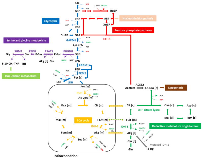
Cancer metabolism has emerged as an indispensable part of contemporary cancer research. During the past 10 years, the use of stable isotopic tracers and network analysis have unveiled a number of metabolic pathways activated in cancer cells. Here, we review such pathways along with the particular tracers and labeling observations that led to the discovery of their rewiring in cancer cells. The list of such pathways comprises the reductive metabolism of glutamine, altered glycolysis, serine and glycine metabolism, mutant isocitrate dehydrogenase (IDH) induced reprogramming and the onset of acetate metabolism. Additionally, we demonstrate the critical role of isotopic labeling and network analysis in identifying these pathways. The alterations described in this review do not constitute a complete list, and future research using these powerful tools is likely to discover other cancer-related pathways and new metabolic targets for cancer therapy.

Keywords: Cancer metabolism; Isotopic labeling analysis.


Figures

Figure 1
Figure 1. A global overview of the metabolic network critical in cancer cells
Glucose is the most highly consumed carbon substrate, and its entry initiates glycolysis, which contains several anabolic arms. The pentose phosphate pathway (PPP) directs glucose-derived glucose-6-phosphate (G6P) toward oxidative PPP where NADPH is generated. From ribulose-5-phosphate (Ru5P), the non-oxidative PPP continues, supplying building blocks for nucleotide biosynthesis and reversibly exchanging metabolites with glycolysis via fructose-6-phosphate (F6P) and glyceraldehyde-3-phosphate (GAP). Serine and glycine metabolism branches from 3-phosphoglycerate (3PG) and connects central carbon metabolism with one-carbon metabolism. The end product pyruvate can be converted to lactate or can enter the TCA cycle in the form of acetyl-CoA, which can also be derived from acetate under certain conditions. Glutamine is metabolized to glutamate and then α-ketoglutarate, which can enter the TCA cycle for replenishing anaplerosis or be reductively metabolized via a carboxylation reaction to fuel de novo lipogenesis. Various pathways are altered to sustain the uncontrolled proliferation exhibited by cancer cells. G6P, glucose-6-phosphate; F6P, fructose-6-phosphate; GAP, glyceraldehyde-3-phosphate; 3PG, 3-phosphoglycerate; R5P, ribose-5-phosphate; Akg, α-ketoglutarate; Cit, citrate; Ac-CoA, acetyl-CoA; Oxa, oxaloacetate.
Figure 2
Figure 2. The PKM2-mediated glycolytic pathway
The M2 isoform of pyruvate kinase (PKM2), which is preferentially expressed by cancer cells, has been hypothesized to promote an alternative catalytic activity that does not produce ATP, in contrast to PKM1. This potential mechanism would likely contribute to continual activation of phosphofructokinase (PFK), which is inhibited by ATP. Consequently, glycolytic flux may be enhanced. This alternative mechanism involves transfer of the phosphate group from phosphoenolpyruvate (PEP) to phosphoglycerate mutase 1 (PGAM1), which activates it. This positive feedback mechanism may drive the enhanced production of 2-phosphoglycerate (2PG), PEP and eventually enhance the overall PGAM1 activity. Through this mechanism, the generation of pyruvate may become partially decoupled from ATP production, which could enable a high rate of glycolysis. Activation and inhibition of enzyme activity are denoted by arrows and blunt arrows, respectively. The relative strengths of metabolic pathways and activation/inhibition effects are shown by the thickness of the arrows.
Figure 3
Figure 3. The GAPDH-mediated reversal of glycolytic flux
Under hypoxic condition, GAPDH activity is inhibited while the expression of other upper glycolytic enzymes is elevated. Due to the accumulation of high concentrations of upper glycolytic metabolites and the reversibility of the non-oxidative PPP, more ribose-5-phosphate (R5P) is synthesized to promote nucleotide biosynthesis. G6P, glucose-6-phosphate, F6P, fructose-6-phosphate; GAP, glyceraldehyde-3-phosphate; DHAP, dihydroxyacetone phosphate; 1,3-BPG, 1,3-bisphosphoglycerate. The relative strengths of metabolic pathways and activation/inhibition effects are shown by the thickness of the arrows. The figure is adapted from Ahn et al. (under review).
Figure 4
Figure 4. The isotopic labeling pattern generated by 1,2-13C2-glucose
(a) Following exclusive use of the glycolytic pathway, the M2-labeled pattern retains until fructose-1,6-bisphosphate (FBP). The aldolase-catalyzed cleavage reaction produces M2-labeled dihydroxyacetone (DHAP) and unlabeled glyceraldehyde-3-phosphate (GAP). The labeling pattern is eventually inherited by pyruvate, yielding an equimolar mixture of M2-labeled and unlabeled pyruvate. (b) This labeling pattern can be best explained by analyzing three molecules of 1,2-13C2-glucose going through the oxidative PPP. The first step in glycolysis transforms all 1,2-13C2-glucose into M2-labeled glucose-6-phosphate (G6P). The oxidative PPP releases one molecule of heavy labeled carbon dioxide per molecule of 1,2-13C2 G6P. The resulting three molecules of M1-labeled ribulose-5-phosphate (Ru5P) have equal chances of going through three distinct biochemical reactions. The transketolation and transaldolation reactions lead to one molecule of M2-labeled fructose-6-phosphate (F6P), one molecule of M1-labeled F6P, and one molecule of unlabeled glyceraldehyde-3-phosphate (GAP). These metabolite intermediates are then diverted back to the glycolytic pathway. One molecule of M2-labeled G6P generates equimolar mixture of M2-labeled and unlabeled pyruvate. Similarly, one molecule of M1-labeled G6P generates an equimolar mixture of M1-labeled and unlabeled pyruvate. The unlabeled GAP molecule yields one molecule of unlabeled pyruvate. Therefore, theoretically, if all 1,2-13C2-glucose goes through the oxidative PPP exclusively, there would be 20% of M2-labeled pyruvate, 20% of M1-labeled pyruvate and 60% of unlabeled pyruvate.
Figure 5
Figure 5. The isotopic labeling pattern generated by U-13C5- glutamine
(a) The isotopically labeled U-13C5- glutamine is converted to glutamate via glutaminase. Transamination or deamination reactions then take place to transform glutamate to α-ketoglutarate. M5-labeled citrate is formed in the mitochondria and cytosol through reductive carboxylation by isocitrate dehydrogenases (IDHs). This citrate is cleaved into oxaloacetate and acetyl-CoA, and this heavy labeled acetyl-CoA is employed for de novo lipogenesis. The reaction network in cytosol further generates M3-labeled oxaloacetate, aspartate, malate and fumarate. The figure is adapted from Metallo et al. (2011). (b) In the first cycle of oxidative metabolism of U-13C5- glutamine, the uniform labeling pattern no longer holds at citrate, which is generated by consolidating unlabeled acetyl-CoA with M4 labeled oxaloacetate. The beginning of the second cycle primes at M3-labeled α-ketoglutarate (in the blue box) after the release of a heavy labeled carbon dioxide. When there is a strong influx of glutamine, the labeling pattern of the first cycle dominates. (c) Starting from the M3 labeled α-ketoglutarate (in the blue box), the release of another heavy labeled carbon dioxide further reduces the labeling pattern to generate M2-labeled succinate and fumarate. Due to the molecular asymmetry of malate and oxaloacetate, two different forms of M2-labeled compounds are generated in equimolar ratio. After the consolidation of unlabeled acetyl-CoA and M2-labeled oxaloacetate, M2-labeled citrate is generated. The labeling pattern of the second cycle is less important when there is a strong glutamine influx.
Figure 6
Figure 6. Serine and glycine metabolism
The de novo serine biosynthesis pathway branches from glycolysis at 3-phosphoglycerate (3PG). The enzyme phosphoglycerate dehydrogenase (PHGDH) oxidizes 3PG to phosphohydroxypyruvate (P-pyruvate) in the presence of cofactor NAD+. P-pyruvate is then converted to phosphoserine (P-ser) by phosphoserine aminotransferase 1 (PSAT1). Serine is eventually synthesized after the last step of dephosphorylation by phosphoserine phosphatase (PSPH). Glycine metabolism is linked to serine metabolism through serine hydroxymethyltransferase (SHMT). This reversible reaction is critical in bridging glycolytic substrates with compounds in one-carbon metabolism. The figure is adapted from Possemato et al. (2011) and Pacold et al. (2016).
Figure 7
Figure 7. Detailed summary of rewired metabolic pathways in cancer cells
Metabolic reprogramming along glycolysis, reductive carboxylation of glutamine, serine and glycine metabolism, IDH mutations and TKTL1-mediated acetate metabolism are shown in a detailed depiction. Glc, glucose; G6P, glucose-6-phosphate; F6P, fructose-6-phosphate; FBP, fructose-1,6-bisphosphate; GAP, glyceraldehyde-3-phosphate; DHAP, dihydroxyacetone phosphate; 1,3-BPG, 1,3-bisphosphoglycerate; 3PG, 3-phosphoglycerate; 2PG, 2-phosphoglycerate; PEP, phosphoenolpyruvate; Pyr, pyruvate; Ru5P, ribulose-5-phosphate; R5P, ribose-5-phosphate; Xu5P, xylulose-5-phosphate; P-Pyr, 3-phosphohydroxypyruvate; P-Ser, phosphoserine; Ser, serine; Gly, glycine; 5,10-CH2-THF, 5,10-methylenetetrahydrofolate; THF, tetrahydrofolate; Ac-CoA, acetyl-CoA; Cit, citrate; I-Cit, isocitrate; Akg, α-ketoglutarate; Suc, succinate; Fum, fumarate; Mal, malate; Oxa, oxaloacetate; Gln, glutamine; Glu, glutamate; Asp, aspartate; 2-Hg, 2-hydroxyglutarate. GAPDH, glyceraldehyde-3-phosphate dehydrogenase; PKM2, pyruvate kinase M2 isoform; PGAM1, phosphoglycerate mutase 1; TKTL1, transketolase-like 1; PHGDH, phosphoglycerate dehydrogenase; PSAT1, phosphoserine aminotransferase 1; PSPH, phosphoserine phosphatase; SHMT, serine hydroxymethyl transferase; PDH, pyruvate dehydrogenase; IDH1, isocitrate dehydrogenase 1; IDH2, isocitrate dehydrogenase 2.

References

    1. Ahn WS, Zhang Z, Cantor JR, Iliopoulos O, Stephanopoulos G. Glyceraldehyde 3-phosphate dehydrogenase modulates non-oxidative pentose phosphate pathway to provide anabolic precursors in hypoxic tumor cells. n.d Under Review.
    1. Amelio I, Cutruzzolá F, Antonov A, Agostini M, Melino G. Serine and glycine metabolism in cancer. Trends in Biochemical Sciences. 2014;39(4):191–198. http://doi.org/10.1016/j.tibs.2014.02.004. - DOI - PMC - PubMed
    1. Barbosa MJ, Rocha JMS, Tramper J, Wijffels RH. Acetate as a carbon source for hydrogen production by photosynthetic bacteria. Journal of Biotechnology. 2001;85(1):25–33. http://doi.org/10.1016/S0168-1656(00)00368-0. - DOI - PubMed
    1. Basan M, Hui S, Okano H, Zhang Z, Shen Y, Williamson JR, Hwa T. Overflow metabolism in Escherichia coli results from efficient proteome allocation. Nature. 2015;528(7580):99–104. http://doi.org/10.1038/nature15765. - DOI - PMC - PubMed
    1. Birsoy K, Wang T, Chen WW, Freinkman E, Abu-Remaileh M, Sabatini DM. An Essential Role of the Mitochondrial Electron Transport Chain in Cell Proliferation Is to Enable Aspartate Synthesis. Cell. 2015;162(3):540–551. http://doi.org/10.1016/j.cell.2015.07.016. - DOI - PMC - PubMed

Publication types

LinkOut - more resources