. 2017 Feb 13;11(2):e0005390.
doi: 10.1371/journal.pntd.0005390. eCollection 2017 Feb.

Species dependent impact of helminth-derived antigens on human macrophages infected with Mycobacterium tuberculosis: Direct effect on the innate anti-mycobacterial response

Affiliations

Species dependent impact of helminth-derived antigens on human macrophages infected with Mycobacterium tuberculosis: Direct effect on the innate anti-mycobacterial response

Naomi Aira et al. PLoS Negl Trop Dis. .

Abstract

Background: In countries with a high prevalence of tuberculosis there is high coincident of helminth infections that might worsen disease outcome. While Mycobacterium tuberculosis (Mtb) gives rise to a pro-inflammatory Th1 response, a Th2 response is typical of helminth infections. A strong Th2 response has been associated with decreased protection against tuberculosis.

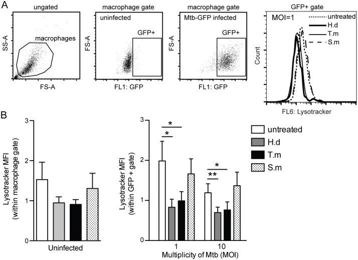
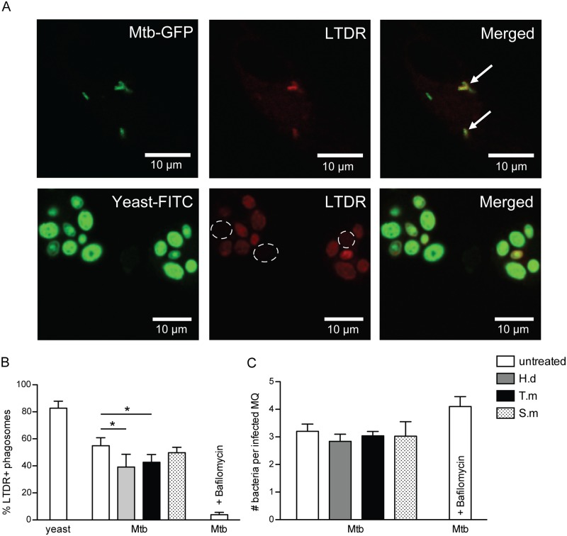
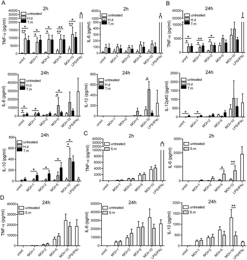
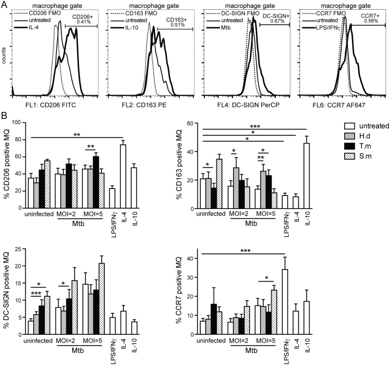
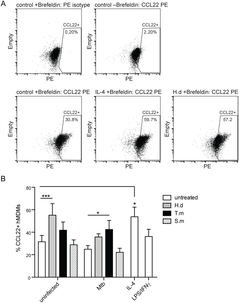
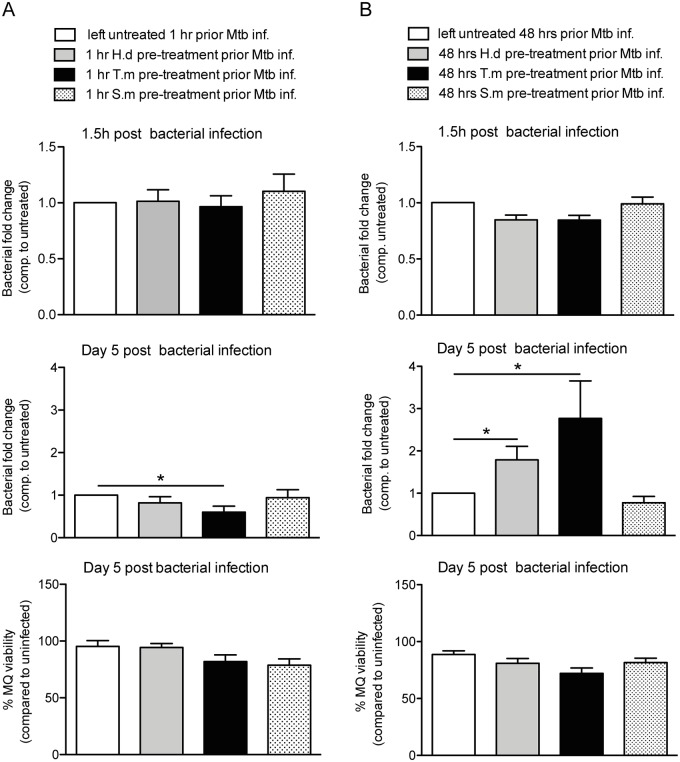
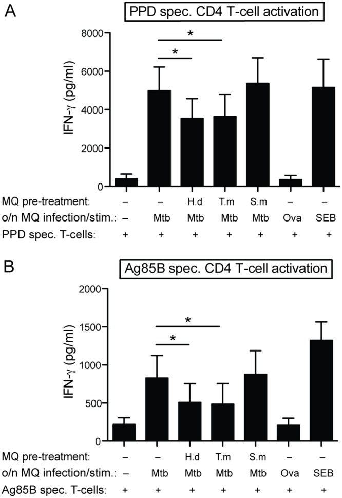
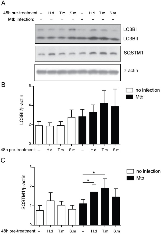
Principal findings: We investigated the direct effect of helminth-derived antigens on human macrophages, hypothesizing that helminths would render macrophages less capable of controlling Mtb. Measuring cytokine output, macrophage surface markers with flow cytometry, and assessing bacterial replication and phagosomal maturation revealed that antigens from different species of helminth directly affect macrophage responses to Mtb. Antigens from the tapeworm Hymenolepis diminuta and the nematode Trichuris muris caused an anti-inflammatory response with M2-type polarization, reduced macrophage phagosome maturation and ability to activate T cells, along with increased Mtb burden, especially in T. muris exposed cells which also induced the highest IL-10 production upon co-infection. However, antigens from the trematode Schistosoma mansoni had the opposite effect causing a decrease in IL-10 production, M1-type polarization and increased control of Mtb.

Conclusion: We conclude that, independent of any adaptive immune response, infection with helminth parasites, in a species-specific manner can influence the outcome of tuberculosis by either enhancing or diminishing the bactericidal function of macrophages.


Conflict of interest statement

The authors have declared that no competing interests exist.

Figures

Fig 1
Fig 1. H. diminuta and T. muris antigens decrease acidification in Mtb infected macrophages.
Human monocyte-derived macrophages (hMDMs) were treated for 1h ± antigens from H. diminuta (H.d; 100 μg/ml), T. muris (T.m; 50 μg/ml), or S. mansoni soluble egg antigen (S.m; 50 μg/ml), before being infected with green fluorescence protein (GFP)-expressing Mtb at different multiplicity of infection (MOI) for 2h. After infection, hMDMs were stained with LysoTracker Deep Red (LTDR). (A) Shows the gating strategy and representative LTDR-histograms for infected hMDMs ± antigens. (B) Results show the MFI-values of the LTDR signal in uninfected to the left, and infected macrophages (GFP-positive macrophages) to the right using flow cytometry. Data are presented as means ± SEM from 6 independent hMDM donors. p*<0.05, p**<0.01 using One-way ANOVA.
Fig 2
Fig 2. Co-localization of LysoTracker and Mtb-phagosomes is decreased by H. diminuta and T. muris antigen treatment.
hMDMs were treated for 1h ± antigens from H. diminuta (H.d; 100 μg/ml), T. muris (T.m; 50 μg/ml), or S. mansoni soluble egg antigen (S.m; 50 μg/ml), before being infected with green fluorescence protein (GFP)-expressing Mtb (MOI = 1) for 4h. hMDMs were stained with LysoTracker Deep Red (LTDR). FITC-labeled yeast was used as a positive control. Bafilomycin (100 nM) was used as an inhibitor of acidification (negative control). (A) Representative micrographs displaying co-localization of LTDR and Mtb-GFP, shown by arrows. Since LTDR co-localized to most FITC-yeast phagosomes, absence of LTDR-staining is instead indicated by dashed circles. (B) Percentage LTDR+ phagosomes, counting 50–100 phagosomes/stimuli per donor, data expressed as means ± SEM from n independent hMDM donors (n = 5 for yeast and n = 7 for Mtb ± bafilomycin). (C) Shows number (#) of bacteria per infected macrophage (MQ) (n = 7). p*<0.05 using One-way ANOVA.
Fig 3
Fig 3. H. diminuta and T. muris antigen exposure stimulates early pro-inflammatory and late anti-inflammatory cytokine release.
hMDMs were treated for 1h ± antigens from H. diminuta (H.d; 100 μg/ml), T. muris (T.m; 50 μg/ml), or S. mansoni soluble egg antigen (S.m; 50 μg/ml), before being infected with Mtb for 2h or 24h at the indicated MOIs. LPS/IFN-γ treatment was used as a positive pro-inflammatory stimulus. (A) TNF-α and IL-6 secretion at 2h with/without H.d or T.m. (B) TNF-α, IL-6, IL-1β, IL-12p40 and IL-10 at 24h with/without H.d or T.m. Data are presented as means ± SEM. n = 7 independent hMDM donors for 2h measurements and n = 6 for 24h measurements, and n = 5–7 for IL-10. p*<0.05, p**<0.01 using One-way ANOVA. S.m data in (C; 2h) and (D; 24h) are presented as means ± SEM from 7 independent donors that are different from the donors presented in (A) and (B), therefore separate graphs. p*≤0.05, p**≤0.01 using paired Student t-test.
Fig 4
Fig 4. Helminth antigen species-specific polarization of hMDMs upon co-exposure with Mtb.
hMDMs were treated for 1h ± antigens from H. diminuta (H.d; 100 μg/ml), T. muris (T.m; 50 μg/ml), or S. mansoni soluble egg antigen (S.m; 50 μg/ml), before being infected with Mtb at the indicated MOI. After 24h hMDMs were detached and stained with a combination of CD206-FITC (M2a marker), CD163-PE (M2c marker), CCR7-AF647 (M1 marker), or DC-SIGN-PerCP (regulatory macrophage phenotype). LPS/IFN-γ was used as a positive control for M1-stimuli, IL-4 as M2a-stimuli, and IL-10 as M2c-stimuli. (A) Representative histograms showing the individual fluorescence minus one (FMO) controls (with indicated %) that each positive gate was based on, and a showing signal profile from untreated together with either a positive stimulus or Mtb infection. (B) Data are expressed as means ± SEM from 8 independent hMDM donors. p*<0.05, p**<0.01, p***<0.001 using One-way ANOVA.
Fig 5
Fig 5. H. diminuta antigen and T. muris antigen co-exposure with Mtb stimulates increased CCL22 protein production in hMDMs.
hMDMs were treated for 1h ± antigens from H. diminuta (H.d; 100 μg/ml), T. muris (T.m; 50 μg/ml), or S. mansoni soluble egg antigen (S.m; 50 μg/ml), before being infected with Mtb at MOI = 5 for 24h. Brefeldin A was added 2h after Mtb infection to retain intracellularly produced proteins. After 24h of infection cytofix/cytoperm treated cells were stained intracellularly for flow cytometry using anti-CCL22 PE or a PE isotype control antibody to set the CCL22+ gate for data shown in (B). (A) Representative dot plots of the entire macrophage population (macrophage gate depicted in Fig 1A) showing the PE-profile of control cells treated with brefeldin A and stained with PE isotype control antibody (upper left) that is used to set the gate for % CCL22+ hMDMs, also illustrating PE-profile for CCL22 stained control (untreated), IL-4 and H.d treated hMDMs. (B) Data are expressed as means ± SEM from 5 independent hMDM donors. p*<0.05, p***<0.001 using One-way ANOVA.
Fig 6
Fig 6. Mimicking chronic helminth infection by long-term antigen exposure results in lost hMDM control of Mtb.
hMDMs were treated with helminth antigens from H. diminuta (H.d; 1.5 μg/ml), T. muris (T.m; 1.5 μg/ml), or S. mansoni soluble egg antigen (S.m; 1.5 μg/ml) for 1h (A) or 48h (B) prior to infection for 1.5h with luciferase expressing Mtb (MOI = 2). Extracellular bacteria were washed away and Mtb phagocytosis was evaluated (upper horizontal panels), or washed hMDMs were incubated for 5 days at 37°C before the bacterial fold change was determined (middle horizontal panels). hMDMs pretreated with antigens for 1h prior Mtb infection had continuous presence of antigens throughout the experiment (A), whereas hMDMs pretreated for 48h received antigens prior Mtb infection only (B). A and B show the total bacteria (combined luciferase signal from hMDM lysate and supernatant) compared to untreated (only Mtb), data expressed as means ± SEM from 4 independent hMDM donors. Bottom horizontal panel, show hMDM viability after 5 days post Mtb infection measured using calcein AM uptake before lysates were generated for middle horizontal panels. Viability data are normalized against uninfected hMDMs set to 100%. p*<0.05 using One-way ANOVA.
Fig 7
Fig 7. H. diminuta and T. muris antigen treated hMDMs reduce Mtb-specific antigen presentation to CD4+ T cells.
hMDMs were left untreated, or treated for 48h with 10 μg/ml of H. diminuta (H.d), T. muris (T.m), or S. mansoni soluble egg antigen (S.m). Thereafter hMDMs were either infected with Mtb (MOI = 5) or stimulated with PPD or SEB (positive controls) or ovalbumin (Ova; background control) for 24h, before being co-cultured with autologous PPD-specific (A) or Ag85B-specific (B) CD4+ T cells (1:5 DC:T cell ratio). Cell free culture supernatants were collected 48h later, and assayed for IFN-γ, data expressed as means ± SEM from 7 independent hMDM donors. p*<0.05 using One-way ANOVA.
Fig 8
Fig 8. H. diminuta and T. muris antigen treated hMDMs infected with Mtb show accumulation of autophagy proteins.
hMDMs were left untreated, or treated for 48h with 10 μg/ml of H. diminuta (H.d), T. muris (T.m), or S. mansoni soluble egg antigen (S.m), before being infected with Mtb (MOI = 2). After 4.5h of infection the cellular lysates were subjected to western blot using anti-LC3B, or anti-SQSTM1 antibodies, and anti-β-actin antibody as a loading control. Representative western blot images of LC3B, SQSTM1 and β-actin (A). Densitometry analysis of LC3BII (B) and SQSTM1(C), normalized against β-actin. Data are expressed as mean ± SEM of 4 independent hMDM donors. p*<0.05 using One-way ANOVA.

References

    1. Gause WC, Wynn TA, Allen JE. Type 2 immunity and wound healing: evolutionary refinement of adaptive immunity by helminths. Nat Rev Immunol. 2013;13: 607–14. 10.1038/nri3476 - DOI - PMC - PubMed
    1. Kreider T, Anthony RM, Urban JF, Gause WC. Alternatively activated macrophages in helminth infections. Curr Opin Immunol. 2007;19: 448–53. 10.1016/j.coi.2007.07.002 - DOI - PMC - PubMed
    1. Rafi W, Ribeiro-Rodrigues R, Ellner JJ, Salgame P. “Coinfection-helminthes and tuberculosis”. Curr Opin HIV AIDS. 2012;7: 239–44. 10.1097/COH.0b013e3283524dc5 - DOI - PubMed
    1. du Plessis N, Walzl G. Helminth-M. tb co-infection. Adv Exp Med Biol. 2014;828: 49–74. 10.1007/978-1-4939-1489-0_3 - DOI - PubMed
    1. Chatterjee S, Nutman TB. Helminth-induced immune regulation: implications for immune responses to tuberculosis. PLoS Pathog. 2015;11: e1004582 10.1371/journal.ppat.1004582 - DOI - PMC - PubMed