. 2017 Feb 13;12(2):e0171448.
doi: 10.1371/journal.pone.0171448. eCollection 2017.

The efficacy of a scaffold-free Bio 3D conduit developed from human fibroblasts on peripheral nerve regeneration in a rat sciatic nerve model

Affiliations

The efficacy of a scaffold-free Bio 3D conduit developed from human fibroblasts on peripheral nerve regeneration in a rat sciatic nerve model

Hirofumi Yurie et al. PLoS One. .

Abstract

Background: Although autologous nerve grafting is the gold standard treatment of peripheral nerve injuries, several alternative methods have been developed, including nerve conduits that use supportive cells. However, the seeding efficacy and viability of supportive cells injected in nerve grafts remain unclear. Here, we focused on a novel completely biological, tissue-engineered, scaffold-free conduit.

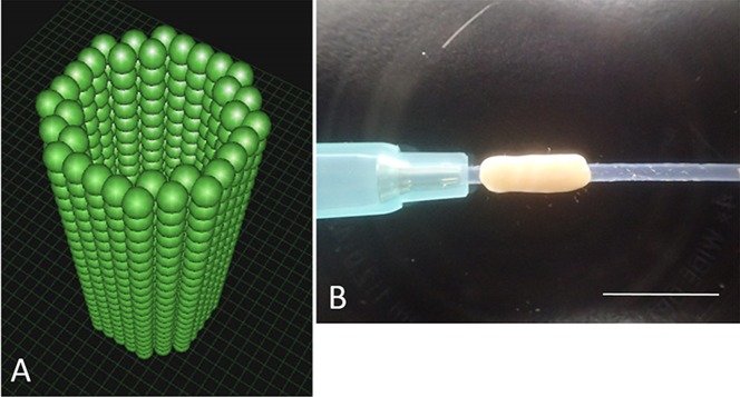
Methods: We developed six scaffold-free conduits from human normal dermal fibroblasts using a Bio 3D Printer. Twelve adult male rats with immune deficiency underwent mid-thigh-level transection of the right sciatic nerve. The resulting 5-mm nerve gap was bridged using 8-mm Bio 3D conduits (Bio 3D group, n = 6) and silicone tube (silicone group, n = 6). Several assessments were conducted to examine nerve regeneration eight weeks post-surgery.

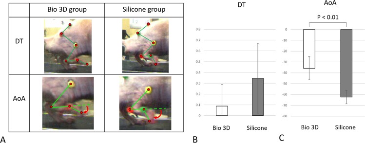
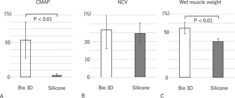
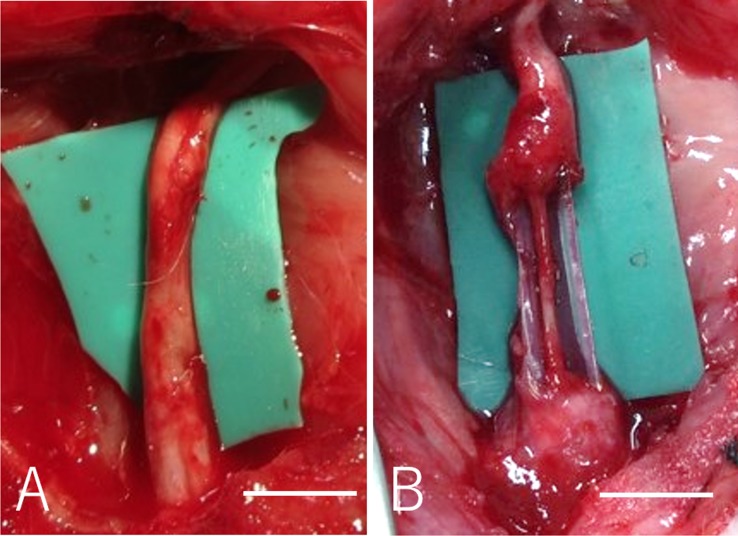
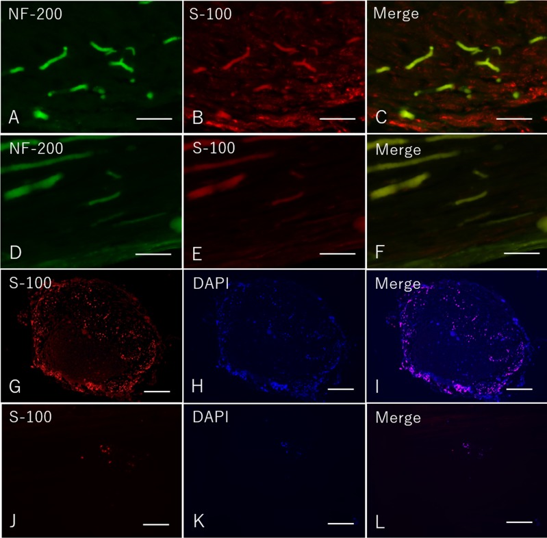
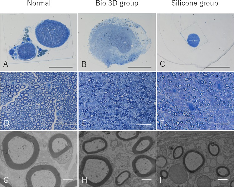
Results: Kinematic analysis revealed that the toe angle to the metatarsal bone at the final segment of the swing phase was significantly higher in the Bio 3D group than the silicone group (-35.78 ± 10.68 versus -62.48 ± 6.15, respectively; p < 0.01). Electrophysiological studies revealed significantly higher compound muscle action potential in the Bio 3D group than the silicone group (53.60 ± 26.36% versus 2.93 ± 1.84%; p < 0.01). Histological and morphological studies revealed neural cell expression in all regions of the regenerated nerves and the presence of many well-myelinated axons in the Bio 3D group. The wet muscle weight of the tibialis anterior muscle was significantly higher in the Bio 3D group than the silicone group (0.544 ± 0.063 versus 0.396 ± 0.031, respectively; p < 0.01).

Conclusions: We confirmed that scaffold-free Bio 3D conduits composed entirely of fibroblast cells promote nerve regeneration in a rat sciatic nerve model.


Conflict of interest statement

There are no patents or marketed products to declare. KN is the co-founder and shareholder of Cyfuse Biomedical K.K., Tokyo, Japan (Cyfuse). SA and MT, who are employees of Cyfuse, contributed to the manufacturing of 3D conduits and Cyfuse provided the bioprinter to manufacture the conduit. The company has the industrial rights related to the bioprinting method used to construct the 3D conduit in this work. Cyfuse provided support in the form of salaries for authors SA, KN, and MT and provided research grants to RI, TA, KN and SM. These competing interests do not alter the authors' adherence to PLOS ONE policies on sharing data and materials.

Figures

Fig 1
Fig 1
A: The pre-designed 3D tube-like structure. The green spheres represent homogeneous multicellular spheroids that were developed using only human normal dermal fibroblasts. B: The Bio 3D conduit according to the pre-designed 3D model. The conduit was cannulated via a 18-gauge intravenous catheter (SURFLO: NIPRO, Osaka, Japan). Scale bar = 10 mm.
Fig 2
Fig 2
A: In the Bio 3D group, an 8-mm Bio 3D conduit was interposed into the nerve defect, and the proximal and distal nerve stumps were secured 1.5 mm into the tube to create a 5-mm interstump gap in the conduit. B: In the silicone group, the silicone tube with 8 mm length was interposed in the same procedure.
Fig 3
Fig 3. Kinematic studies.
A: The photographs demonstrate drag toe (DT) and angle of attack (AoA) in both Bio 3D and silicone groups. In the silicone group, the rat’s toe was not off the ground. The red curved arrows represent the AoA. B: Regarding the DT, there was no significant difference among two groups. C: AoA was significantly different between the two groups (p < 0.01). Error bars represent the standard deviation.
Fig 4
Fig 4. Electrophysiological studies and wet muscle weight of the tibialis anterior muscle eight weeks after surgery.
A: CMAP was significantly higher in the Bio 3D group than the silicone group (p < 0.01). B: Regarding the NCV, there was no significant difference among two groups. C: Wet muscle weight was significantly higher in the Bio 3D group than in the silicone group (p < 0.01). All values are expressed as the percentage of those from the left hind limb. Error bars represent standard deviations.
Fig 5
Fig 5
A: Regenerated sciatic nerve eight weeks after surgery in the Bio 3D group. B: In the silicone group, the nerve gap was bridged, however the regenerated nerve was very thin in the silicone tube. Scale bar = 5mm.
Fig 6
Fig 6. Immunohistochemistry of the mid portion of the regenerated nerve eight weeks after surgery in both groups.
A-C: Longitudinal sections in the Bio 3D group. D-F: Longitudinal sections in the silicone group. G-I: Transverse sections in the Bio 3D group. J-L: Transverse sections in the silicone group. A-F: scale bar = 100 μm. G-L: scale bar = 500 μm.
Fig 7
Fig 7
A—F: Semi-thin transverse sections (toluidine blue staining) of the regenerated nerve eight weeks after surgery. A—C: scale bar = 1000 μm. D—F: scale bar = 50 μm. G—I: Transmission electron microscopy of the regenerated nerve eight weeks after surgery. scale bar = 2 μm.

References

    1. Kim DH, Han K, Tiel RL, Murovic JA, Kline DG. Surgical outcomes of 654 ulnar nerve lesions. J Neurosurg. 2003; 98: 993–1004. 10.3171/jns.2003.98.5.0993 - DOI - PubMed
    1. Lundborg G. A 25-year perspective of peripheral nerve surgery: evolving neuroscientific concepts and clinical significance. J Hand Surg Am. 2000; 25: 391–414. 10.1053/jhsu.2000.4165 - DOI - PubMed
    1. Milleisi H. Techniques for nerve grafting. Hand Clin. 2000; 16: 73–91. - PubMed
    1. Battiston B, Geuna S, Ferrero M, Tos P. Nerve repair by means of tubulization: Literature review and personal clinical experience comparing biological and synthetic conduits for sensory nerve repair. Microsurgery. 2005; 25: 258–267. 10.1002/micr.20127 - DOI - PubMed
    1. Konofaos P, Ver Halen JP. Nerve repair by means of tubulization: past, rresent, future. J Reconstr Microsurg. 2013; 28: 149–164. - PubMed

MeSH terms