. 2017 Feb 13;13(2):e1006196.
doi: 10.1371/journal.ppat.1006196. eCollection 2017 Feb.

CD8+ T cell cytotoxicity mediates pathology in the skin by inflammasome activation and IL-1β production

Affiliations

CD8+ T cell cytotoxicity mediates pathology in the skin by inflammasome activation and IL-1β production

Fernanda O Novais et al. PLoS Pathog. .

Abstract

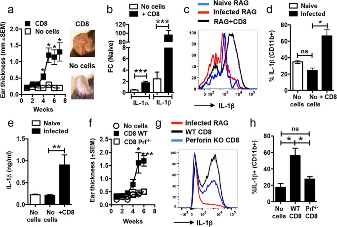
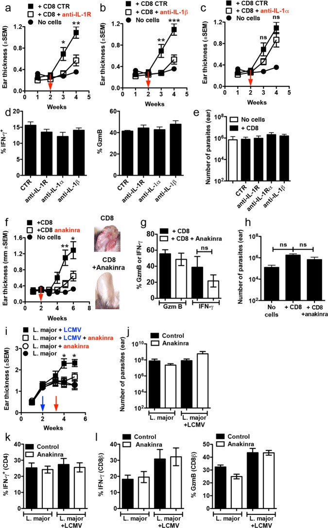
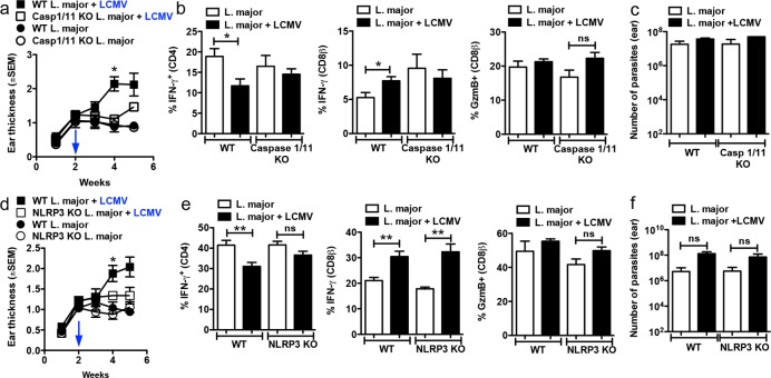
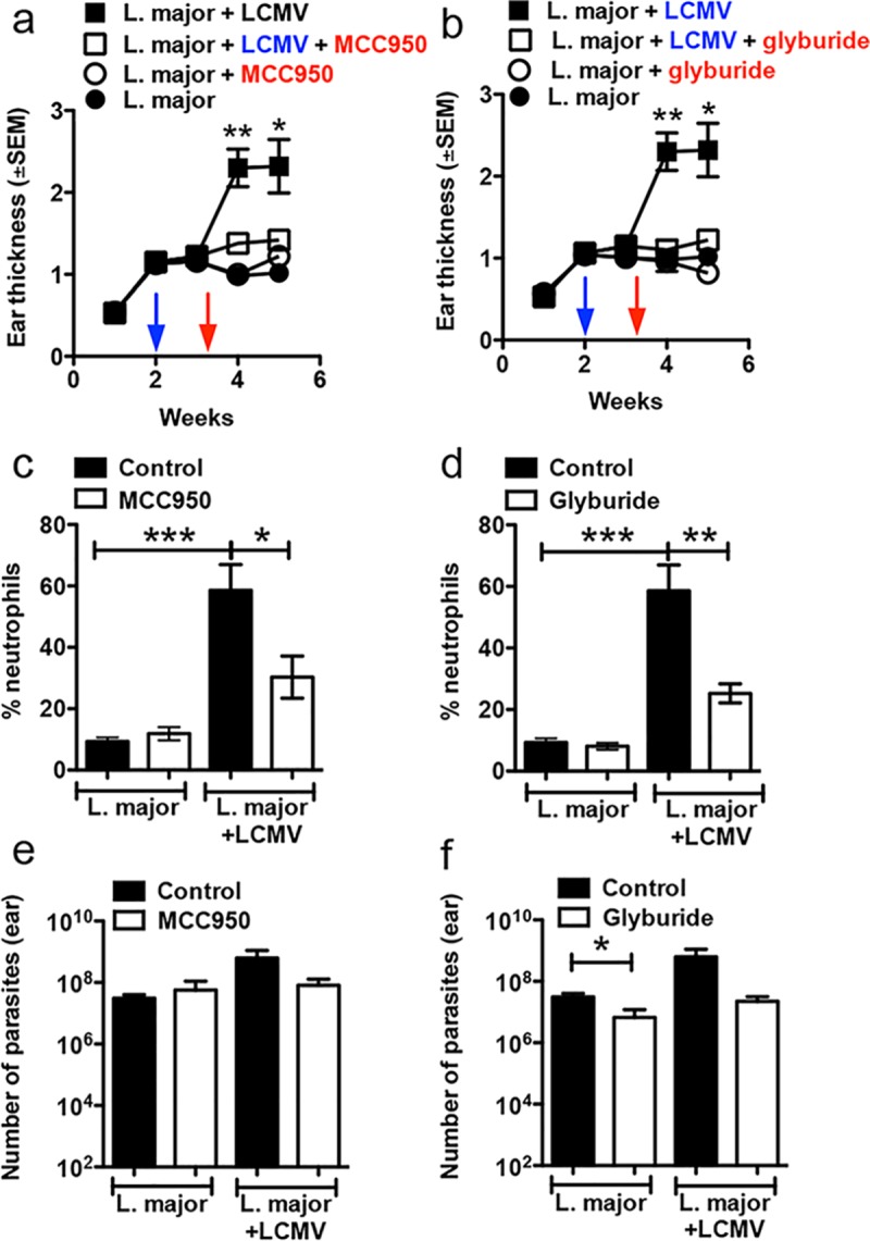
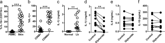
Deregulated CD8+ T cell cytotoxicity plays a central role in enhancing disease severity in several conditions. However, we have little understanding of the mechanisms by which immunopathology develops as a consequence of cytotoxicity. Using murine models of inflammation induced by the protozoan parasite leishmania, and data obtained from patients with cutaneous leishmaniasis, we uncovered a previously unrecognized role for NLRP3 inflammasome activation and IL-1β release as a detrimental consequence of CD8+ T cell-mediated cytotoxicity, ultimately resulting in chronic inflammation. Critically, pharmacological blockade of NLRP3 or IL-1β significantly ameliorated the CD8+ T cell-driven immunopathology in leishmania-infected mice. Confirming the relevance of these findings to human leishmaniasis, blockade of the NLRP3 inflammasome in skin biopsies from leishmania-infected patients prevented IL-1β release. Thus, these studies link CD8+ T cell cytotoxicity with inflammasome activation and reveal novel avenues of treatment for cutaneous leishmaniasis, as well as other of diseases where CD8+ T cell-mediated cytotoxicity induces pathology.


Conflict of interest statement

The authors have declared that no competing interests exist.

Figures

Fig 1
Fig 1. Increased IL-1β in L. braziliensis lesions is dependent on CD8 T cell cytotoxicity.
RAG-/- mice were infected with L. braziliensis in the ear, and reconstituted with CD8 T cells or did not receive cells and (a) the course of infection was monitored and representative images of lesions are shown. At 7 weeks post infection mice were euthanized and (b) mRNA levels for IL1a and IL1b were assessed. mRNA data is represented as a fold change (FC) over expression in naïve mice. At 7 weeks post infection, lesions were also digested and used for flow cytometric analysis. Depicted are (c) representative histogram and (d) bar graph of intracellular staining for IL-1β. (e) Ears were cultured for 48 hours and IL-1β release was measured in the supernatants by ELISA. RAG-/- mice were infected with L. braziliensis in the ear, and reconstituted with either WT or perforin-/- CD8 T cells or did not receive cells and (f) course of infection was monitored. At 7 weeks post infection mice were euthanized, lesions were digested and used for flow cytometric analysis. Depicted are (g) representative histogram and (h) bar graph of intracellular staining for IL-1β. Representative data from one of three or more independent experiments (n = 3 to 5 mice per group) with similar results are presented. *p ≤ 0.05 or ***p ≤ 0.001; ns, non-significant
Fig 2
Fig 2. Immunopathology caused by CD8 T cells is IL-1β-dependent.
RAG-/- mice were infected with L. braziliensis in the ear, and reconstituted with CD8 T cells or did not receive cells and at 2 weeks post infection mice were treated with (a) anti-IL-1R mAb, (b) anti-IL-1β mAb (c) anti-IL-1α mAb or (f) anakinra; ear thickness was assessed weekly. (d) 4 weeks or (g) 6 weeks post infection mice were euthanized and lesions were digested and used for flow cytometric analysis of intracellular IFN-γ and GzmB on CD8 T cells. (e and h) Parasite burden in the lesions. Graphs are data combined from 2 independent experiments (n = 3 to 5 mice per group in each experiment). C57BL/6 mice were infected with L. major in the ear, and 2 weeks later mice were co-infected with 2×10⁵ PFU of LCMV Armstrong strain by i.p. injection. Ten days post LCMV infection mice were treated with anakinra or were left untreated; (i) ear thickness was assessed weekly. Five weeks post infection with L. major, mice were euthanized and the (j) number of parasites in the skin was determined and lesions were digested and used for flow cytometric analysis of intracellular IFN-γ, and GzmB on (k) CD4 T cells or (l) CD8 T cells. Graphs are data from 2 independent experiments (n = 5 mice per group) with similar results are presented. *p ≤ 0.05, **p ≤ 0.01 or ***p ≤ 0.001; ns, non-significant
Fig 3
Fig 3. Immunopathology caused by CD8 T cells is dependent on the NLRP3 inflammasome.
WT, caspase-1/11-/- or NLRP3-/- C57BL/6 mice were infected with L. major in the ear, and 2 weeks later mice were co-infected with 2×10⁵ PFU of LCMV Armstrong strain by i.p. injection; (a and d) ear thickness was assessed weekly. Five weeks post infection with L. major, mice were euthanized and the lesions were digested and used for flow cytometric analysis of intracellular IFN-γ and GzmB on (b and e) CD4 T cells or CD8 T cells. (c and f) Number of parasites in the skin was determined at 5 weeks post infection with L. major. Graphs are data from 2 to 3 independent experiments (n = 5 mice per group) with similar results. **p<0.01
Fig 4
Fig 4. Treatment of mice with NLRP3 inhibitors dampens the immunopathology caused by CD8 T cells.
WT C57BL/6 mice were infected with L. major in the ear, and 2 weeks later mice were co-infected with 2×10⁵ PFU of LCMV Armstrong strain by i.p. injection. Ten days post LCMV infection mice were treated with MCC950, glyburide or vehicle; (a and b) ear thickness was assessed weekly. Five weeks post infection with L. major, mice were euthanized and the lesions were digested and the (c and d) frequency of neutrophils in the skin was determined directly ex vivo by flow cytometry. (e and f) Number of parasites in the skin was determined at 5 weeks post infection with L. major. Results in mice are data from one experiment with 5 mice per group. *p0.05, **p<0.01 or ***p0.001
Fig 5
Fig 5. IL-1β is highly expressed in human lesions and blockade of NLRP3 prevents IL-1β release from human skin biopsies.
Cells isolated from lesions or PBMC obtained from L. braziliensis patients were stained for flow cytometry directly ex vivo and depicted are (a) IL-1β+ CD11b+ cells (b) IL-1β expression within CD68+ (macrophages and monocytes) or CD66b+ (granulocytes) in the skin. Data obtained from (a) 19 PBMC or 18 skin lesions or (b) 13 skin lesions. (c) Punch biopsies from normal skin or L. braziliensis lesions were cultured for 48h and IL-1β was measured in the supernatants by ELISA. Data obtained from 5 normal skin samples and 14 lesions. L. braziliensis patients’ skin biopsies were cut in half and one half was cultured in media+DMSO and the other half with media+glyburide; 48 hours later, the presence of (d) IL-1β, (e) IL-6 and (f) IL-10 in the supernatants was determined by ELISA. Data obtained from 9 skin samples from two independent experiments. PBMC, peripheral blood mononuclear cells. **p<0.01 or ***p<0.001

References

    1. Voskoboinik I, Whisstock JC, Trapani JA (2015) Perforin and granzymes: function, dysfunction and human pathology. Nat Rev Immunol 15: 388–400. 10.1038/nri3839 - DOI - PubMed
    1. Haque A, Best SE, Unosson K, Amante FH, de Labastida F, et al. (2011) Granzyme B expression by CD8+ T cells is required for the development of experimental cerebral malaria. J Immunol 186: 6148–6156. 10.4049/jimmunol.1003955 - DOI - PubMed
    1. Nitcheu J, Bonduelle O, Combadiere C, Tefit M, Seilhean D, et al. (2003) Perforin-dependent brain-infiltrating cytotoxic CD8+ T lymphocytes mediate experimental cerebral malaria pathogenesis. J Immunol 170: 2221–2228. - PubMed
    1. Silverio JC, Pereira IR, Cipitelli Mda C, Vinagre NF, Rodrigues MM, et al. (2012) CD8+ T-cells expressing interferon gamma or perforin play antagonistic roles in heart injury in experimental Trypanosoma cruzi-elicited cardiomyopathy. PLoS Pathog 8: e1002645 10.1371/journal.ppat.1002645 - DOI - PMC - PubMed
    1. Gebhard JR, Perry CM, Harkins S, Lane T, Mena I, et al. (1998) Coxsackievirus B3-induced myocarditis: perforin exacerbates disease, but plays no detectable role in virus clearance. Am J Pathol 153: 417–428. 10.1016/S0002-9440(10)65585-X - DOI - PMC - PubMed

Publication types

MeSH terms

Substances