. 2017 Feb 13;13(2):e1006217.
doi: 10.1371/journal.ppat.1006217. eCollection 2017 Feb.

Multi-layered control of Galectin-8 mediated autophagy during adenovirus cell entry through a conserved PPxY motif in the viral capsid

Affiliations

Multi-layered control of Galectin-8 mediated autophagy during adenovirus cell entry through a conserved PPxY motif in the viral capsid

Charlotte Montespan et al. PLoS Pathog. .

Abstract

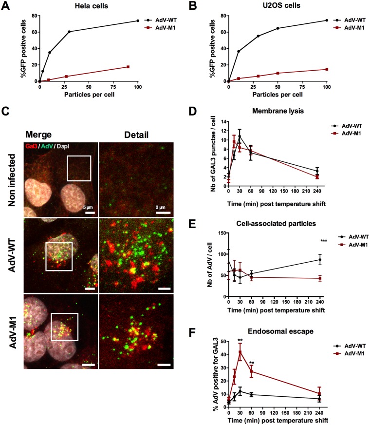
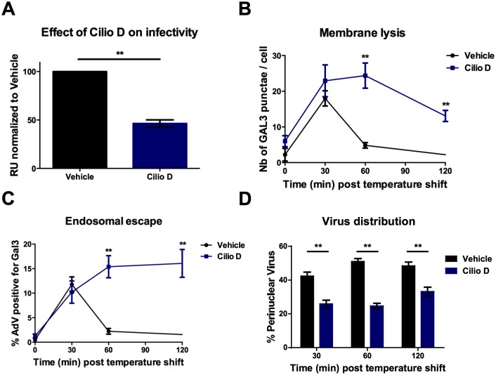
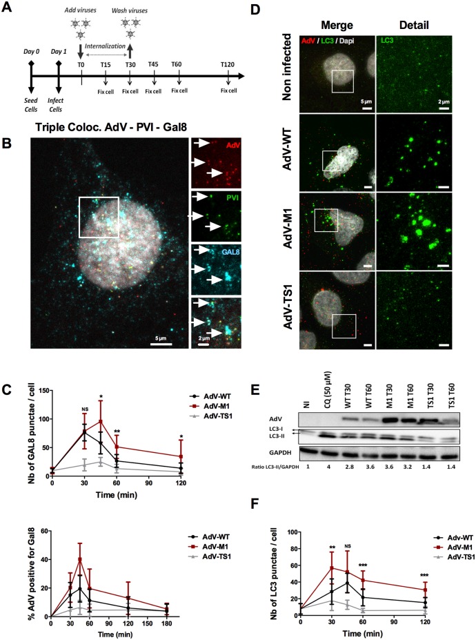
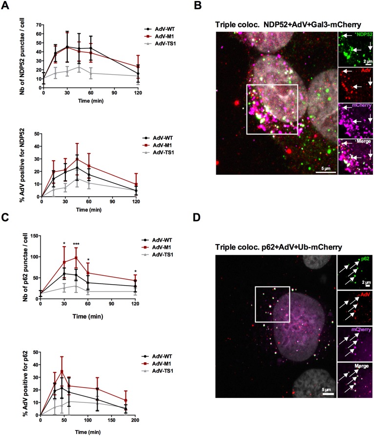
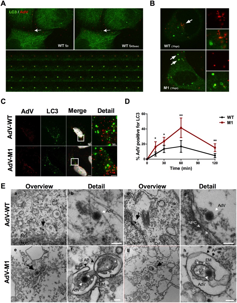
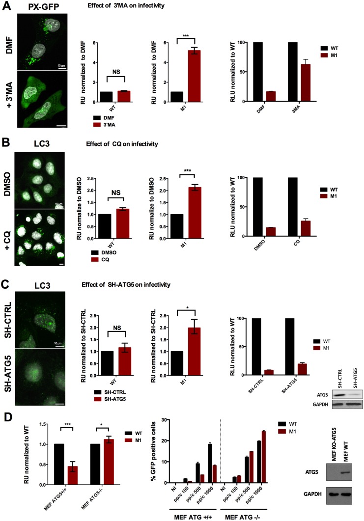
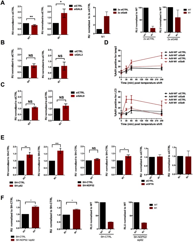
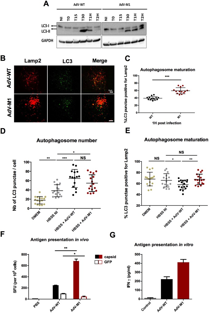
Cells employ active measures to restrict infection by pathogens, even prior to responses from the innate and humoral immune defenses. In this context selective autophagy is activated upon pathogen induced membrane rupture to sequester and deliver membrane fragments and their pathogen contents for lysosomal degradation. Adenoviruses, which breach the endosome upon entry, escape this fate by penetrating into the cytosol prior to autophagosome sequestration of the ruptured endosome. We show that virus induced membrane damage is recognized through Galectin-8 and sequesters the autophagy receptors NDP52 and p62. We further show that a conserved PPxY motif in the viral membrane lytic protein VI is critical for efficient viral evasion of autophagic sequestration after endosomal lysis. Comparing the wildtype with a PPxY-mutant virus we show that depletion of Galectin-8 or suppression of autophagy in ATG5-/- MEFs rescues infectivity of the PPxY-mutant virus while depletion of the autophagy receptors NDP52, p62 has only minor effects. Furthermore we show that wildtype viruses exploit the autophagic machinery for efficient nuclear genome delivery and control autophagosome formation via the cellular ubiquitin ligase Nedd4.2 resulting in reduced antigenic presentation. Our data thus demonstrate that a short PPxY-peptide motif in the adenoviral capsid permits multi-layered viral control of autophagic processes during entry.


Conflict of interest statement

The authors have declared that no competing interests exist.

Figures

Fig 1
Fig 1. AdV endosomal escape is PPxY dependent.
HeLa cells (A) or U2OS cells (B) were infected with varying amounts of adenoviral vector particles expressing GFP (WT in black, M1 in red) and the percentage of GFP positive cells was determined 24hpi by FACS analysis. (C) U2OS cells expressing Gal3-mCherry (red signal) were infected with WT or M1 viruses and fixed at 30 minutes post infection and stained for AdV (green signal). Note that colocalization (yellow signal) indicates membrane rupture. (D) U2OS cells were infected with fluorescent viruses fixed at different time points and stained for endogenous GAL3. At indicated time points Gal3 positive signals where quantified. The total number of Gal3 punctae is indicated. (E) U2OS cells were infected as in D and cell-associated virus was quantified at indicated time points. (F) Colocalization between Gal3 and AdV signals was quantified and is displayed as percentage colocalization of total AdV signal. **: P<0.01 and errors bars are standard error. (See also S1 Fig).
Fig 2
Fig 2. AdV endosomal escape is dynein dependent.
(A) Cells were pretreated with 100 μM of Ciliobrevin D for 30 minutes or DMSO and transduced with GFP expressing WT vector in presence of drugs. Twenty four hours later transduction levels were determined by FACS and normalized for the vehicle control (B) Cells were infected with fluorescent viruses in presence and absence of 100μM CilioD and fixed at different time points. The number of Gal3 punctae per cell was determined (C) Cells were infected as in B. Colocalization between Gal3 and AdV signals was quantified and is displayed as percentage of colocalization. (D) Cells were infected as in B. Fluorescent viruses in control or drug treated or control cells were quantified at indicated time points in the perinuclear region vs. the rest of the cytosol. Error bars (SE) show cell-to-cell variations.
Fig 3
Fig 3. AdV induced membrane damage causes selective autophagy.
(A) Schematic representation of the experimental setup for quantitative immune fluorescence analysis. Cells grown on coverslips were infected with virus at 37°C followed by inoculum removal after 30min. Coverslips were collected and fixed at indicated time points for quantitative immunofluorescence analysis as detailed in SI. (B) U2OS cells were infected with WT fluorescently labeled viruses (red signal) and fixed at 30min post infection and stained with anti PVI antibodies (green signal) and anti Gal8 antibodies (cyan signal). The insets to the right show triple colocalization (AdV+VI+Gal8) indicated by arrows (white signal). (C) U2OS cells were infected with fluorescently labeled WT (black line), M1 (red line) or TS1 viruses (gray line) as outlined in (A). At each time points cells were fixed and stained for Gal8. The top panel shows the absolute number of Gal8 punctae per cell, the bottom panel the percentage of Gal8 positive particles. Error bars show cell-to-cell variations (n>10 cells; NS: no significant; *: P<0.05; **: P<0.01; ***: P<0.005 and errors bars are standard deviation). (D) Representative confocal images of cells infected with different AdV showing virus induced autophagy. Conditions are indicated to the left of each row. Cells were fixed at 30 minutes post infection and stained with anti-AdV (first column, red signal) and anti-LC3 (first and second columns, green signal). (E) Infected U2OS cells were analyzed by western blot at 30 min (T30) and 60 min (T60) post infection using LC3 and GAPDH specific antibodies. As a positive control cells were treated with 50μM of CQ during 3hours. The LC3 signal depicts unconjugated (LC3-I) or PE-conjugated (LC3-II) LC3. Note that upon PE conjugation LC3 has a higher mobility in SDS-PAGE. The ratio of LC3II/GAPDH normalized to the non-infected condition was determined and is given below the panel. (F) Experiment as in (C) stained with anti-LC3 depicting the absolute number of LC3 punctae per cell.
Fig 4
Fig 4. Recruitment of adaptor proteins NDP52 and p62 upon entry.
(A) U2OS cells were infected with WT (black line), M1 (red line) or TS1 viruses (gray line) as outlined in Fig 3A. At each time points cells were fixed and stained for NDP52. The top panel shows the absolute number of NDP52 punctae per cell, the bottom panel the percentage of NDP52 positive particles. Error bars show cell-to-cell variations (n>10 cells; NS: no significant; *: P<0.05; **: P<0.01; ***: P<0.005 and errors bars are standard deviation). (B) U2OS cells stably expressing Gal3-mCherry (depicted as magenta signal) were infected with WT viruses and fixed at 30min post infection and stained with anti-AdV (red signal) and anti-NDP52 (green signal). The insets to the right show triple colocalization (AdV+Gal3mCh+NDP52) indicated by arrows (white signal). (C) U2OS cells were infected as in (A) and stained for p62. The top panel shows is the absolute number of p62 punctae per cell, the bottom panel the percentage of p62 positive particles. (D) U2OS cells transfected with a plasmid encoding Ubiquitin-mCherry (depicted as magenta signal) were infected with WT viruses and fixed at 30min post infection and stained with anti-AdV (red signal) and anti-p62 (green signal). The insets to the right show triple colocalization (AdV+UbmCh+p62) indicated by arrows (white signal).
Fig 5
Fig 5. The PPxY-mutant M1 associates with autophagosomes.
(A) Live-cell imaging showing LC3 acquisition of WT upon entry. Stable expressing U2OS-LC3-GFP cells were infected with Alexa594 coupled WT and imaged using spinning-disk confocal microscopy. The top two images show individual frames separated by ~45 seconds from S1 Movie. The arrow points to an LC3 negative virus (left panel) becoming LC3-positive (right panel). The bottom panel shows a higher magnification and frame resolution of the same event. (See also S1 Movie). (B) The panel shows single frames of cells infected as in (A) either with WT (top) or M1 virus (bottom) at 1 hpi. The arrow at the top panel points to the microtubule organizing center where WT viruses accumulate (shown at higher magnification to the right). The arrows at the bottom panel point to autophagosomes engulfing mutant M1 viruses (shown at higher magnification to the right). (C) Representative confocal images of cells at 1hpi infected with WT (top row) and M1 (bottom row) and stained for AdV (red signal) and LC3 (green signal). Virus association with LC3 appears as yellow signal (see detail). (D) Experiment as in (C). The percentage of each AdV colocalizing with LC3 was quantified over time and is given as percentage of total virus. Error bars show cell to cell variation (n>10 cells; *: P<0.05; **: P<0.01). (E) TEM analysis of U2OS cells infected for 30 minutes with WT (a-d, top row) or M1 (e-h, bottom row). The overview images show cytosolic WT viruses (a, c) and vesicle associated M1 viruses (e, g) depicted by a black arrow. At higher magnification WT (b, d) and M1 (f, h) particles (AdV) are depicted by white arrowheads. The nuclear pore complex (NPC in d) and the endosomal membrane (EM in f, g) are indicated with grey arrowheads and autophagosomes (AP) by black arrowheads (f, h). The * indicates the putative location for autophagy receptors between EM and AP. Error bars are 100 nm.
Fig 6
Fig 6. PPxY-mediated endosomal escape prevents autophagic degradation of incoming virions.
(A) Left panel: U2OS cells expressing the Pi3P in cellulo binding probe PX-GFP were treated with vehicle (top) or with 5mM of the Pi3K inhibitor 3’MA (bottom). Middle panel: U2OS cells pre-treated with vehicle alone (black bars) or 3’MA (red bars) were transduced with WT or M1. Transgene expression was determined and normalized to vehicle treated controls to show the fold induction of infectivity upon treatment. Right panel: The same data as in the middle panel showing the level of M1 infectivity rescue compared to the normalized WT infectivity upon treatment. (B) Left panel: U2OS cells were treated with vehicle (top) or with chloroquine (CQ, 50μM, bottom) to block the autophagic flux, fixed and stained for LC3. Middle panel: Cells were treated with vehicle alone (black bars) or chloroquine (red bars) and transduced with WT or M1. Transgene expression was determined and normalized to vehicle treated controls to show the fold induction of infectivity. Right panel: The same data as in the middle panel showing the level of M1 infectivity rescue compared to normalized WT infectivity. (C) Left panel: U2OS cells depleted for ATG5 (SH-ATG5) or control depleted cells (SH-CTRL) and starved using HBSS during 4h, fixed and stained for LC3. Middle panel: Cells were transduced with WT or M1 and the relative transduction efficiency in SH-CTRL cells (black bars) and SH-ATG5 cells (red bars) was determined. Right panel: The same data as in the middle panel showing the level of M1 infectivity rescue compared to normalized WT infectivity. ATG5 expression levels were determined by western blot. (D) Left panel: Control MEFs (ATG5 +/+) and KO MEFs (ATG5 -/-) were transduced with WT or M1 as indicated and the relative transduction efficiency for the M1 (red bars) compared to the WT (black bars) was determined. Right panel: The panel shows the absolute number of transduced cells at indicated amounts of physical particles added to the cell (pp/c) for the WT (black bars) and the M1 (red bars) in ATG5 control (left) and KO (right) MEFs. ATG5 expression levels were determined by western blot.
Fig 7
Fig 7. M1 restriction is mediated by Gal8.
(A) Left panel: U2OS cells were depleted with siRNAs specific for galectin8. Cells were transduced with WT or M1 as indicated and the relative transduction efficiency was calculated for control depleted cells (black bars) or galectin depleted cells (red bars). Middle panel: Relative transduction efficiency of the M1 virus following two rounds of siRNA depletion. Right panel: The same data as in the middle panel showing the level of M1 infectivity rescue compared to the normalized WT infectivity upon treatment. Each experiment was done in triplicate (B) Experiment essentially done as in (A) except that galectin 3 was depleted. (C) Experiment essentially done as in (A) except that galectin 9 was depleted. (D) HeLa cells were transfected twice with control or galectin8 specific siRNAs followed by infection with WT or M1 viruses and fixed at different time points after infection. Quantification of AdV colocalizing with LC3 (top panel.) and Lamp1 (bottom panel) was performed and is shown as percentage of colocalization for each virus and condition according to the legend. (E) Left panel: Cells were depleted with SH-RNA specific for p62. Specific or control depleted cells were transduced with WT or M1 as indicated and the relative transduction efficiency was calculated for control depleted cells (black bars) or p62 depleted cells (red bars). Middle panel: Experiment essentially done as for p62 except that SH-RNA was directed against NDP52. Right panel: Experiment essentially done as for p62 except that SH-RNA was directed against optineurin (OPTN). (F) Left panel: Cells were depleted with SH-RNA specific for NDP52 followed by transfection with siRNA against p62. Double depleted or control depleted cells were transduced with WT or M1 as indicated and the relative transduction efficiency was calculated for control depleted cells (black bars) or p62 depleted cells (red bars). Middle panel: The same data as in the left panel showing the level of M1 infectivity rescue compared to the normalized WT infectivity upon treatment.
Fig 8
Fig 8. AdV-WT limits autophagosome maturation and antigen presentation.
(A) Cells were infected with WT and M1 viruses for indicated time points and cell lysates were analyzed by western blot with LC3 specific antibodies. Specific LC3 bands and GAPDH loading control are indicated. (NI = non infected). (B) The panel shows representative confocal images of U2OS cells infected for 1h with WT or M1 as indicated to left of each row and stained with Lamp2 (red signal) and LC3 (green signal) specific antibodies. (C) Quantification of autolysosomes from the experiment shown in (B) comparing the percentage of LC3 punctae positive for Lamp2 in WT vs. M1 infected cells as indicated below the graph. (D) Cells were starved overnight in HBSS then infected for one hour with AdV as indicated below the graph. Samples were fixed and stained for LC3 and Lamp2. The total number of LC3 punctae per cell at 1hpi is shown. (NI = non infected, DMEM = non starved control cells). (E) Experiment as in (D) showing the percentage of LC3 punctae also positive for Lamp2 (n>13 cells; NS: no significant; *: P<0.05; **: P<0.01; ***: P<0.001). (F) Mice were infected with 1010 GFP expressing vector particles of WT, M1 or PBS control. 10 days post infection mice were sacrificed and splenocytes were stimulated with AdV-luc or GFP purified from E.coli. IFNγ was determined by ELIspot. (G) CD4+ T-cell clones recognizing a conserved AdV hexon epitope were incubated 3:1 with syngeneic APCs transduced with either control media or WT or M1 vectors for 24 hours. IFNγ secretion was quantified by ELISA. (see also S5 Fig).
Fig 9
Fig 9. Nedd4.2 controls autophagy upon AdV infection.
(A) Nedd4.2 or control depleted cells were infected with WT virus. Cell lysates were analyzed at indicated time points by western blot using LC3 and GAPDH specific antibodies as shown to the left. (NI = non infected). The ratio of LC3II/GAPDH normalized to the non-infected condition in siCTRL depleted cells was determined and is given below the panel. (B) Representative panel of confocal images from Nedd4.2 or control depleted cells (indicated to the left) stained with LC3 and Lamp2 specific antibodies. (C) Nedd4.2 or control depleted cells non-infected (NI) or infected with WT or M1 virus for 1h were fixed and stained as in (B). The percentage of LC3 and Lamp2 positive autolysosomes is indicated for each condition. (n>15 cells; NS: no significant; *: P<0.05; **: P<0.01). (D) Representative panel of confocal images from Nedd4.2 or control depleted cells infected with WT virus for 1h and stained with LC3 and pericentrin specific antibodies. (E) Quantification of the distribution of LC3 dots in cells. Localization was determined by calculating LC3 punctae distribution in concentric circles positioned around pericentrin stain as detailed in SI. The graph shows the color coded relative abundance within the 3 regions. The error bar represents cell-to-cell variation (n>15 cells; NS: no significant; **: P<0.01). (see also S6 Fig).
Fig 10
Fig 10. Nuclear transport of AdV involves the autophagic machinery.
(A) Representative panel of WT or M1 infected cells at 1hpi stained with LC3 (red signal) and pericentrin (green signal) specific antibodies. (B) Quantification of the relative distribution of autophagosomes for experiment shown in (A) essentially analyzed as described for Fig 9E. (n>12 cells; NS: no significant; *: P<0.05; **: P<0.01) (C) Representative panel of WT infected cells at 1hpi depleted for ATG5 or control depleted (as indicated) and stained with AdV (red signal) and pericentrin (green signal) specific antibodies. (D) Quantification of the relative virus distribution as in (B) for the experiment shown in (C) including the distribution of M1 and WT viruses. (n>12 cells; NS: no significant, **: P<0.01) (E) Representative panel of WT infected cells at 1hpi depleted for ATG5 or control depleted (as indicated) and stained with AdV (red signal) and PVI (green signal) specific antibodies to mark PVI separation from the virus. (F) Infection time course analysis of PVI release from M1 (red line) vs. WT (black line) viruses in ATG5 depleted (dotted line) vs. control depleted cells (solid line). Shown is the percentage of PVI positive AdV at indicated time points. The errors bars are cell-to-cell variation (10 cells were analyzed for each conditions). (G) Representative panel of WT infected cells at 1hpi and stained with specific antibodies against AdV (red signal) and specific antibodies against PVII (green signal) to mark nuclear genomes. (H) Quantification of nuclear genome delivery. ATG5 and control depleted cells were infected with WT and M1 and fixed at 1 and 2hpi and stained for AdV and PVII. The number of nuclear PVII dots was calculated and normalized for virus particles at each condition as indicated below the graph (n>16 cells; **: P<0.01; ***: P<0.0001). (See also S7 Fig).
Fig 11
Fig 11. Model for adenovirus control of autophagic processes upon entry.
AdV enter cells by receptor-mediated endocytosis (1) followed by partial disassembly to release the internal membrane lytic capsid protein PVI (2). PVI release initiates membrane rupture and intralumenal glycans are recognized via galectins and the autophagic machinery is recruited through Gal8 and LC3 to the damaged endosome mediated by yet to clarify adapter molecules (3). The recruitment of Nedd4.2 via the PPxY motif in PVI prevents formation of autophagosomes via an unknown mechanisms and facilitates endosomal escape (4). Endosomal escape involves the autophagic machinery because ATG5 depletion affects dissociation of virus from damaged vesicle. Dynein motor complexes are also required to access cytosolic microtubule mediated transport towards the MTOC which may occur in association with LC3 (5). Subsequent genome release occurs at the nuclear pore complex (6). Genome delivery is delayed upon ATG5 depletion. If PVI is not released (AdV-TS1), no membrane damage occurs and viruses are degraded via lysosomal sorting (7). If PVI is released and membrane damage occurs but the virus does not escape (AdV-M1), capsids are degraded via autophagy (8). This degradation is limited in absence of functional autophagy (e.g. upon ATG5 depletion).

References

    1. Koyama S, Ishii KJ, Coban C, Akira S. Innate immune response to viral infection. Cytokine. 2008. September;43(3):336–41. 10.1016/j.cyto.2008.07.009 - DOI - PubMed
    1. Nemerow GR, Stewart PL. Role of alpha(v) integrins in adenovirus cell entry and gene delivery. Microbiol Mol Biol Rev MMBR. 1999. September;63(3):725–34. - PMC - PubMed
    1. Wickham TJ, Mathias P, Cheresh DA, Nemerow GR. Integrins αvβ3 and αvβ5 promote adenovirus internalization but not virus attachment. Cell. 1993. April 23;73(2):309–19. - PubMed
    1. Greber UF, Willetts M, Webster P, Helenius A. Stepwise dismantling of adenovirus 2 during entry into cells. Cell. 1993. November 5;75(3):477–86. - PubMed
    1. Maier O, Marvin SA, Wodrich H, Campbell EM, Wiethoff CM. Spatiotemporal dynamics of adenovirus membrane rupture and endosomal escape. J Virol. 2012. October;86(19):10821–8. 10.1128/JVI.01428-12 - DOI - PMC - PubMed

Publication types

MeSH terms