. 2017 Apr:124:106-115.
doi: 10.1016/j.biomaterials.2017.01.042. Epub 2017 Feb 2.

Direct 3D bioprinting of prevascularized tissue constructs with complex microarchitecture

Affiliations

Direct 3D bioprinting of prevascularized tissue constructs with complex microarchitecture

Wei Zhu et al. Biomaterials. 2017 Apr.

Abstract

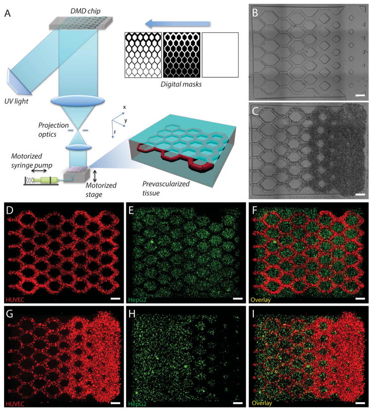
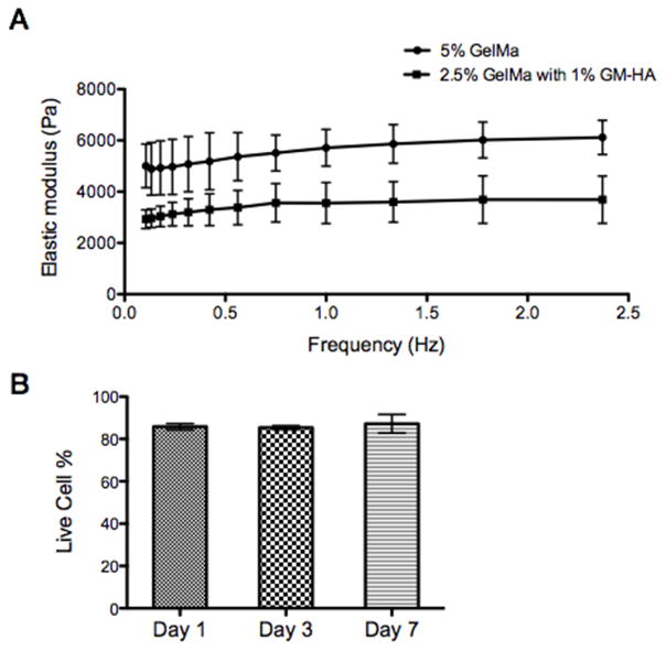
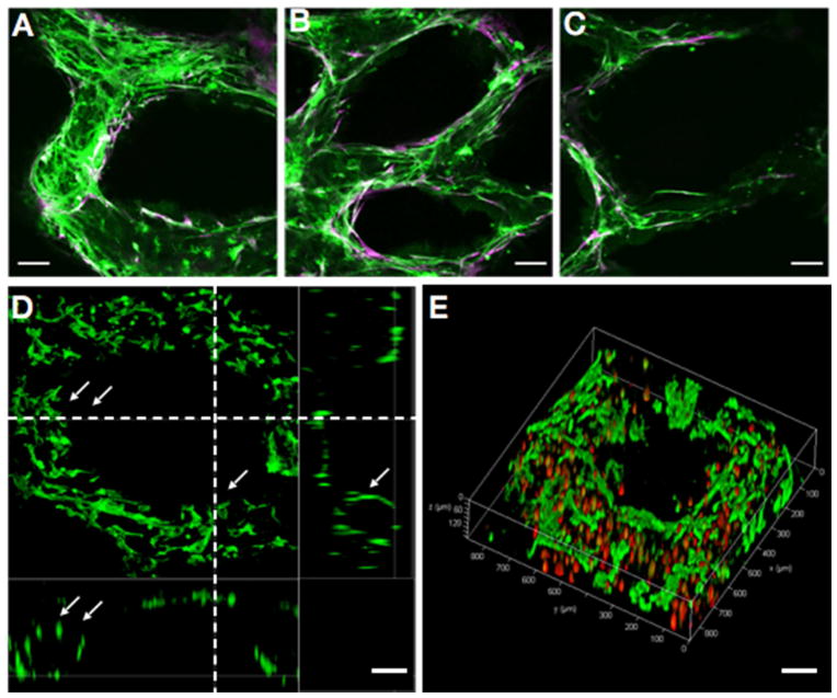
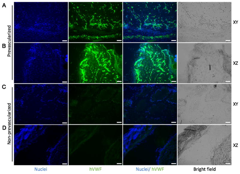
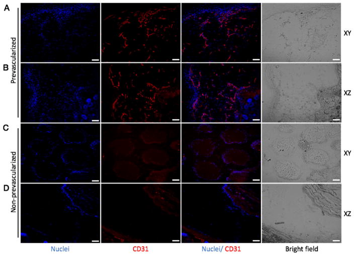
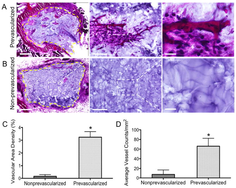
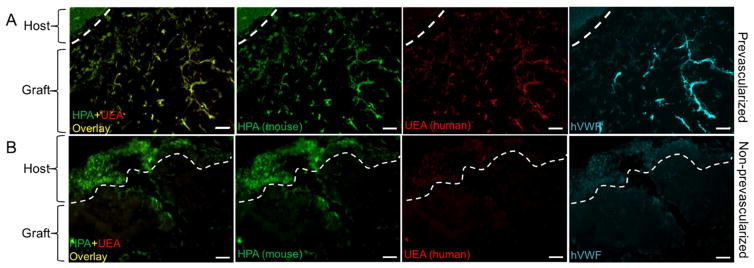
Living tissues rely heavily on vascular networks to transport nutrients, oxygen and metabolic waste. However, there still remains a need for a simple and efficient approach to engineer vascularized tissues. Here, we created prevascularized tissues with complex three-dimensional (3D) microarchitectures using a rapid bioprinting method - microscale continuous optical bioprinting (μCOB). Multiple cell types mimicking the native vascular cell composition were encapsulated directly into hydrogels with precisely controlled distribution without the need of sacrificial materials or perfusion. With regionally controlled biomaterial properties the endothelial cells formed lumen-like structures spontaneously in vitro. In vivo implantation demonstrated the survival and progressive formation of the endothelial network in the prevascularized tissue. Anastomosis between the bioprinted endothelial network and host circulation was observed with functional blood vessels featuring red blood cells. With the superior bioprinting speed, flexibility and scalability, this new prevascularization approach can be broadly applicable to the engineering and translation of various functional tissues.

Keywords: 3D bioprinting; Biomaterials; Complex microarchitecture; Tissue engineering; Vasculature.


Conflict of interest statement

Competing interests. The authors declare no competing interests.

Figures

Fig. 1
Fig. 1. 3D bioprinting of the prevasularized tissue constructs
(A) Schematic of the bioprinting platform. (B) Bioprinted acellular construct featuring intended channels with gradient widths. (C) Bioprinted cellular construct with HUVECs and 10T1/2 (50:1) encapsulated in the intended channels. (DF) Fluorescent images demonstrating the bioprinting of heterogeneous cell-laden tissue constructs with uniform channel width. HUVECs (red) are encapsulated in the intended channels and HepG2 (green) are encapsulated in the surrounding area. (GI) Fluorescent images demonstrating the bioprinting of heterogeneous cell-laden tissue constructs with gradient channel widths. Scale bars, 250 μm.
Fig. 2
Fig. 2. In vitro characterization of the bioprinted tissue
(A) Elastic modulus of the biomaterials used to encapsulate cells measured by DMA: 2.5% GelMa with 1% HA for the channel region and 5% GelMa for the surrounding region. (B) Results of cell-viability assay for the bioprinted tissue constructs encapsulated with HUVECs demonstrating over 85% cell viability. Error bars represent SEM, n=3 for all data points.
Fig. 3
Fig. 3. Endothelial network formation after 1-week culture of the prevascularized tissue construct in vitro
(AC) Confocal microscopy images show HUVECs (Green, CD31-positive) and supportive mesenchymal cells (10T1/2, Purple, alpha-smooth muscle actin (α-SMA)-positive) aligned within the patterned gradient channel regions with different vessel sizes. (D) Cross-section view shows the endothelial cells (CD31-positive) form lumen-like structures (highlighted by arrows) along the bioprinted channels. (E) 3D view of the endothelial cells lining along the printed microchannel walls by confocal microscopy. Endothelial cells were labeled by fluorescent cell tracker (red) and stained by CD31 (green). Scale bars: 100 μm.
Fig. 4
Fig. 4. Endothelial network formation in the prevascularized tissues (A, B) and non-prevascularized tissues (C, D) after 2-week subcutaneous implantation shown by hVWF staining
In the prevascularized tissues, a mixture of HUVECs and supportive 10T1/2 cells were printed into the gradient channels (Fig. 1C presents the prevascularized tissue before implantation). In the non-prevascularized tissues, no cells were printed into the samples, only biomaterials were used (Fig. 1B presents the non-prevascularized tissue before implantation). The bioprinted tissues were implanted under the dorsal skin of SCID mice for two weeks. The samples harvested from the implantation were cryosectioned in both xy and xz planes (xyz axises were designated in Fig. 1A). DAPI was used to stain the nuclei (blue) and hVWF was used to stain the endothelial network (green). Scale bars, 100 μm.
Fig. 5
Fig. 5. Endothelial network formation in the prevascularized tissues (A, B) and non-prevascularized tissues (C, D) after 2-week subcutaneous implantation shown by CD31 staining
In the prevascularized tissues, a mixture of HUVECs and supportive 10T1/2 cells were printed into the gradient channels (Fig. 1C presents the prevascularized tissue before implantation). In the non-prevascularized tissues, no cells were printed into the samples, only biomaterials were used (Fig. 1B presents the non-prevascularized tissue before implantation). The bioprinted tissues were implanted under the dorsal skin of SCID mice for two weeks. The samples harvested from the implantation were cryosectioned in both xy and xz planes (xyz axises were designated in Fig. 1A). DAPI was used to stain the nuclei (blue) and CD31 was used to stain the endothelial network (red). Scale bars, 100 μm.
Fig. 6
Fig. 6. H&E staining of the grafted tissues after 2-week subcutaneous implantation and quantification of vasculature parameters
(A) Representative H&E stained images of the prevascularized tissues showing significant amount of endothelial vessels with red blood cells were found. Yellow dash line marks the interface between the graft and host tissue. (B) Representative H&E stained images of the non-prevascularized tissues, showing limited endothelial vessels. Yellow dash line marks the interface between the graft and host tissue. (C) Quantification of vascular area density in the grafted tissues. (D) Quantification of average vessel counts per area in the grafted tissue. Error bars represent SEM, n=6 for all data points. * indicates significant difference between the prevascularized group and the nonprevascularized group, p < 0.05. Scale bars: (A) 500μm (left), 100μm (middle), 25μm (right); (B) 500μm (left), 100μm (middle), 25μm (right).
Fig. 7
Fig. 7. Perfusion of mouse and human specific lectins after two-week subcutaneous implantation
(A) In the prevascularized tissue, mouse-specific lectin (HPA) and human-specific lectin (UEA) are chimeric in the graft area, and the host tissue is only stained with mouse-specific lectin. Further staining of hVWF confirms the endothelial network formed by human-origin HUVECs. (B) In the nonprevascularized tissue, HPA stains the host tissue and minor regions of the graft area, no UEA or hVWF staining is observed. Scale bars: 100μm.

References

    1. Miller JS, Stevens KR, Yang MT, Baker BM, Nguyen D-HT, Cohen DM, Toro E, Chen Aa, Galie Pa, Yu X, Chaturvedi R, Bhatia SN, Chen CS. Rapid casting of patterned vascular networks for perfusable engineered three-dimensional tissues. Nat Mater. 2012;11:768–74. doi: 10.1038/nmat3357. - DOI - PMC - PubMed
    1. Baranski JD, Chaturvedi RR, Stevens KR, Eyckmans J, Carvalho B, Solorzano RD, Yang MT, Miller JS, Bhatia SN, Chen CS. Geometric control of vascular networks to enhance engineered tissue integration and function. Proc Natl Acad Sci. 2013;110:7586–7591. doi: 10.1073/pnas.1217796110. - DOI - PMC - PubMed
    1. Kolesky DB, Truby RL, Gladman aS, Busbee Ta, Homan Ka, Lewis Ja. 3D bioprinting of vascularized, heterogeneous cell-laden tissue constructs. Adv Mater. 2014;26:3124–3130. doi: 10.1002/adma.201305506. - DOI - PubMed
    1. Lee KY, Peters MC, Anderson KW, Mooney DJ. Controlled growth factor release from synthetic extracellular matrices. Nature. 2000;408:998–1000. doi: 10.1038/35050141. - DOI - PubMed
    1. Fischbach C, Mooney DJ. Polymers for pro- and anti-angiogenic therapy. Biomaterials. 2007;28:2069–2076. doi: 10.1016/j.biomaterials.2006.12.029. - DOI - PubMed

Publication types

MeSH terms