Review
. 2017 Jan 27;12(1):37.
doi: 10.1186/s13014-016-0754-z.

Present state and issues in IORT Physics

Affiliations
Review

Present state and issues in IORT Physics

Frank W Hensley. Radiat Oncol. .

Abstract

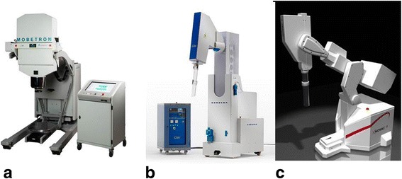
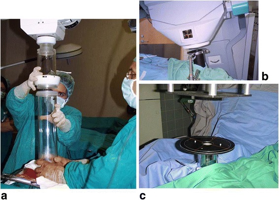
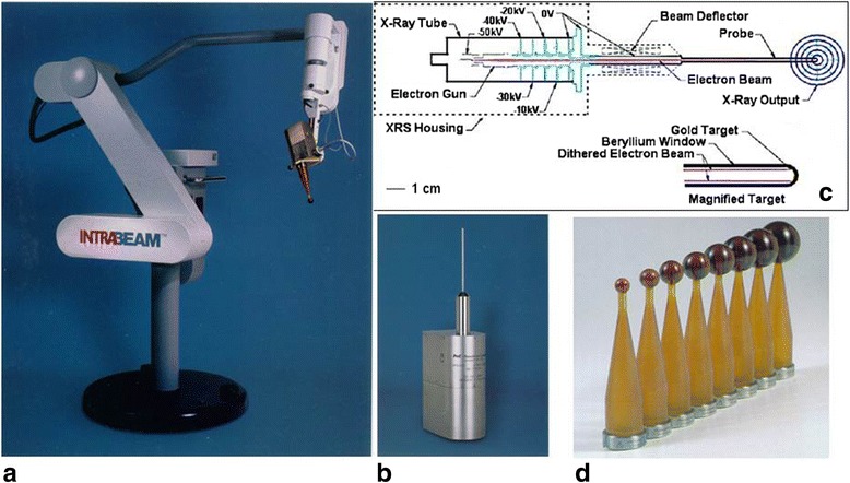
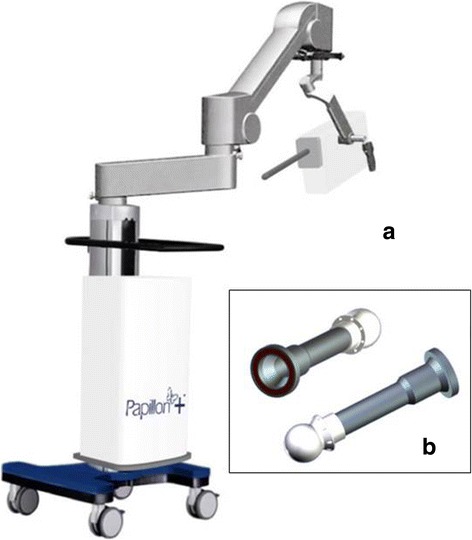
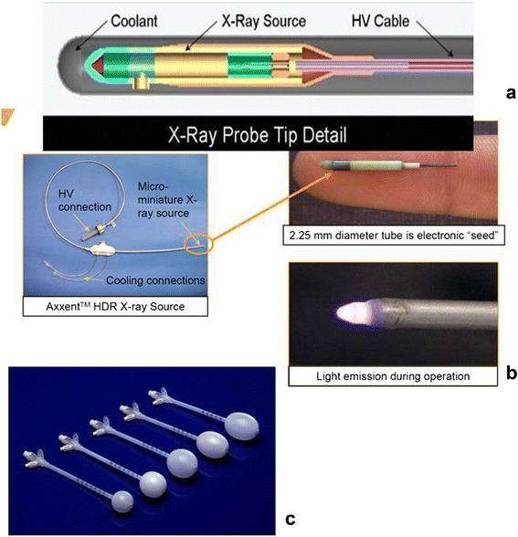
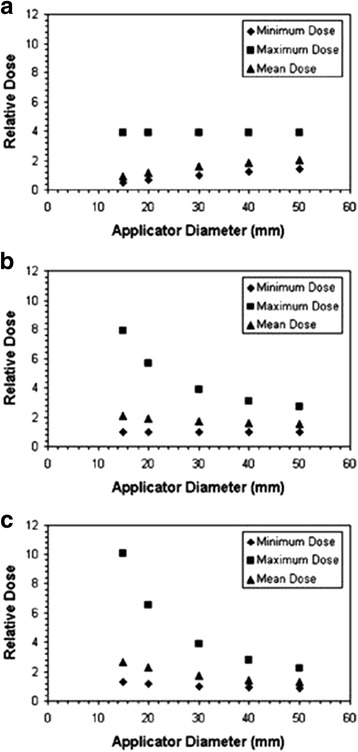
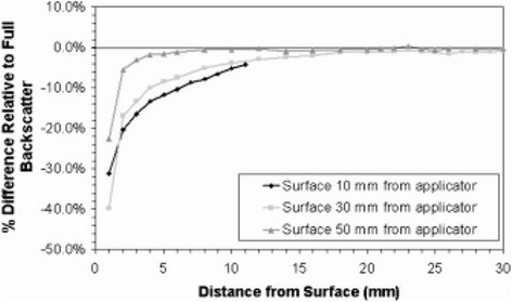
Literature was reviewed to assess the physical aspects governing the present and emerging technologies used in intraoperative radiation therapy (IORT). Three major technologies were identified: treatment with electrons, treatment with external generators of kV X-rays and electronic brachytherapy. Although also used in IORT, literature on brachytherapy with radioactive sources is not systematically reviewed since an extensive own body of specialized literature and reviews exists in this field. A comparison with radioactive sources is made in the use of balloon catheters for partial breast irradiation where these are applied in almost an identical applicator technique as used with kV X-ray sources. The physical constraints of adaption of the dose distribution to the extended target in breast IORT are compared. Concerning further physical issues, the literature on radiation protection, commissioning, calibration, quality assurance (QA) and in-vivo dosimetry of the three technologies was reviewed. Several issues were found in the calibration and the use of dosimetry detectors and phantoms for low energy X-rays which require further investigation. The uncertainties in the different steps of dose determination were estimated, leading to an estimated total uncertainty of around 10-15% for IORT procedures. The dose inhomogeneity caused by the prescription of electrons at 90% and by the steep dose gradient of kV X-rays causes additional deviations from prescription dose which must be considered in the assessment of dose response in IORT.


Figures

Fig. 1
Fig. 1
Mobile electron accelerators for intraoperative radiation therapy. a The Mobetron (IntraOp Medical Corporation, 570 Del Rey Ave, Sunnyvale, CA 94085, USA), b the Liac, c the Novac 7 (both: Sit Sordina IORT Technologies Spa, Galleria del Pozzo Rosso, 13, 36100 Vicenza VI, Italy)
Fig. 2
Fig. 2
Hard and soft docking technique in electron IORT. a Hard docking: the applicator is attached to a receptor on the accelerator head by a sterile person while a second sterile person holds the applicator in place. (shown for a Liac accelerator). b Soft docking: the applicator is attached to the couch with a table stand. There is no mechanical connection between applicator and accelerator. (shown for a Mobetron accelerator). c The alignment of accelerator and applicator is adjusted with a laser alignment system. (Shown for a Siemens Mevatron ME accelerator)
Fig. 3
Fig. 3
The Zeiss Intrabeam System (Carl Zeiss Meditec AG, Göschwitzer Str. 51-52, 07745 Jena, Germany). a The X-ray source (XRS) with a spherical breast applicator mounted on the floor stand. b The XRS. c Schematic drawing of the XRS accelerator. d Spherical applicators for breast IORT
Fig. 4
Fig. 4
The Papillon system (Ariane Medical Systems Limited, Derby DE1 3BY UK). a The X-ray source mounted on the floor stand. b Spherical applicators for breast IORT
Fig. 5
Fig. 5
The Xoft Axxent System (Xoft Inc. 345 Potero Ave. Sunnyvale, CA 94085, USA). a Schematic diagram of the X-ray source. b Left: the cooling tube in which the source is guided to the irradiation position. Right top: The source, bottom: light emission during source operation. c Spherical balloon applicators for breast IORT
Fig. 6
Fig. 6
Maximum, mean and minimum doses in 10 mm thick spherical shells of tissue surrounding Intrabeam breast applicators for different depths of prescription of the reference dose with different sizes of applicators. a prescription at the applicator surface. b prescription at 10mm distance from the applicator surface. c prescription at 20mm distance from the applicator surface. The prescription dose is defined as unity for the minimum target dose with the 30mm applicator in each diagram. For small applicators, a prescription at the applicator surface (a) produces smaller overdoses at the applicator surface, however also lower doses at a depth of 10mm which is considered as the margin surrounding the tumor cavity which must be treated. Prescription at 10mm depth ensures that the desired dose arrives at this depth, produces higher overdoses for small applicators, prescription at 20mm depth increases the overdoses. Therefore the authors conclude that a prescription at 10mm depth is the best compromise in order to ensure that desired dose arrives at depth for all applicator sizes. From Ebert and Carruthers (2003) [35]
Fig. 7
Fig. 7
Dose reduction due to missing backscatter at different depths under the tissue surface for three different depths of the applicator surface in tissue (10mm, 30mm and 50mm) for a 30mm diameter intrabeam applicator. For shallow depths of the applicator dose reduction in superficial tissues can amount as much as 25-40%. From Ebert and Carruthers (2003) [35]
Fig. 8
Fig. 8
The O-arm mobile surgical cone beam CT imaging device (Medtronic GmbH, Earl-Bakken-Platz 1,40670 Meerbusch, Germany)
Fig. 9
Fig. 9
The robot-mounted flat panel surgical imaging device Siemens Artis zeego (Siemens Healthcare GmbH, Henkestr. 127, 91052 Erlangen, Germany)
Fig. 10
Fig. 10
The PAIR patient alignment imaging ring (medPhoton G.m.b.H., Müllner Hauptstraße 48, 5020 Salzburg, Austria)

References

    1. Palta JR, Biggs PJ, Hazle JD, Huq MS, Dahl RA, Ochran TG, Soen J, Dobelbower RR, Jr, McCullough EC. Intraoperative electron beam radiation therapy: technique, dosimetry, and dose specification: report of task force 48 of the Radiation Therapy Committee, American Association of Physicists in Medicine. Int J Radiat Oncol Biol Phys. 1995;33:725–46. doi: 10.1016/0360-3016(95)00280-C. - DOI - PubMed
    1. Beddar AS, Biggs PJ, Chang S, Ezzell GA, Faddegon BA, Hensley FW, Mills MD. Intraoperative radiation therapy using mobile electron linear accelerators: Report of AAPM Radiation Therapy Committee Task Group No. 72. Med Phys. 2006;33:1476–89. doi: 10.1118/1.2194447. - DOI - PubMed
    1. Rosi A and Viti V (eds.). Guidelines for quality assurance in intra-operative radiation therapy (English version). Rapporti ISTISAN 03/1 EN 2003 Istituto Superiore di Sanità (Viale Regina Elena, 299 - 00161 Roma)
    1. Soriani A, Felici G, Fantini M, Paolucci M, Borla O, Evangelisti G, Benassi M, Strigari L. Radiation protection measurements around a 12 MeV mobile dedicated IORT accelerator. Med Phys. 2010;37:995–1003. doi: 10.1118/1.3298012. - DOI - PubMed
    1. National Council on Radiation Protection and Measurements . Neutron contamination from medical accelerators. NCRP Report No. 79. Bethesda: NCRP; 1984.