. 2017 Feb 13;14(1):12.
doi: 10.1186/s12977-017-0338-5.

Immune regulator ABIN1 suppresses HIV-1 transcription by negatively regulating the ubiquitination of Tat

Affiliations

Immune regulator ABIN1 suppresses HIV-1 transcription by negatively regulating the ubiquitination of Tat

Shiyou Chen et al. Retrovirology. .

Abstract

Background: A20-binding inhibitor of NF-κB activation (ABIN1), an important immune regulator, was previously shown to be involved in HIV-1 replication. However, the reported studies done with overexpressed ABIN1 provided controversial results.

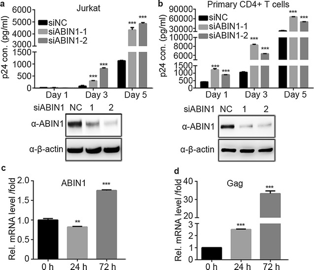
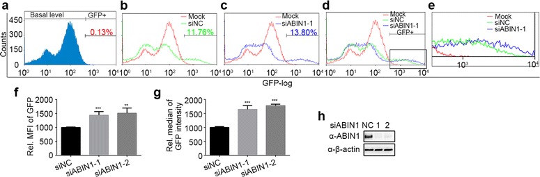
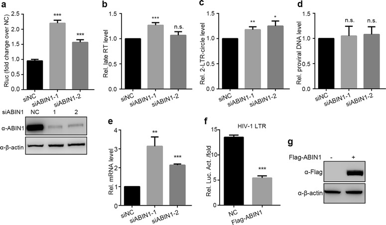
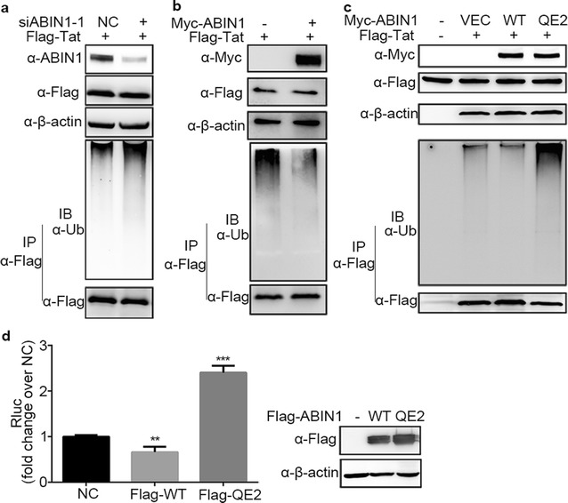
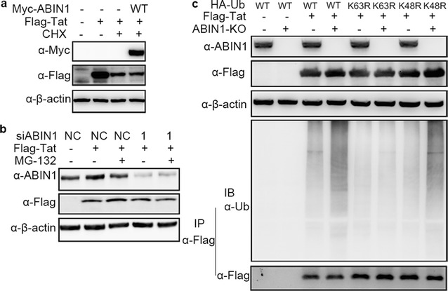
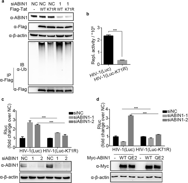
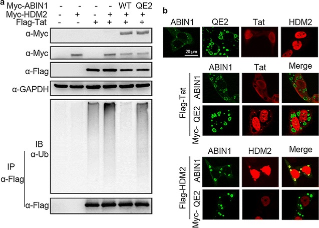
Results: Here we identified ABIN1 as a suppressor of HIV-1 transcription since transient knockdown of ABIN1 led to increased HIV-1 replication both in transformed Jurkat T cell line and in primary human CD4+ T lymphocytes. Depletion of ABIN1 specifically enhanced the HIV-1 transcription from the integrated genome during viral life cycle, but not the earlier steps such as reverse transcription or integration. Immunoprecipitation assays revealed that ABIN1 specifically inhibits the proto-oncogene HDM2 catalyzed K63-linked polyubiquitination of Tat at Lys71, which is critical for the transactivation activity of Tat. The ubiquitin chain binding activity of ABIN1 carried by UBAN domain turned out to be essential for the inhibitory role of ABIN1. The results of immunofluorescence localization experiments suggested that ABIN1 may obstruct Tat ubiquitination by redistributing some of HDM2 from the nucleus to the cytoplasm.

Conclusions: Our findings have revealed ABIN1 as intrinsic suppressor of HIV-1 mRNA transcription by regulating the ubiquitination of Tat.

Keywords: ABIN1; HDM2; HIV-1; Tat; Transcription; Ubiquitination.


Figures

Fig. 1
Fig. 1
ABIN1 knockdown increases HIV-1 replication. a Jurkat T cells were transfected with siRNAs targeting ABIN1 (siABIN1-1 or siABIN1-2) or scramble siRNA (siNC) as described in “Methods”. 36 h post transfection (hpt), cells were counted and challenged with HIV-1 strain NL4-3. At 1, 3, 5 days post infection, the cells and supernatants were harvested to measure viral production by p24 ELISA of the supernatants, against a standard curve. Cells were lysed and subjected to Western Blots to determine the knockdown efficiency of ABIN1. The lower panel showed the ABIN1 knockdown efficiency at day 5 as the representative. b The experiments were conducted similarly as in (a), except that human primary CD4+ T lymphocytes were used. c, d Determination of ABIN1 mRNA level after viral infection. Jurkat cells were challenged with HIV(NL4-3) as in a, b, at 0, 24, 72 hpi, cells were harvested for RNA extraction. The mRNA levels of ABIN1 and HIV-1 gag was measured by real time PCR, normalized to cellular GAPDH. Data are shown as mean ± SD of triplicate samples and are representative of at least three independent experiments. p values were calculated based on unpaired t test and significant changes relative to siNC transfected cells or samples collected at 0 hpi. *p < 0.05; **p < 0.01; ***p < 0.001
Fig. 2
Fig. 2
ABIN1 depletion stimulates the activity of HIV-1 promoter (Ghost-CXCR4 (X4) cells). ae Ghost-X4 cells stably transfected a HIV-1 LTR driving GFP expression were treated with siNC, siABIN1-1, or siABIN1-2 before challenged with HIV-1(NL4-3). 48 hpi, cells were harvested and subjected to flowcytometry analysis. The effect of ABIN1 depletion on HIV-1 replication was determined by calculating the percentage of GFP positive cells, the relative mean fluorescence intensity (MFI) and relative median of GFP intensity of these GFP positive cells, over 10,000 cells were measured for each sample. a Indicates the basal level of GFP in non-infected Ghost-X4 cells. bd Comparison of the cell counts at different GFP intensity after viral infection following ABIN1 knockdown. e The emphasis of the comparison. f, g Changing fold of MFI (f) and GFP intensity median (g) in infected cells following transfection of siRNAs targeting ABIN1 relative to siNC. The basal MFI and median of GFP intensity in siNC transfected cells were both set to 1000. h Representative of ABIN1 knockdown efficiency in Ghost-X4 cells. Data are shown as mean ± SD of triplicate samples and are representative of at least three independent experiments. p values were calculated based on unpaired t test and significant changes relative to siNC indicated. *p < 0.05; **p < 0.01; ***p < 0.001
Fig. 3
Fig. 3
ABIN1 depletion stimulates the activity of HIV-1 promoter (HeLa cells). ae HeLa cells were transfected with siNC, siABIN1-1, or siABIN1-2 for 36 h, then cells were challenged with VSV-G pseudotyped luciferase reporter HIV-1(Luc). Cells were harvested at the indicated time points post infection for luciferase activity analysis, DNA or RNA extraction. a Luciferase activities of the infected cells were monitored at 48 hpi (upper panel) and ABIN1 knockdown efficiency was determined by Western Blots as the representative (lower panel). bd Total DNA of the infected cells were extracted and quantified for subsequent real-time PCR at b 12 hpi for late RT levels, c 24 hpi 2-LTR-circle levels, d 48 hpi proviral DNA levels. e Total RNA was extracted at 48 hpi, and used for quantitation of HIV-1 mRNA. f The effect of overexpressed ABIN1 on LTR promoter activity were determined by luciferase assays after co-transfection of Flag-ABIN1 expressing plasmid or vector control together with pNL4-3.Luc.R-.E-, and pRL-TK in HEK-293T cells for 24 h. g Expression of Flag-ABIN1 was determined by Western Blots. The real-time PCR analysis of DNA and RNA extract were normalized to cellular β-globin and GAPDH, respectively. The primer pairs used were as described in “Methods”. Data are represented as mean ± SD of triplicate samples and are representative of at least three independent experiments. p values were calculated based on unpaired t test and significant changes relative to siNC or NC indicated. *p < 0.05; **p < 0.01; ***p < 0.001, ns not significant
Fig. 4
Fig. 4
ABIN1 suppresses HIV-1 Tat ubiquitination via its ubiquitin binding property. a HEK-293T cells were transfected with siRNAs targeting ABIN1 (siABIN1-1, siABIN1-2) or siNC for 24 h, then a second round transfection of Flag-Tat expressing constructs was performed for another 24 h, and cells were then harvested and assayed as described in “Methods”. b After transfection with Myc-ABIN1 and Flag-Tat expressing plasmids for 24 h  as described in “Methods”, the ubiquitination of Tat in HEK-293T cells was analyzed by Flag IP under denaturing conditions as in (a). c HEK-293T cells were co-transfected with plasmids encoding Myc tagged wild-type ABIN1 or ABIN1-QE2 mutant and Flag-Tat for 24 h, cells were then harvested and analyzed as in (a). d The effect of overexpressed ABIN1 and QE2 mutant were determined by luciferase assays after HIV-1(Luc) infection following ABIN1, QE2 or vector control (NC) transfection in HeLa cells. The panel below indicates the expression of ABIN1 or its mutant. β-actin was detected as sample loading control. Data are represented as mean ± SD of triplicate samples, all data and Western Blots are representative of at least three independent experiments. p values were calculated based on unpaired t test and significant changes relative to NC indicated. *p < 0.05; **p < 0.01; ***p < 0.001
Fig. 5
Fig. 5
ABIN1 negatively regulates the K63-linked polyubiquitination of HIV-1 Tat. a To determine the effect of ABIN1 on the stability of Tat, HEK-293T cells were transfected with Myc-ABIN1 and Flag-Tat encoding plasmids as indicated for 24 h, followed by treatment of CHX at the final concentration of 10 μg/ml for 6 h to arrest cell translation. The levels of Tat protein were detected by Western Blot. b To elucidate whether ABIN1 targeting Tat for proteasome-dependent degradation, HEK-293T cells were transfected with siABIN1-1 or siNC for 24 h, followed by a second round transfection of Flag-Tat encoding plasmids, cells were then treated with MG-132 at the final concentration of 10 ng/ml for 2 h to block proteasomal degradation. The accumulation of Flag-Tat were detected by IB. c The type of ubiquitination regulated by ABIN1 was explored in HEK-293T or HEK-293TABIN1-KO cells by immunoprecipitation as in Fig. 4a after transfected with Flag-Tat, together with wild-type HA–Ub, HA–UbK63R or HA–UbK48R mutant expression vectors for 24 h. β-actin was detected as sample loading control. The blots were representatives of at least three independent experiments achieving similar results
Fig. 6
Fig. 6
ABIN1 specifically decreases the ubiquitination of Tat at Lys71. a HEK-293T cells were treated with siRNAs and plasmids similar as in Fig. 4a as indicated, except that Flag-Tat-K71R mutant was used to assess the ubiquitination of wild-type Tat and TatK71R mutant in the presence or absence of ABIN1. b HeLa cells were infected with HIV-1(Luc) or HIV-1(Luc-K71R) at the same MOI of 0.2, replication activity (Repl. activity) was assessed by luciferase assays 48 hpi to evaluate the importance of Lys71 in Tat. c, d HEK-293T cells were treated with siRNAs targeting ABIN1 (siABIN1-1, siABIN1-2) or siNC (c) or transfected with wild-type ABIN1 or QE2 mutant encoding vectors and challenged with HIV-1(Luc) or HIV-1(Luc-K71R) (d), then subjected to luciferase assays 24 hpi to monitor the replication efficiency, and the cell lysates were collected and cleared for detection of knockdown efficiency and overexpression level of ABIN1/mutant. The blots are representatives of at least three independent experiments achieving similar results
Fig. 7
Fig. 7
ABIN1 regulates the ubiquitination of Tat by modulating the distribution of HDM2. a HEK-293TABIN1-KO cells were transfected with plasmids encoding Myc-ABIN1 or Myc-QE2, together with Myc-HDM2, Flag-Tat or plasmid vectors as indicated, and subjected to IP as in Fig. 4a. b HeLa cells were transfected with expression vectors encoding or Myc-ABIN1, Myc-QE2, Myc-HDM2, Flag-HDM2 or Flag-Tat individually (upper panel) or in combination (middle and lower panel) as indicated for 24 h. Then cells were washed with PBS and harvested for Immunofluorescence analysis. Mouse anti-Myc and rabbit anti-Flag antibodies were used as primary antibodies, FITC-conjugated goat anti-rabbit IgG and Rhodamine-conjugated goat anti-mouse IgG were used to detect the two proteins. The consensus scale bar was 20 μm

Similar articles

Cited by

References

    1. Verstrepen L, Carpentier I, Verhelst K, Beyaert R. ABINs: A20 binding inhibitors of NF-kappa B and apoptosis signaling. Biochem Pharmacol. 2009;78(2):105–114. doi: 10.1016/j.bcp.2009.02.009. - DOI - PubMed
    1. Oshima S, Turer EE, Callahan JA, Chai S, Advincula R, Barrera J, et al. ABIN-1 is a ubiquitin sensor that restricts cell death and sustains embryonic development. Nature. 2009;457(7231):906–909. doi: 10.1038/nature07575. - DOI - PMC - PubMed
    1. Nanda SK, Venigalla RK, Ordureau A, Patterson-Kane JC, Powell DW, Toth R, et al. Polyubiquitin binding to ABIN1 is required to prevent autoimmunity. J Exp Med. 2011;208(6):1215–1228. doi: 10.1084/jem.20102177. - DOI - PMC - PubMed
    1. Zhang S, Fukushi M, Hashimoto S, Gao C, Huang L, Fukuyo Y, et al. A new ERK2 binding protein, Naf1, attenuates the EGF/ERK2 nuclear signaling. Biochem Biophys Res Commun. 2002;297(1):17–23. doi: 10.1016/S0006-291X(02)02086-7. - DOI - PubMed
    1. Gurevich I, Aneskievich BJ. Liganded RARalpha and RARgamma interact with but are repressed by TNIP1. Biochem Biophys Res Commun. 2009;389(3):409–414. doi: 10.1016/j.bbrc.2009.08.159. - DOI - PMC - PubMed

MeSH terms

Substances

LinkOut - more resources