. 2017 May;16(5):956-965.
doi: 10.1158/1535-7163.MCT-16-0637. Epub 2017 Feb 13.

Safe and Effective Sarcoma Therapy through Bispecific Targeting of EGFR and uPAR

Affiliations

Safe and Effective Sarcoma Therapy through Bispecific Targeting of EGFR and uPAR

Antonella Borgatti et al. Mol Cancer Ther. 2017 May.

Abstract

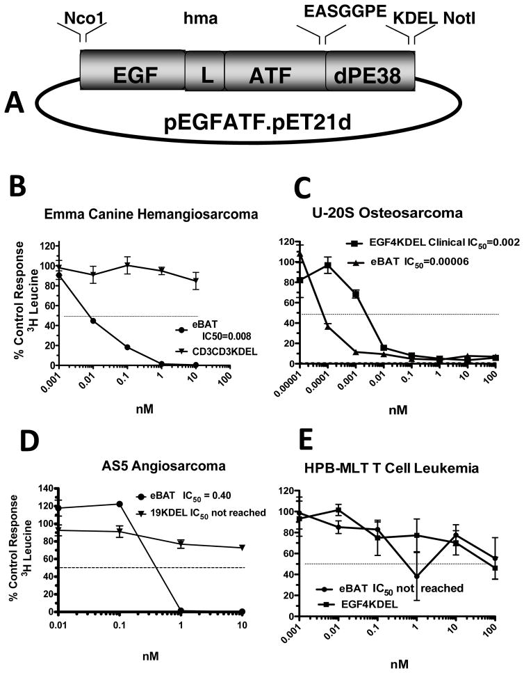
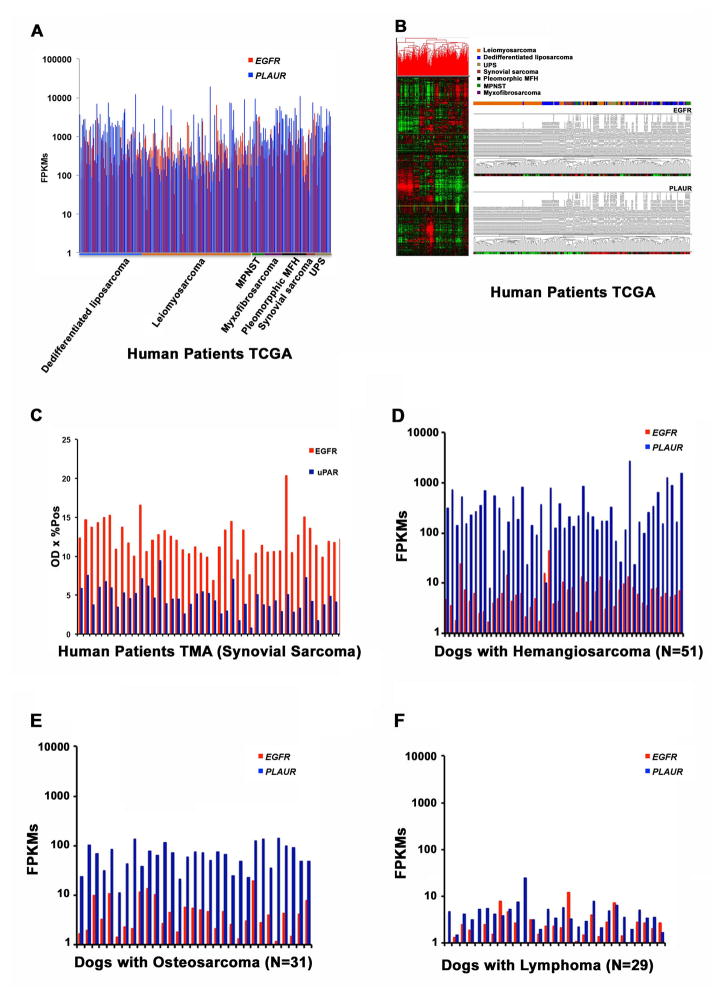
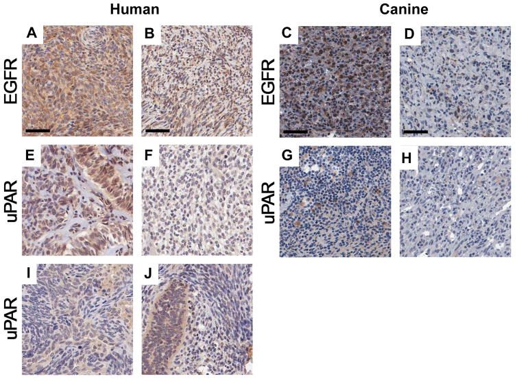
Sarcomas differ from carcinomas in their mesenchymal origin. Therapeutic advancements have come slowly, so alternative drugs and models are urgently needed. These studies report a new drug for sarcomas that simultaneously targets both tumor and tumor neovasculature. eBAT is a bispecific angiotoxin consisting of truncated, deimmunized Pseudomonas exotoxin fused to EGF and the amino terminal fragment of urokinase. Here, we study the drug in an in vivo "ontarget" companion dog trial as eBAT effectively kills canine hemangiosarcoma and human sarcoma cells in vitro We reasoned the model has value due to the common occurrence of spontaneous sarcomas in dogs and a limited lifespan allowing for rapid accrual and data collection. Splenectomized dogs with minimal residual disease were given one cycle of eBAT followed by adjuvant doxorubicin in an adaptive dose-finding, phase I-II study of 23 dogs with spontaneous, stage I-II, splenic hemangiosarcoma. eBAT improved 6-month survival from <40% in a comparison population to approximately 70% in dogs treated at a biologically active dose (50 μg/kg). Six dogs were long-term survivors, living >450 days. eBAT abated expected toxicity associated with EGFR targeting, a finding supported by mouse studies. Urokinase plasminogen activator receptor and EGFR are targets for human sarcomas, so thorough evaluation is crucial for validation of the dog model. Thus, we validated these markers for human sarcoma targeting in the study of 212 human and 97 canine sarcoma samples. Our results support further translation of eBAT for human patients with sarcomas and perhaps other EGFR-expressing malignancies. Mol Cancer Ther; 16(5); 956-65. ©2017 AACR.


Conflict of interest statement

Conflict of Interest: The authors declare that patent “Reduction of EGFR therapeutic toxicity,” related to this work and listing JFM, DV, and AB as inventors, has been filed by the University of Minnesota Office of Technology Commercialization.

Figures

Figure 1
Figure 1. Construction and in vitro activity of eBAT
Bispecific eBAT was studied for its activity against canine and human sarcoma cells. A) Expression vector for eBAT, human EGF and the high affinity amino terminal fragment of urokinase linked to a deimmunized PE38KDEL molecule. The fusion gene (from 5′ end to 3′ end) consisted of an NcoI restriction site, the genes for human EGF, an ATG initiation codon, the downstream 135-amino terminal fragment (ATF) from uPA linked by a 20 amino-acid segment of human muscle aldolase (HMA), the 7 amino-acid EASGGPE linker, the first 362 amino acids of the pseudomonas exotoxin (PE) molecule with KDEL at the C terminus, and a NotI restriction site at the 3′ end of the construct. B) Canine EMMA cells were treated with various concentrations of eBAT and control CD3CD3KDEL and then protein synthesis was measured 3 days later using a tritiated leucine uptake assay. Experimental variability is shown as quadruplicate samples ± SED. C) Human U-20S Osteosarcoma cells were treated with various concentrations of eBAT tested against EGF4KDEL and then leucine incorporation was measured. D) Human AS5 angiosarcoma cells were treated with various concentrations of eBAT tested against CD19KDEL as negative control. Leucine incorporation was measured. E) eBAT was tested against HPB-MLT cells to test specificity. eBAT, EGF4KDEL and 2219KDEL showed no significant cytotoxicity.
Figure 2
Figure 2. EGFR and PLAUR gene expression analysis in human sarcomas and spontaneous canine tumors
(A) EGFR and PLAUR gene expression analysis was done in 212 tumor tissue samples extracted from the TCGA database. The X-axis represents the patients supervised by tumor type and the Y-axis is the expression intensity as fragments per kilobase of transcript per million (FPKM) mapped reads. (B) Unsupervised hierarchical cluster and heat map highlighting EGFR and PLAUR expression in the human TCGA dataset. (C) EGFR and uPAR protein expression is shown in TMAs constructed from human synovial sarcoma tissue samples. The X-axis represents patient TMAs and the Y-axis represents optical density of EGFR and uPAR on immunohistochemistry. D) EGFR and PLAUR gene expression analysis in an independent data set of canine hemangiosarcoma samples. (E) EGFR and PLAUR gene expression analysis in canine osteosarcoma samples. (F) EGFR and PLAUR gene expression analysis in canine lymphoma samples. Tumor-bearing dogs are on the X-axis and fragments per kilobase of transcript per million mapped reads on the Y-axis, illustrating the levels of EGFR and PLAUR expression from the individual tumors. The following detailed values pertain to gene expression in TCGA samples of EGFR and PLAUR, respectively: Count: 212, 212; Mean (FPKM):653.4, 1,713; Mean (FPKM) lower confidence limit: 548.7, 1,387; Mean (FPKM) upper confidence limit: 758.0, 2,040; Variance: 600,273, 5,844,287; Standard Deviation: 774.8, 2,418; Mean Standard Error: 53.1, 165; Coefficient of Variation: 1.2, 1.4; Minimum (FPKM): 3.1, 40.9; Minimum (FPKM): 6,575.1, 19,171.7; Median (FPKM): 410.0, 757.9; Median Error: 4.56, 14.2; Percentile 25%(Q1): 215.4, 250.4; Percentile 75% (Q3): 752.9, 2,149.
Figure 3
Figure 3. EGFR and uPAR expression in human synovial sarcomas and canine HSA TMA from 15 dogs in the SRCBST study
Synovial cell sarcoma TMA spots immunohistochemically stained for EGFR and uPAR. Representative highly and lowly stained spots for EGFR are shown (A–B human) (C–D canine). Representative highly and lowly stained spots for uPAR are shown (E–F human) (G–H canine). An example of heterogeneous expression of uPAR is shown in the human synovial TMA where uPAR expression is much higher in the glandular cells staining darkly brown and forming elongated glands, sometimes with compressed slit-like spaces between the gland cells (I). An admixture of spindled and glandular cells imparting a marbled-like appearance is also shown (J).
Figure 4
Figure 4. Effect of eBAT on survival of dogs with splenic HSA treated with adjuvant doxorubicin chemotherapy
(A) Kaplan-Meier Curve for all 23 dogs in the SRCBST-1 study versus the comparison dogs. (B) Kaplan-Meier Curve for the 17 dogs treated at the biologically active dose versus the comparison dogs. Curves illustrate prolongation of survival in dogs treated with eBAT compared to the comparison group.

References

    1. Cassier PA, Polivka V, Judson I, Soria JC, Penel N, Marsoni S, et al. Outcome of patients with sarcoma and other mesenchymal tumours participating in phase I trials: a subset analysis of a European Phase I database. Ann Oncol. 2014;25:1222–8. - PubMed
    1. Linch M, Miah AB, Thway K, Judson IR, Benson C. Systemic treatment of soft-tissue sarcoma-gold standard and novel therapies. Nat Rev Clin Oncol. 2014;11:187–202. - PubMed
    1. Ng VY, Scharschmidt TJ, Mayerson JL, Fisher JL. Incidence and survival in sarcoma in the United States: a focus on musculoskeletal lesions. Anticancer Res. 2013;33:2597–2604. - PubMed
    1. Borden EC, Baker LH, Bell RS, Bramwell V, Demetri VGD, Eisenberg BL, et al. Soft tissue sarcomas of adults: state of the translational science. Clin Cancer Res. 2003;9:1941–1956. - PubMed
    1. Frith AE, Hirbe AC, Van Tine BA. Novel pathways and molecular targets for the treatment of sarcoma. Curr Oncol Rep. 2013;15(4):378–85. - PubMed

MeSH terms