Review
. 2017 Apr 5;372(1717):20160074.
doi: 10.1098/rstb.2016.0074.

The evolution of rod photoreceptors

Affiliations
Review

The evolution of rod photoreceptors

Ala Morshedian et al. Philos Trans R Soc Lond B Biol Sci. .

Abstract

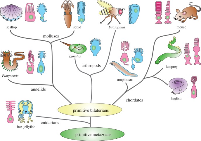
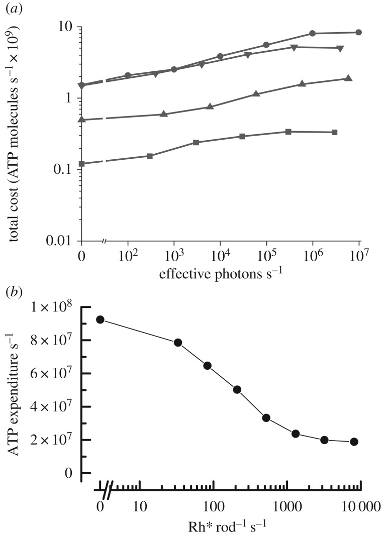
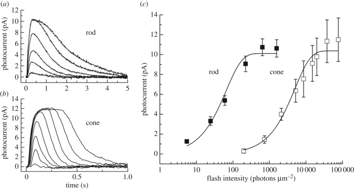
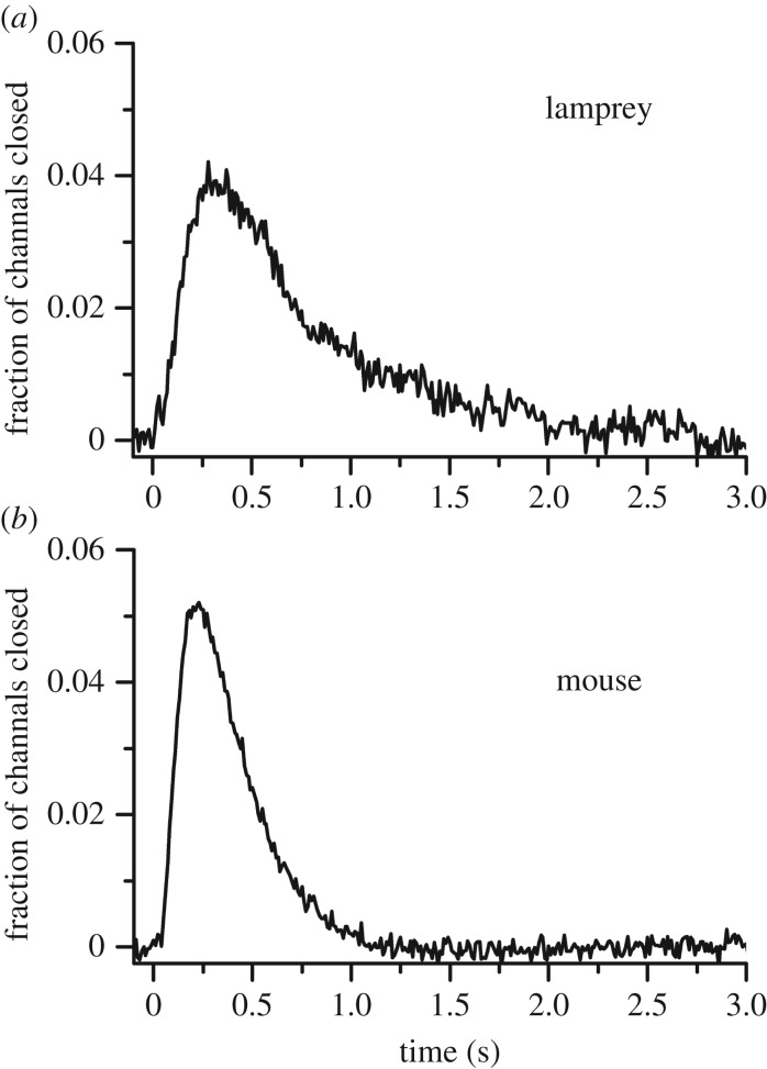
Photoreceptors in animals are generally of two kinds: the ciliary or c-type and the rhabdomeric or r-type. Although ciliary photoreceptors are found in many phyla, vertebrates seem to be unique in having two distinct kinds which together span the entire range of vision, from single photons to bright light. We ask why the principal photoreceptors of vertebrates are ciliary and not rhabdomeric, and how rods evolved from less sensitive cone-like photoreceptors to produce our duplex retina. We suggest that the principal advantage of vertebrate ciliary receptors is that they use less ATP than rhabdomeric photoreceptors. This difference may have provided sufficient selection pressure for the development of a completely ciliary eye. Although many of the details of rod evolution are still uncertain, present evidence indicates that (i) rods evolved very early before the split between the jawed and jawless vertebrates, (ii) outer-segment discs make no contribution to rod sensitivity but may have evolved to increase the efficiency of protein renewal, and (iii) evolution of the rod was incremental and multifaceted, produced by the formation of several novel protein isoforms and by changes in protein expression, with no one alteration having more than a few-fold effect on transduction activation or inactivation.This article is part of the themed issue 'Vision in dim light'.

Keywords: cone; evolution; lamprey; photoreceptor; rod; vision.


Figures

Figure 1.
Figure 1.
Phylogenetic tree of metazoans showing only representative animal groups or species with photoreceptor types in principal eyes illustrated as ciliary (red) or microvillar (blue). (Modified and reproduced with permission from Fain et al. [1]).
Figure 2.
Figure 2.
Comparison of ATP utilization by rhabdomeric and ciliary photoreceptors. (a) Mean rate of hydrolysis of ATP molecules per second calculated from membrane conductance and rate of ion pumping, as a function of rate of photon absorption for R1–6 rhabdomeric photoreceptors from four species of flies: Calliphora vicina (filled circles), Sarcophaga carnaria (filled downside triangles), Drosophila virilis (filled upside triangles), and Drosophila melanogaster (filled squares). (Reprinted with permission from Niven et al. [17]). (b) Mean rate of hydrolysis of ATP molecules per second from all sources. Calculated as a function of photon absorption for mouse ciliary rod photoreceptors. (Adapted and reprinted with permission from Okawa et al. [18]).
Figure 3.
Figure 3.
Current responses and sensitivity of lamprey rod and cone photoreceptors to brief light stimuli. (a) Mean responses of 11 rods to 20 ms, 500 nm flashes given at t = 0 for the following intensities (in photons μm−2): 5, 24, 60, 222, 642 and 1576. (b) Mean responses of eight cones to 20 ms, 600 nm flashes given at t = 0 for the following intensities (in photons μm−2): 735, 2120, 5210, 1.98 × 10⁴, 7.70 × 10⁴, 2.28 × 10⁵, 6.96 × 10⁵ and 2.03 × 10⁶. (c) Mean peak current response amplitudes plotted against flash intensity for 11 rods (filled squares) and eight cones (open squares). Error bars are standard errors of the mean. Cells are the same as in (a) and (b). The data for both cell types were fitted with the equation r = rmax [1 − exp(−kI)]. The best-fitting values of rmax and k were 10.1 pA and 1.52 × 10⁻² photons−1 µm2 for rods and 10.4 pA and 2 × 10⁻⁴ photons−1 µm2 for cones. (Reproduced with permission from Morshedian & Fain [23]).
Figure 4.
Figure 4.
Single-photon responses of lamprey rods and mouse rods. Responses were calculated from the squared mean and variance (as in [27]) for 10 lamprey rods (a) and 41 mouse rods (b), normalized rod by rod to circulating current and averaged to give the mean fractional closure of channels as a function of time. (Reprinted with permission from Morshedian & Fain [23]).
Figure 5.
Figure 5.
Transduction cascade of vertebrate photoreceptor. Steps contributing to rate of activation are indicated by green arrows; steps contributing to rate of inactivation by red arrows. Arr, arrestin; cGMP, 3′, 5′-cyclic guanosine monophosphate; GAP, GTPase-activating protein; GDP, guanosine diphosphate; GCAP, guanylyl cyclase-activating proteins; GMP, guanosine monophosphate; GRK, G-protein receptor kinase (rhodopsin kinase); GTP, guanosine triphosphate; hν, light; PDE, phosphodiesterase 6; Pi, inorganic phosphate; Rh, rhodopsin; Rh*, light-activated rhodopsin.

References

    1. Fain GL, Hardie R, Laughlin SB. 2010. Phototransduction and the evolution of photoreceptors. Curr. Biol. 20, R114–R124. (10.1016/j.cub.2009.12.006) - DOI - PMC - PubMed
    1. Garm A, Coates MM, Gad R, Seymour J, Nilsson DE. 2007. The lens eyes of the box jellyfish Tripedalia cystophora and Chiropsalmus sp. are slow and colorblind. J. Comp. Physiol. A Neuroethol. Sens Neural Behav. Physiol. 193, 547–557. (10.1007/s00359-007-0211-4) - DOI - PubMed
    1. Suga H, Schmid V, Gehring WJ. 2008. Evolution and functional diversity of jellyfish opsins. Curr. Biol. 18, 51–55. (10.1016/j.cub.2007.11.059) - DOI - PubMed
    1. Porter ML, Blasic JR, Bok MJ, Cameron EG, Pringle T, Cronin TW, Robinson PR. 2012. Shedding new light on opsin evolution. Proc. R. Soc. B 279, 3–14. (10.1098/rspb.2011.1819) - DOI - PMC - PubMed
    1. Nordstrom K, Wallen R, Seymour J, Nilsson D. 2003. A simple visual system without neurons in jellyfish larvae. Proc. R. Soc. Lond. B 270, 2349–2354. (10.1098/rspb.2003.2504) - DOI - PMC - PubMed

MeSH terms