. 2017 Apr 1;198(7):2834-2843.
doi: 10.4049/jimmunol.1601281. Epub 2017 Feb 13.

Monocyte Subsets Coregulate Inflammatory Responses by Integrated Signaling through TNF and IL-6 at the Endothelial Cell Interface

Affiliations

Monocyte Subsets Coregulate Inflammatory Responses by Integrated Signaling through TNF and IL-6 at the Endothelial Cell Interface

Myriam Chimen et al. J Immunol. .

Abstract

Two major monocyte subsets, CD14+CD16- (classical) and CD14+/dimCD16+ (nonclassical/intermediate), have been described. Each has different functions ascribed in its interactions with vascular endothelial cells (EC), including migration and promoting inflammation. Although monocyte subpopulations have been studied in isolated systems, their influence on EC and on the course of inflammation has been ignored. In this study, using unstimulated or cytokine-activated EC, we observed significant differences in the recruitment, migration, and reverse migration of human monocyte subsets. Associated with this, and based on their patterns of cytokine secretion, there was a difference in their capacity to activate EC and support the secondary recruitment of flowing neutrophils. High levels of TNF were detected in cocultures with nonclassical/intermediate monocytes, the blockade of which significantly reduced neutrophil recruitment. In contrast, classical monocytes secreted high levels of IL-6, the blockade of which resulted in increased neutrophil recruitment. When cocultures contained both monocyte subsets, or when conditioned supernatant from classical monocytes cocultures (IL-6hi) was added to nonclassical/intermediate monocyte cocultures (TNFhi), the activating effects of TNF were dramatically reduced, implying that when present, the anti-inflammatory activities of IL-6 were dominant over the proinflammatory activities of TNF. These changes in neutrophil recruitment could be explained by regulation of E-selectin on the cocultured EC. This study suggests that recruited human monocyte subsets trigger a regulatory pathway of cytokine-mediated signaling at the EC interface, and we propose that this is a mechanism for limiting the phlogistic activity of newly recruited monocytes.


Figures

FIGURE 1.
FIGURE 1.
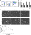
Comparative adhesion and migration of classical and nonclassical/intermediate monocytes on EC. (A) Levels of expression of CD14 and CD16 on circulating monocytes and subsets isolated from PBMC measured by flow cytometry (plots representative of >10 preparations). (B) Total adhesion of monocyte subsets on unstimulated EC or EC treated with cytokines (n = 3–4). (C) Representative images of monocyte subsets adhered and transmigrated across unstimulated and TNF stimulated EC. Original magnification ×100. (D) Time course of transmigration of monocytes across TNF-stimulated EC (n = 3). Data are mean ± SEM. *p ≤ 0.05, **p ≤ 0.01 using a two-ANOVA and Bonferroni posttest (B) and comparison between classical and nonclassical/intermediate monocytes using a two-way repeated measures ANOVA and Bonferroni posttest (D). C, classical monocyte; NC/I, nonclassical/intermediate monocyte.
FIGURE 2.
FIGURE 2.
Comparative abilities of classical and nonclassical/intermediate monocyte subsets to activate EC: role of TNF. (A) Representative images of neutrophils adhered from flow on EC in coculture with monocyte subsets for 24 h (n = 3). Original magnification ×200. (B) Recruitment of neutrophils from flow on EC in coculture with monocyte subsets (Mix monocytes indicates unfractionated monocytes; classical plus nonclassical/intermediate monocytes indicates 90% classical plus 10% nonclassical/intermediate monocytes) for 24 h (n = 3). (C) Concentration of TNF-α (picograms per milliliter) in supernatants of monocyte subsets in coculture with EC for 24 h measured using a Luminex assay (n = 3). (D) mRNA expression of E-selectin on EC alone or in coculture with monocyte subsets for 24 h (n = 7–9). (E) Surface expression of E-selectin on EC following coculture with monocyte subsets for 24 h, measured by flow cytometry (n = 4–5). TNF was neutralized using an Ab at 10 μg/ml during the 24-h coculture with nonclassical/intermediate monocytes. Data were normalized to expression of E-selectin on EC alone. (F) Recruitment of neutrophils from flow on EC cocultured with monocyte subsets for 24 h, with or without 10 μg/ml anti-TNF Ab (n = 3). Data are mean ± SEM from n experiments. *p ≤ 0.05, **p ≤ 0.01, ***p ≤ 0.001 comparison between classical and nonclassical/intermediate monocytes using repeated measures ANOVA and a Bonferroni posttest (A), ANOVA and a Bonferroni posttest (B, C, and E), an unpaired t test (D), and a two-way ANOVA and a Bonferroni posttest (F). C, classical monocyte; NC/I, nonclassical/intermediate monocyte.
FIGURE 3.
FIGURE 3.
Expression of VCAM-1 and ICAM-1 in EC in coculture with monocyte subsets. (A and B) mRNA expression of VCAM-1 (A) and ICAM-1 (B) on EC in coculture with monocyte subsets for 24 h (n = 7–9). (C and D) Surface expression of VCAM-1 (C) and ICAM-1 (D) on EC following coculture with monocyte subsets for 24 h, measured by flow cytometry (n = 4–5). Data are normalized to classical monocyte mean fluorescence intensity (MFI). Data are mean ± SEM from n experiments. *p ≤ 0.05, **p ≤ 0.01, NS, not significant using unpaired t test (A and B) and ANOVA and a Dunnet posttest to EC alone (C and D). C, classical monocyte; NC/I, nonclassical/intermediate monocyte.
FIGURE 4.
FIGURE 4.
Role of IL-6 in regulating EC activation by monocyte subsets. (A) Concentration of IL-6 (picograms per milliliter) in supernatants of monocyte subsets in coculture with EC for 24 h measured using a Luminex assay (n = 3). (B) Recruitment of neutrophils from flow on EC cocultured with classical monocytes, with or without neutralizing Ab against IL-6 (10 μg/ml) (n = 4). (C) Recruitment of neutrophils on EC cocultured with nonclassical/intermediate monocytes, with or without rIL-6 (1 ng/ml) (n = 4). (D) Surface expression of E-selectin on EC following coculture with monocyte subsets for 24 h, measured by flow cytometry. Data were normalized to expression of E-selectin on cocultures with classical monocytes (n = 4–5). (E) Recruitment of neutrophils from flow on EC cocultured with monocyte subsets for 24 h: effect of adding supernatants from cocultures with either subset, and of neutralizing Ab against IL-6 (10 μg/ml) (n = 3). (F) mRNA expression of SOCS3 on EC alone or in coculture with monocyte subsets for 24 h (n = 7–9). Data are shown as mean ± SEM. *p ≤ 0.05, ***p ≤ 0.001, ****p ≤ 0.0001, comparison between classical and nonclassical/intermediate monocytes using an unpaired t test (D and F), a paired t test (B and C), and ANOVA and a Bonferroni posttest (A and E). C, classical monocyte; NC/I, nonclassical/intermediate monocyte; PMN, polymorphonuclear neutrophil.
FIGURE 5.
FIGURE 5.
Cytokines secreted by monocyte subsets balance EC activation. Nonclassical/intermediate monocytes generate high levels of TNF once in proximity with EC, which causes upregulation of E-selectin expression at the EC surface. This in turn promotes secondary leukocyte recruitment. In normal inflammatory conditions, classical monocytes regulate the extent of nonclassical/intermediate monocyte–induced EC activation by secreting IL-6, which reduces surface expression of E-selectin via upregulation of SOCS3, a negative regulator of the JAK/STAT signaling pathway. In disease, potential changes in classical and nonclassical/intermediate monocyte recruitment may alter the balanced control of inflammation mediated by the regulation mediated by both subsets. C, classical monocyte; NC/I, nonclassical/intermediate monocyte.

References

    1. Yang J., Zhang L., Yu C., Yang X. F., Wang H. 2014. Monocyte and macrophage differentiation: circulation inflammatory monocyte as biomarker for inflammatory diseases. Biomark. Res. 2: 1–9. - PMC - PubMed
    1. Bobryshev Y. V. 2006. Monocyte recruitment and foam cell formation in atherosclerosis. Micron 37: 208–222. - PubMed
    1. Ross R. 1995. Cell biology of atherosclerosis. Annu. Rev. Physiol. 57: 791–804. - PubMed
    1. Baeten D., Boots A. M., Steenbakkers P. G., Elewaut D., Bos E., Verheijden G. F., Berheijden G., Miltenburg A. M., Rijnders A. W., Veys E. M., De Keyser F. 2000. Human cartilage gp-39+,CD16+ monocytes in peripheral blood and synovium: correlation with joint destruction in rheumatoid arthritis. Arthritis Rheum. 43: 1233–1243. - PubMed
    1. Kawanaka N., Yamamura M., Aita T., Morita Y., Okamoto A., Kawashima M., Iwahashi M., Ueno A., Ohmoto Y., Makino H. 2002. CD14+,CD16+ blood monocytes and joint inflammation in rheumatoid arthritis. Arthritis Rheum. 46: 2578–2586. - PubMed

Publication types