. 2017 Mar 31;292(13):5392-5404.
doi: 10.1074/jbc.M116.773416. Epub 2017 Feb 13.

Experimental Arthritis Is Dependent on Mouse Mast Cell Protease-5

Affiliations

Experimental Arthritis Is Dependent on Mouse Mast Cell Protease-5

Richard L Stevens et al. J Biol Chem. .

Abstract

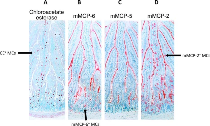
The constitutive heparin+ (HP) mast cells (MCs) in mice express mouse MC protease (mMCP)-5 and carboxypeptidase A (mMC-CPA). The amino acid sequence of mMCP-5 is most similar to that of human chymase-1, as are the nucleotide sequences of their genes and transcripts. Using a homologous recombination approach, a C57BL/6 mouse line was created that possessed a disrupted mMCP-5 gene. The resulting mice were fertile and had no obvious developmental abnormality. Lack of mMCP-5 protein did not alter the granulation of the IL-3/IL-9-dependent mMCP-2+ MCs in the jejunal mucosa of Trichinella spiralis-infected mice. In contrast, the constitutive HP+ MCs in the tongues of mMCP-5-null mice were poorly granulated and lacked mMC-CPA protein. Bone marrow-derived MCs were readily developed from the transgenic mice using IL-3. Although these MCs contained high levels of mMC-CPA mRNA, they also lacked the latter exopeptidase. mMCP-5 protein is therefore needed to target translated mMC-CPA to the secretory granule along with HP-containing serglycin proteoglycans. Alternately, mMCP-5 is needed to protect mMC-CPA from autolysis in the cell's granules. Fibronectin was identified as a target of mMCP-5, and the exocytosis of mMCP-5 from the MCs in the mouse's peritoneal cavity resulted in the expression of metalloproteinase protease-9, which has been implicated in arthritis. In support of the latter finding, experimental arthritis was markedly reduced in mMCP-5-null mice relative to wild-type mice in two disease models.

Keywords: arthritis; carboxypeptidase; heparin; mast cell; serine protease.


Conflict of interest statement

The authors declare that they have no conflicts of interest with the contents of this article

Figures

FIGURE 1.
FIGURE 1.
Targeted disruption of the mMCP-5 gene in B6 mice. A, in the targeting vector, a portion of the mMCP-5 gene (including part of intron 2 and exon 3) was deleted. The Neor gene was then placed at the disrupted locus. Although the HSV-TK gene also was placed 3′ of the targeting construct in case negative selection had to be used to obtain ES cell clones with a disrupted allele of the mMCP-5 gene, only positive selection was used to obtain ES cell clones that underwent correct homologous recombination. B, a PCR approach was devised to expedite the genotyping of mMCP-5-null mice. Depicted are representative PCR analyses carried out on genomic DNA isolated from the tail of mMCP-5 knock-out (lane 2), heterozygous (lane 3), and WT mice (lane 4) using primers that reside 5′ and 3′of exon 3 of the mMCP-5 gene. The presence of the Neor gene in the mutated mMCP-5 gene causes the generation of a larger DNA fragment. Molecular weight markers are indicated in lane 1.
FIGURE 2.
FIGURE 2.
Analysis of the MCs in the jejunum of helminth-infected mice. A–D, sections of jejunum from T. spiralis-infected mMCP-5-null mice at day 14 were evaluated cytochemically for the presence of CE+ MCs (A) or immunohistochemically for the presence of mMCP-6+ (B), mMCP-5+ (C), and mMCP-2+ (D) MCs. Arrows point to muscle/submucosa mMCP-6+ MCs (B) and mucosal CE+ (A)/mMCP-2+ (D) MCs present in mMCP-5-null mice (C).
FIGURE 3.
FIGURE 3.
Histochemical and immunohistochemical evaluation of tongue MCs in WT and mMCP-5-null mice. a–j, serial sections (a–c, d–f, and g–j) of tongue from mMCP-5-null (a–f) or WT (g–j) mice were stained with methylene blue (a, d, and g), anti-mMCP-5 antibody (b and h), anti-mMCP-6 antibody (c, f, and i), or anti-MC-CPA antibody (e and j). The MCs in the tongue of WT mice have granules filled with HP (g), mMCP-5 (h), mMCP-6 (i), and mMC-CPA (j). As expected, disruption of the mMCP-5 gene resulted in MCs unable to express mMCP-5. Although these MCs contained variable amounts of mMCP-6 (c and f), they were unable to store HP (a and d) and mMC-CPA (e) in their granules.
FIGURE 4.
FIGURE 4.
Analysis of WT and mMCP-5-null mBMMCs. A, cytospins of mMCP-5-null (−/−) and WT mBMMCs (+/+) were stained with toluidine blue. B, RNA blot analyses were carried out to evaluate the levels of the transcripts that encode mMCP-1, mMCP-2, mMCP-4, mMCP-5, mMCP-6, mMC-CPA, and β-actin in mMCP-5+/+ (data not shown), mMCP-5−/+, and mMCP-5−/− mBMMCs. C, lysates of these three populations of mBMMCs also were evaluated for their mMCP-5 and mMC-CPA protein levels by SDS-PAGE/immunoblot analysis.
FIGURE 5.
FIGURE 5.
mMC-CPA protein levels in mBMMCs before and after co-culture with fibroblasts. mMCP-5+/− (lanes 1 and 2) and mMCP-5−/− (lanes 3 and 4) were cultured in the absence (lanes 1 and 3) or presence (lanes 2 and 4) of 3T3 fibroblasts for 2 weeks. Lysates of the resulting cell suspensions were then evaluated for their levels of mMC-CPA protein by SDS-PAGE/immunoblot analysis.
FIGURE 6.
FIGURE 6.
Fibronectin is highly susceptible to naturally occurring mMCP-5 but not mMCP-6. Lysates of mMCP-5-null (lanes 1–3), mMCP-6-null (lanes 4–6), and WT (data not shown) B6 mBMMCs were incubated with fibronectin (Fib.) for 30 (lanes 1 and 4), 60 (lanes 2 and 5), and 120 (lanes 3 and 6) min. The resulting digests were then subjected to SDS-PAGE. Undigested fibronectin (lane 7) was incubated 120 min in the absence of mBMMC lysate as a negative control.
FIGURE 7.
FIGURE 7.
2D-DIGE analysis of protein expression in mBMMC·fibroblast co-cultures. Mouse 3T3 fibroblasts were co-cultured with WT or mMCP-5-null mBMMCs. The proteins in the resulting lysates of the two co-cultures were differentially labeled by Cy2 and Cy3, pooled, and subjected to 2D-DIGE. The green-labeled proteins were only found in the co-cultures that contained WT mBMMCs, whereas the red-labeled proteins were only found in the co-cultures that contained mMCP-5-null mBMMCs. Yellow-labeled proteins were found in both. Those differentially expressed proteins selected for MALDI-ToF MS proteomic analysis (also see supplemental Table S1) are indicated.
FIGURE 8.
FIGURE 8.
Expression of biologically active proteins in the peritoneal cavities of WT and mMCP-5-null mice after MC activation. A, RayBiotech mouse G series 1000 antibody arrays III and IV were used to evaluate the presence of 96 biologically active proteins in the peritoneal cavity exudates of WT (samples 1 and 2) and mMCP-5-null (samples 3 and 4) B6 mice 30 min (samples 1 and 3) and 120 min (samples 2 and 4) after these animals were given C48/80. B, MMP-9 levels were selectively increased in the peritoneal cavity of WT mice 2 h after given C48/80 (also see supplemental Table S2).
FIGURE 9.
FIGURE 9.
Inflammatory K/BxN arthritis in WT and mMCP-5-null mice. A–D, WT (●) (n = 15) and mMCP-5-null (▵) (n = 15) B6 mice were subjected to K/BxN arthritis. Arthritis was significantly reduced in mMCP-5-null B6 mice relative to WT B6 as assessed by clinical index (A), ankle thickness (B), inflammation (C), bone erosion (C), cartilage destruction (C), and aggrecan proteoglycan loss from the diseased cartilage (D).
FIGURE 10.
FIGURE 10.
Inflammatory meBSA/IL1β arthritis in WT and mMCP-5-null mice. A–C, meBSA/IL-1β arthritis were induced in 10 knees of 5 WT B6 mice and 10 knees of 5 mMCP-5-null B6 mice. A, representative histochemistry data are shown. B, cellular inflammation of the joint space, pannus size, and the extent of cartilage and bone erosion were all significantly reduced in the diseased mMCP-5-null mice relative to the diseased WT mice. C, the mean total scores for the respective groups were 17.8 versus 12.0 (WT versus 5-KO). *, p < 0.05; **, p < 0.01.

References

    1. Caughey G. H., Zerweck E. H., and Vanderslice P. (1991) Structure, chromosomal assignment, and deduced amino acid sequence of a human gene for mast cell chymase. J. Biol. Chem. 266, 12956–12963 - PubMed
    1. Urata H., Kinoshita A., Perez D. M., Misono K. S., Bumpus F. M., Graham R. M., and Husain A. (1991) Cloning of the gene and cDNA for human heart chymase. J. Biol. Chem. 266, 17173–17179 - PubMed
    1. Urata H., Kinoshita A., Misono K. S., Bumpus F. M., and Husain A. (1990) Identification of a highly specific chymase as the major angiotensin II-forming enzyme in the human heart. J. Biol. Chem. 265, 22348–22357 - PubMed
    1. Semaan W., Desbiens L., Houde M., Labonté J., Gagnon H., Yamamoto D., Takai S., Laidlaw T., Bkaily G., Schwertani A., Pejler G., Levesque C., Desjardins R., Day R., and D'Orléans-Juste P. (2015) Chymase inhibitor-sensitive synthesis of endothelin-1 (1–31) by recombinant mouse mast cell protease 4 and human chymase. Biochem. Pharmacol. 94, 91–100 - PubMed
    1. Mizutani H., Schechter N., Lazarus G., Black R. A., and Kupper T. S. (1991) Rapid and specific conversion of precursor interleukin 1β to an active IL-1 species by human mast cell chymase. J. Exp. Med. 174, 821–825 - PMC - PubMed

Publication types