. 2017 Feb 28;114(9):E1587-E1596.
doi: 10.1073/pnas.1616097114. Epub 2017 Feb 13.

Vulnerability of primitive human placental trophoblast to Zika virus

Affiliations

Vulnerability of primitive human placental trophoblast to Zika virus

Megan A Sheridan et al. Proc Natl Acad Sci U S A. .

Abstract

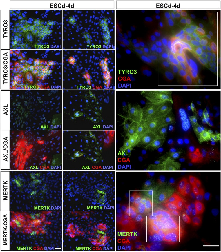
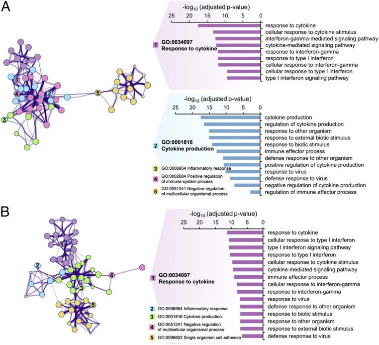
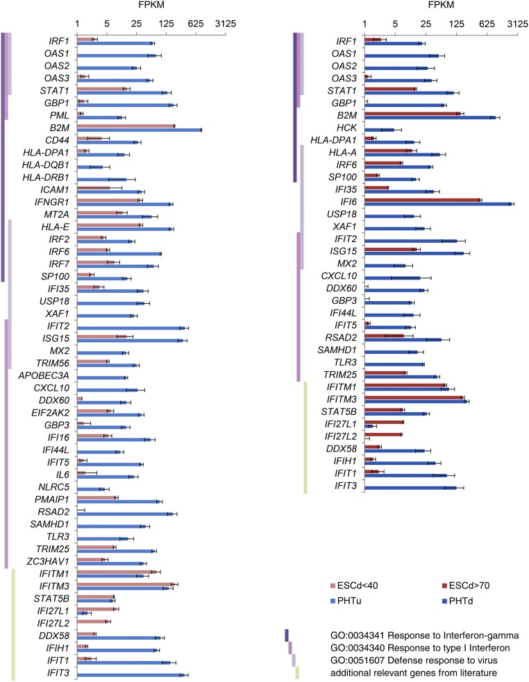
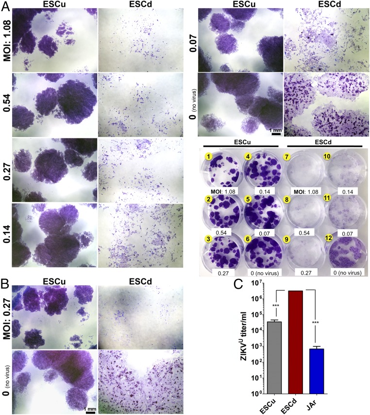
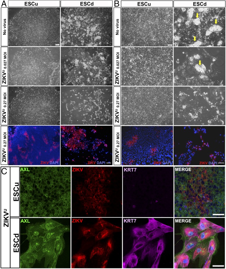
Infection of pregnant women by Asian lineage strains of Zika virus (ZIKV) has been linked to brain abnormalities in their infants, yet it is uncertain when during pregnancy the human conceptus is most vulnerable to the virus. We have examined two models to study susceptibility of human placental trophoblast to ZIKV: cytotrophoblast and syncytiotrophoblast derived from placental villi at term and colonies of trophoblast differentiated from embryonic stem cells (ESC). The latter appear to be analogous to the primitive placenta formed during implantation. The cells from term placentas, which resist infection, do not express genes encoding most attachment factors implicated in ZIKV entry but do express many genes associated with antiviral defense. By contrast, the ESC-derived trophoblasts possess a wide range of attachment factors for ZIKV entry and lack components of a robust antiviral response system. These cells, particularly areas of syncytiotrophoblast within the colonies, quickly become infected, produce infectious virus and undergo lysis within 48 h after exposure to low titers (multiplicity of infection > 0.07) of an African lineage strain (MR766 Uganda: ZIKVU) considered to be benign with regards to effects on fetal development. Unexpectedly, lytic effects required significantly higher titers of the presumed more virulent FSS13025 Cambodia (ZIKVC). Our data suggest that the developing fetus might be most vulnerable to ZIKV early in the first trimester before a protective zone of mature villous trophoblast has been established. Additionally, MR766 is highly trophic toward primitive trophoblast, which may put the early conceptus of an infected mother at high risk for destruction.

Keywords: Zika virus; embryonic stem cell; placenta; pregnancy; trophoblast.


Conflict of interest statement

The authors declare no conflict of interest.

Figures

Fig. 1.
Fig. 1.
Expression of candidate attachment factors and bridging proteins implicated in ZIKV infection of human cells. (A) Expression levels (in FPKM) of genes for five candidate receptors in ESCu; in CTB (ESCd < 40) and STB (ESCd > 70) fractions from BAP-treated H1 ESCs; in trophoblasts from BMP4-treated H1 ESC (Roadmap Epigenomics Project H1 BMP4 hTBCs) (40); in human chorionic villi collected from late first and early second trimester [samples N05–N10 from (41) in human term placenta-derived CTB (PHTu) and STB (PHTd)]; and in in vitro-derived human NPC, mock-infected samples) (10). (B) Western blots showing expression of TAM receptors (TYRO3, AXL, MERTK) and IFN response proteins (MX2 and IFIT3) in ESCu, ESCd after 4 d and 8 d of differentiation with BAP, and STB derived from term placenta at 24 h and 48 h of in vitro differentiation (PHTd). “ESCd-8d” correlates with the samples in A, consisting of both ESCd < 40 and ESCd > 70 fractions combined. “ESCd-4d” correlates with samples (see Figs. 5 and 6), as it is the time point of initial ZIKV infections. “PHTd-48h” correlates with the sample in A named PHTd. The loading control is TUBA. All data are from the same blot. (C) Expression levels of PROS1 and GAS6 in the same cell types as in A. For TYRO3, AXL, MERTK, PROS1, and GAS6, the differences between ESCd < 40 and PHTu and between ESCd > 70 and PHTd cells were statistically significant (two-tailed t test, equal variance, P < 0.01). Error bars show SD.
Fig. 2.
Fig. 2.
Putative ZIKV attachment expression in ESCd-4d and its correlation with CGA, a marker for emerging STB. Two representative images have been selected for each receptor at low magnification (Left panels) and one representative image at higher magnification (Right panels). The sample “ESCd-4d” correlates with other samples (see Figs. 5 and 6) as it is the time point of initial ZIKV infections. Immunofluorescent detection of TYRO3 and MERTK correlates with CGA, whereas AXL expression is mainly outside of the CGA+ areas. The far Right panels further highlight the coexpression of TYRO3 and MERTK with CGA, indicated by white boxes. Red, CGA; green, receptors; blue, DAPI. (Scale bars, 50 μm.)
Fig. 3.
Fig. 3.
GO terms of genes associated with responses to pathogens and down-regulated in ESC-derived trophoblast cells relative to placenta-derived trophoblast cells. The figures represent clusters of related, enriched Biological Process GO terms of genes that showed strong significant down-regulation (q-value < 0.01, fold change > 4) in ESCd < 40 versus PHTu cells (A) and in ESCd > 70 versus PHTd cells (B). Note that these GO terms are related to responses to pathogens. The network view on thte Left represents GO categories by nodes; shared genes between nodes are linked by lines. The number of genes in a given GO category is represented by the size of the corresponding node, and the number of shared genes between two given GO categories is reflected by the thickness of the line that connects the nodes. Shown are the five most significantly enriched clusters, which are named in accordance with the most significantly enriched GO term within each one of the categories. Genes in GO categories Response to IFN gamma, Response to Type I IFN, and Defense response to virus are discussed in the text (see Fig. 4 for representative gene expressions). These genes are located either in cluster 1 or 2 in A, but all belong to cluster 1 in B. The bar graphs on the Right show enrichment significance of all GO categories that belong to these clusters. Enrichments for all GO terms within the shown clusters are presented in SI Appendix, Table S2.
Fig. 4.
Fig. 4.
Expression levels (in FPKM) for representative genes from Response to IFN gamma, Response to type I IFN, and Defense response to virus GO categories. These genes showed strong, significant down-regulation (q-value < 0.01, fold change > 4) in the ESCd < 40 and ESCd > 70 cell fractions relative to PHTu and PHTd cells, respectively. Vertical dark violet, pink, and light-violet bars indicate affiliation of a given gene with the GO categories mentioned in the key at the lower right. Light green vertical bars indicate that a given gene was added based on published data regarding its role in immune response. Error bars show SD.
Fig. 5.
Fig. 5.
Relative susceptibilities of ESCu and ESCd to ZIKVU infection (A and B) and release of infection-competent virus by ESCu and ESCd following ZIKVU infection (C). (A) After passage of ESCu onto a 12-well plate, colonies in six wells (#1–6) were maintained undifferentiated with MEF-conditioned medium supplemented with FGF2 (ESCu). The colonies in the other six wells (wells 7–12) were differentiated into trophoblasts with BAP (ESCd). After 4 d, the cells were infected with ZIKVU (MOIs are presented in the image) for 1 h. Two wells (wells 6 and 12) were respective controls with no virus. At 48 h (A) and 72 h (B) postinfection, cells were stained with crystal violet. Individual wells were imaged by using the Leica M205 FA Stereo Microscope to demonstrate cytopathic effects, and the whole-plate image is featured as a reference. Colonies of ESCu (wells 1–6) showed deeper staining than ESCd because of higher cell density per colony. (B) The images presented are at the median MOI (0.27) to demonstrate the severity of cell lysis in ESCd compared with ESCu 72 h after ZIKVU infection. (Scale bars, 1 mm.) (C) ESCu, ESCd, and JAr cells were infected with ZIKVU at an MOI of 0.27. New medium was placed on the cells 24 h postinfection and then collected 24 h later (48 h postinfection). ZIKV titer was determined by TCID50 by means of three biological replicates of the collected supernatants. The viral titer was higher in ESCd compared with ESCu and JAr cells (***P < 0.0001) (one-way ANOVA).
Fig. 6.
Fig. 6.
Cytopathic effect of ZIKVU on ESCu and ESCd 48 h after infection. Upper three panels of A and B are phase-contrast images of colonies of no virus control (Top) and ZIKVU-infected cells (second and third rows) at low (A) and high (B) magnifications. Although ESCu showed no signs of cell lysis even at the highest virus concentration (0.27 MOI in A and B), a cytopathic effect on ESCd was evident even at the lower virus titer (0.027 MOI in A and B). In ESCd, areas of presumptive STB are selectively lost as virus titer is increased (arrows in B). Bottom panels of A and B are immunofluorescent (IF) images. Cell nuclei were stained with DAPI (blue) and, for ZIKV antigen, with monoclonal antibody 4G2 (red). (C) Confocal images show the presence of AXL (green), ZIKV (red), and KRT7 (magenta) in ESCu and ESCd ZIKVU-infected cells. [Scale bars: 200 μm (A) and 100 μm (B) for both phase-contrast and IF images and 50 μm (C).]

Similar articles

Cited by

References

    1. Doran KS, Banerjee A, Disson O, Lecuit M. Concepts and mechanisms: Crossing host barriers. Cold Spring Harb Perspect Med. 2013;3(7):a010090. - PMC - PubMed
    1. Cardenas I, et al. Viral infection of the placenta leads to fetal inflammation and sensitization to bacterial products predisposing to preterm labor. J Immunol. 2010;185(2):1248–1257. - PMC - PubMed
    1. Delorme-Axford E, Sadovsky Y, Coyne CB. The placenta as a barrier to viral infections. Ann Rev Virol. 2014;1(1):133–146. - PubMed
    1. Adibi JJ, Marques ET, Jr, Cartus A, Beigi RH. Teratogenic effects of the Zika virus and the role of the placenta. Lancet. 2016;387(10027):1587–1590. - PubMed
    1. Brasil P, et al. Zika virus infection in pregnant women in Rio de Janeiro. N Engl J Med. 2016;375(24):2321–2334. - PMC - PubMed

Publication types

LinkOut - more resources