. 2017 Feb 28;114(9):2395-2400.
doi: 10.1073/pnas.1608763114. Epub 2017 Feb 13.

Kv1.1 channelopathy abolishes presynaptic spike width modulation by subthreshold somatic depolarization

Affiliations

Kv1.1 channelopathy abolishes presynaptic spike width modulation by subthreshold somatic depolarization

Umesh Vivekananda et al. Proc Natl Acad Sci U S A. .

Abstract

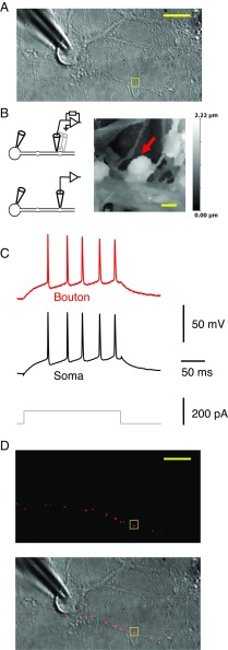
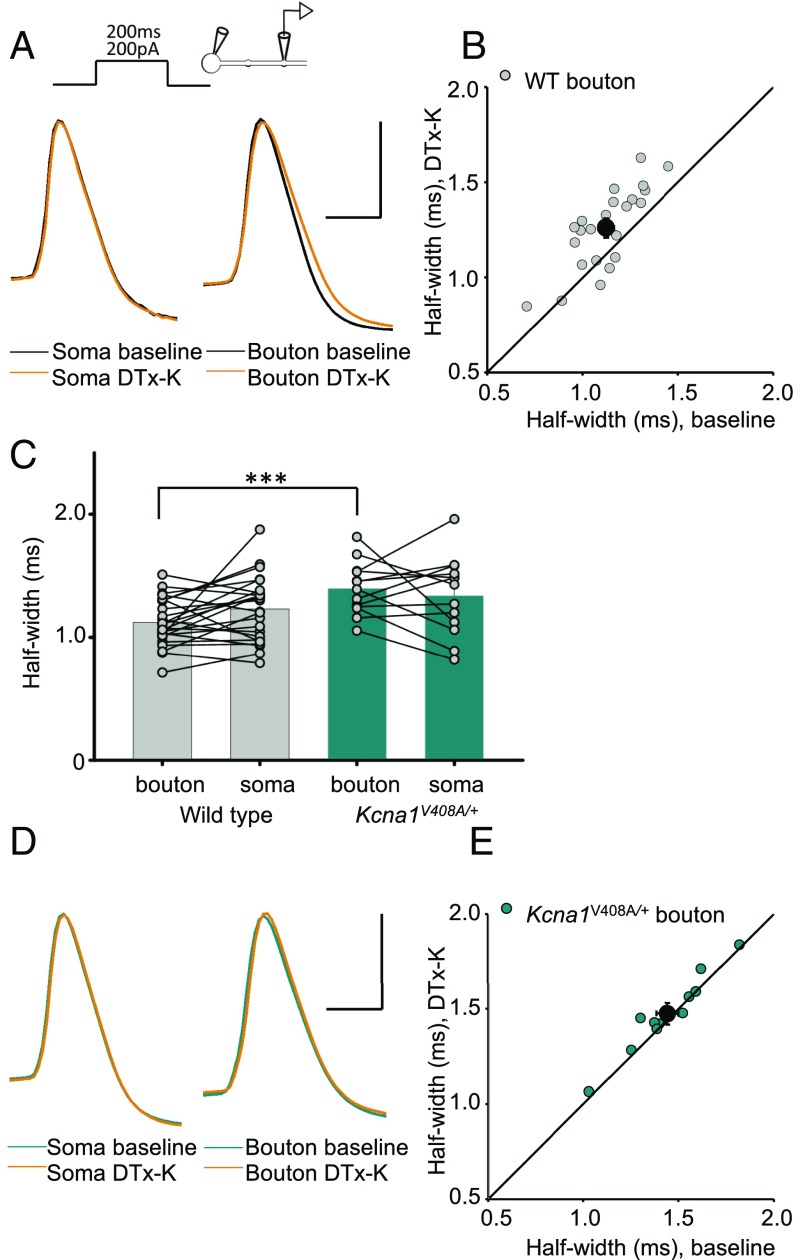
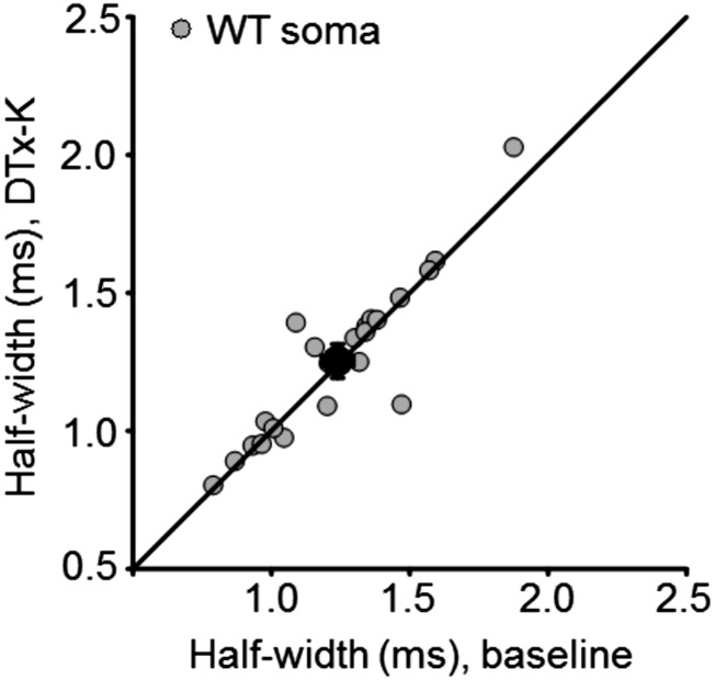
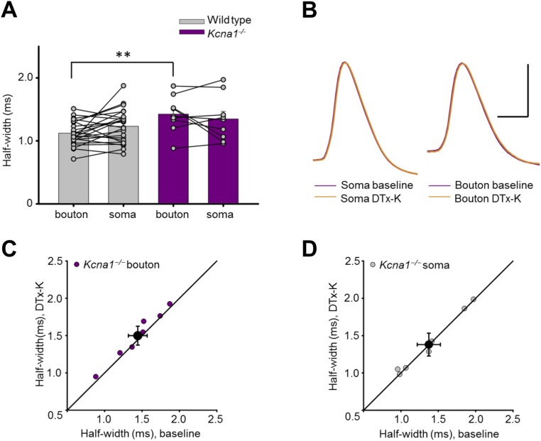
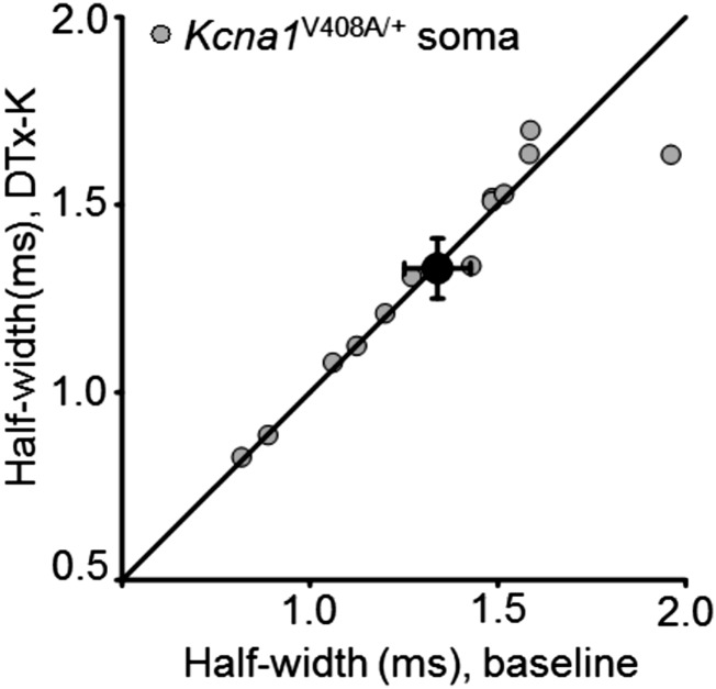
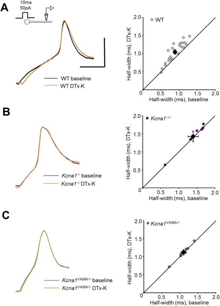
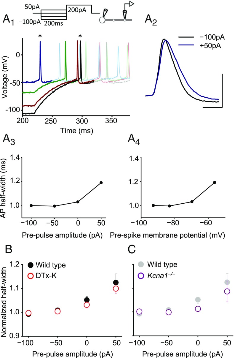
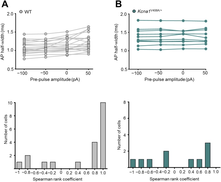
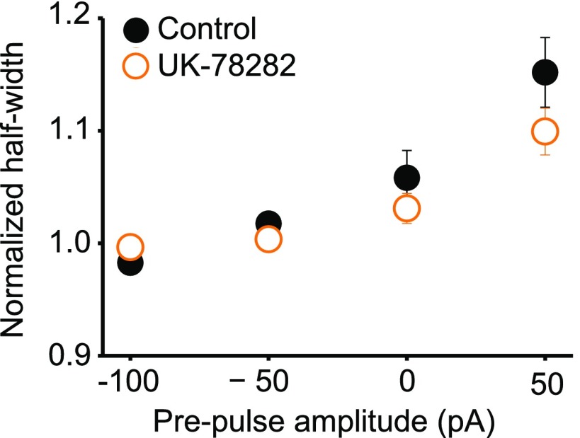
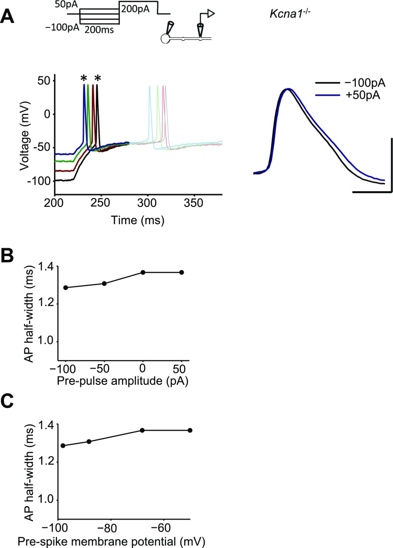
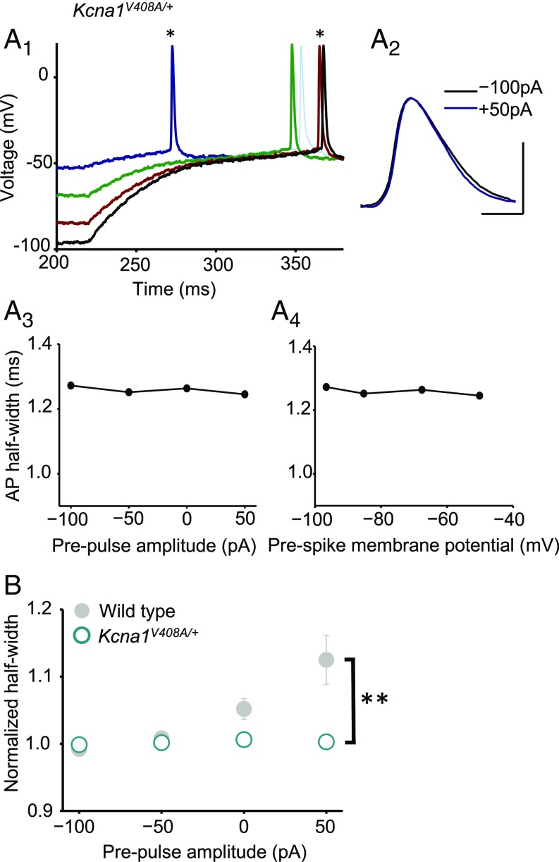
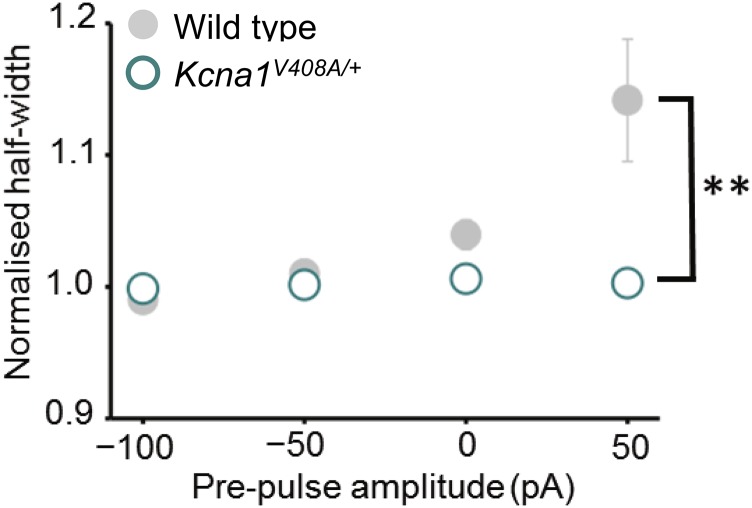
Although action potentials propagate along axons in an all-or-none manner, subthreshold membrane potential fluctuations at the soma affect neurotransmitter release from synaptic boutons. An important mechanism underlying analog-digital modulation is depolarization-mediated inactivation of presynaptic Kv1-family potassium channels, leading to action potential broadening and increased calcium influx. Previous studies have relied heavily on recordings from blebs formed after axon transection, which may exaggerate the passive propagation of somatic depolarization. We recorded instead from small boutons supplied by intact axons identified with scanning ion conductance microscopy in primary hippocampal cultures and asked how distinct potassium channels interact in determining the basal spike width and its modulation by subthreshold somatic depolarization. Pharmacological or genetic deletion of Kv1.1 broadened presynaptic spikes without preventing further prolongation by brief depolarizing somatic prepulses. A heterozygous mouse model of episodic ataxia type 1 harboring a dominant Kv1.1 mutation had a similar broadening effect on basal spike shape as deletion of Kv1.1; however, spike modulation by somatic prepulses was abolished. These results argue that the Kv1.1 subunit is not necessary for subthreshold modulation of spike width. However, a disease-associated mutant subunit prevents the interplay of analog and digital transmission, possibly by disrupting the normal stoichiometry of presynaptic potassium channels.

Keywords: channelopathy; potassium channel; synaptic transmission.


Conflict of interest statement

The authors declare no conflict of interest.

Figures

Fig. 1.
Fig. 1.
Dual recordings from the soma and small presynaptic bouton of the same neuron. (A) Transmitted light image of a neuron with somatic patch pipette, with a neighboring 6-µm × 6-µm region selected for HPICM indicated with the square. (B, Left) Schematics indicating HPICM in voltage-clamp mode (Top) and whole-cell recording from presynaptic bouton (Bottom). (Right) Height-coded image corresponding to highlighted area in A, showing bouton (arrow) supplied by an axon adjacent to a dendrite. (C) Simultaneous somatic and presynaptic recordings of action potential train elicited by somatic current injection from the same cell as in A. (D) Epifluorescence image (Top) and overlaid epifluorescence image on the transmitted light image (Bottom), showing neighboring boutons supplied by the same axon filled with Alexa Fluor 568 in the bouton pipette (after pipette withdrawal). The axon runs approximately horizontally through the image but typically could not be traced back to the soma. [Scale bars: (A) 20 μm, (B) 1 μm, and (D) 20 μm.]
Fig. 2.
Fig. 2.
Kv1.1 channels determine spike width. (A) Example recordings from one neuron before and after 20 nM DTx-K application. This neurotoxin had no effect on somatic spikes but led to broadening of presynaptic action potentials. (B) Presynaptic spike widths elicited by somatic current injection before and after DTx-K perfusion, showing a significant broadening (n = 24 neurons, P < 0.001, paired t test). The diagonal line indicates no change. Filled symbol: mean ± SEM. (C) Action potentials recorded from boutons, but not somata, of Kcna1V408A/+ neurons were wider than in wild-type neurons (WT: n = 29; Kcna1V408A/+, n = 13; ***P < 0.001, unpaired t test). Spikes were also significantly narrower in boutons than somata in wild-type (P < 0.05, paired t test), but not Kcna1V408A/+ neurons. (D) Example traces showing failure of DTx-K to broaden either somatic or presynaptic spikes in a Kcna1V408A/+ neuron. (E) Summary data showing occlusion of presynaptic spike broadening by DTx-K and Kcna1V408A/+ (mean change: 1 ± 1%, n =12; P = 0.33). (Scale bar in A and D: 40 mV/1ms.)
Fig. S1.
Fig. S1.
DTx-K had no effect on somatic spike half-width in wild-type neurons. Mean change ± SEM: 1 ± 2%, n = 24 (P = 0.6, paired t test).
Fig. S2.
Fig. S2.
Genetic deletion of Kcna1 broadened presynaptic spikes and occluded the effect of DTx-K. (A) Action potentials recorded from boutons, but not somata, of Kcna1−/− neurons (n = 9) were broader than in wild-type neurons (replotted from Fig. 2C; **P < 0.01, t test). (B) Sample traces from one Kcna1−/− neuron showing simultaneously recorded somatic and presynaptic spikes before and after DTx-K application. (Scale bar: 40 mV/1 ms.) (C) Summary data showing partial occlusion of presynaptic spike broadening by DTx-K in Kcna1−/− neurons (mean change: 4 ± 2%, n = 7, P = 0.05, paired t test). (D) Summary data showing absence of somatic spike broadening by DTx-K (mean change: 1 ± 1%; P = 0.77, paired t test).
Fig. S3.
Fig. S3.
DTx-K had no effect on somatic spike half-width in Kcna1V408A/+ neurons. Mean change: 0 ± 2% (n = 12; P = 0.75, paired t test).
Fig. S4.
Fig. S4.
Effect of DTx-K on spikes elicited at the bouton, without simultaneous somatic recordings. (A) Recordings from wild-type neurons. (Bottom Left) Sample traces before and after 20 nM DTx-K application. Right, presynaptic spike widths in 24 neurons before and after DTx-K perfusion, showing 18 ± 3% broadening (n = 23, P < 0.001, paired t test). (Top Left) Schematic showing current injection into the recoded bouton. (B) Recordings from Kcna1−/− neurons. (Left) Sample traces from one neuron before and after DTx-K application. (Right) DTx-K failed to broaden bouton spike width in Kcna1−/− neurons (2 ± 1%, n = 7; P = 0.09, paired t test). (C) Recordings from Kcna1V408A/+ neurons. (Left) Sample traces before and after DTx-K. (Right) DTx-K had no effect on bouton spike width in Kcna1V408A/+ neurons (2 ± 1%, n = 8; P = 0.09). (Scale bar in A applies to B and C: 40 mV/2 ms.)
Fig. 3.
Fig. 3.
Blockade or deletion of Kv1.1 does not prevent analog modulation of presynaptic spike width. (A) Trace analysis from one wild-type cell showing bidirectional changes in presynaptic spike width by subthreshold somatic current injections before evoking action potentials. (A1) Superimposed spikes elicited after prepulses ranging between –100 pA and +50 pA. Each trace is shown in bold until after the first spike. (Top) Experimental design showing somatic current injection protocol. (A2) Zoomed presynaptic spikes obtained following –100-pA and +50-pA prepulses (asterisks in A1). (A3) Spike half-width plotted against somatic current injection showing positive dependence (half-width was ranked –50 pA < –100 pA < 0 pA < +50 pA, yielding Spearman rank correlation coefficient ρ = 0.8). (A4) Half-width plotted against membrane potential measured at bouton before +200-pA somatic current injection to elicit spike. (B) Summary data obtained from wild-type neurons before (n = 20, filled symbols) and after DTx-K (n = 18, open symbols), showing persistence of analog modulation of spike width. Error bars indicate SEM. To compare among neurons, spike half-widths recorded in individual experiments were normalized by the average width measured with –100- and –50-pA prepulses. (C) Genetic deletion of Kv1.1 also failed to abolish prepulse-evoked spike broadening (n = 9; wild-type data are replotted from B and shown in gray). (Scale bar in A: 40 mV/1 ms.)
Fig. S5.
Fig. S5.
Analog spike width modulation in WT and Kcna1V408A/+ neurons. (A, Top) Spike width dependence on somatic prepulse current shown for individual WT neurons. (Bottom) Corresponding distribution of Spearman rank correlation coefficients (ρ) demonstrating prevalence of cells that show a positive relationship between the spike half-width and the prepulse (n = 20, P < 0.005, Wilcoxon signed rank test). (B, Top) Spike width dependence on somatic prepulse current shown for individual Kcna1V408A/+ neurons. (Bottom) Corresponding distribution of Spearman rank correlation coefficients (ρ) showing no systematic relationship between half-width and prepulse (n = 10, P < 0.8, Wilcoxon signed rank test, P < 0.02, Mann–Whitney u test comparing to WT).
Fig. S6.
Fig. S6.
Subthreshold modulation of spike width is unaffected by the Kv1.3 and Kv1.4 blocker UK-78282. Data are plotted as for Fig. 3B (n = 5).
Fig. S7.
Fig. S7.
Modulation of spike width in a Kcna1−/− neuron. (A) Sample traces showing spikes evoked by somatic +200-pA current injection following subthreshold somatic prepulses. Each trace is shown in bold until after the first spike. The spikes evoked following –100-pA and +50-pA prepulses (asterisks) are expanded at Right. (Scale bar: 40 mV/1 ms.) (B) Spike half-width plotted against somatic current injection showing positive dependence. (C) Half-width plotted against the membrane potential measured at the bouton before the +200-pA somatic current injection used to elicit spike.
Fig. 4.
Fig. 4.
A heterozygous episodic ataxia mutation abolishes analog modulation of presynaptic spike width. (A) Absence of spike broadening in an example Kcna1V408A/+ neuron. Data are shown as in Fig. 3A. (A1) Superimposed spikes elicited after prepulses ranging between –100 pA and +50 pA. (A2) Zoomed presynaptic spikes following –100-pA and +50-pA prepulses. (A3) Spike half-width plotted against somatic current injection. (A4) Half-width plotted against bouton membrane potential prior to +200-pA somatic current injection. (B) Summary data from Kcna1V408A/+ neurons (n = 10; open symbols) superimposed on wild-type data (gray). Error bars are in some cases smaller than the symbols. **P < 0.01 (t test). (Scale bar in A: 40 mV/1 ms.)
Fig. S8.
Fig. S8.
Subthreshold modulation of spike width for Kcna1V408A/+ and wild-type littermates. Data are plotted as in Fig. 3C (error bars for Kcna1V408A/+ are smaller than the symbols). **P < 0.01 (WT n = 11, Kcna1V408A/+ n = 10, t test).
Fig. S9.
Fig. S9.
Subthreshold modulation of spike width using longer prepulses. (A) Superimposed spikes elicited after –100-pA and +50-pA prepulses lasting 2 s in a wild-type (Left) or a Kcna1V408A/+ (Right) neuron. (B1) Presynaptic spikes following somatic prepulses ranging between –100 pA and +50 pA in a Kcna1V408A/+ neuron [asterisks correspond to spikes in (A)]. (B2) Spike half-width plotted against somatic current injection. (B3) Half-width plotted against membrane potential measured at bouton before +200-pA somatic current injection. (C) Summary data obtained from wild-type (gray symbols) and Kcna1V408A/+ neurons (open symbols). **P < 0.01 (WT n = 5, Kcna1V408A/+ n = 5, t test).
Fig. S10.
Fig. S10.
Kv1 subunit expression in cortical synaptosomes from wild-type and Kcna1V408A/+ mice. (A) Representative immunoblots of Kv1 subunits in synaptosome preparations from WT and Kcna1V408A/+ mice (Kcna1V408A/+). (Top) Immunostaining with subunit-specific anti-Kv1 antibodies (Abs); (Bottom) loading control, immunostaining with anti-SNAP25 Ab. Molecular weight protein markers (in kilodaltons) are shown on the Left of each blot. (B) Quantification. Specific immunoreactive signals for Kv1 subunits in Kcna1V408A/+ synaptosomes in each lane (integral band intensity) were normalized to the corresponding SNAP25 band intensity and then expressed as a percentage of the corresponding WT preparation. Data are mean (± SEM) from three independent synaptosomal preparations from 4 WT and 4 Kcna1V408A/+ mice.

References

    1. Alle H, Geiger JRP. Combined analog and action potential coding in hippocampal mossy fibers. Science. 2006;311(5765):1290–1293. - PubMed
    1. Shu Y, Hasenstaub A, Duque A, Yu Y, McCormick DA. Modulation of intracortical synaptic potentials by presynaptic somatic membrane potential. Nature. 2006;441(7094):761–765. - PubMed
    1. Scott R, Ruiz A, Henneberger C, Kullmann DM, Rusakov DA. Analog modulation of mossy fiber transmission is uncoupled from changes in presynaptic Ca2+ J Neurosci. 2008;28(31):7765–7773. - PMC - PubMed
    1. Debanne D, Bialowas A, Rama S. What are the mechanisms for analogue and digital signalling in the brain? Nat Rev Neurosci. 2013;14(1):63–69. - PubMed
    1. Christie JM, Chiu DN, Jahr CE. Ca(2+)-dependent enhancement of release by subthreshold somatic depolarization. Nat Neurosci. 2011;14(1):62–68. - PMC - PubMed

Publication types

MeSH terms

Supplementary concepts