. 2017 Feb 28;114(9):2218-2223.
doi: 10.1073/pnas.1612927114. Epub 2017 Feb 13.

Structure of a lipid A phosphoethanolamine transferase suggests how conformational changes govern substrate binding

Affiliations

Structure of a lipid A phosphoethanolamine transferase suggests how conformational changes govern substrate binding

Anandhi Anandan et al. Proc Natl Acad Sci U S A. .

Abstract

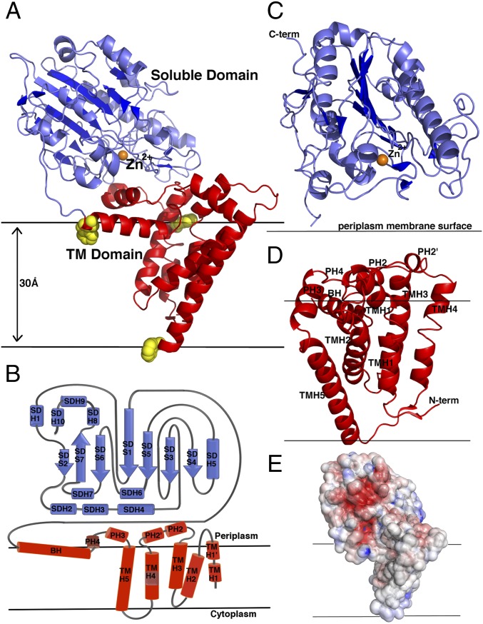
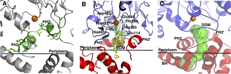
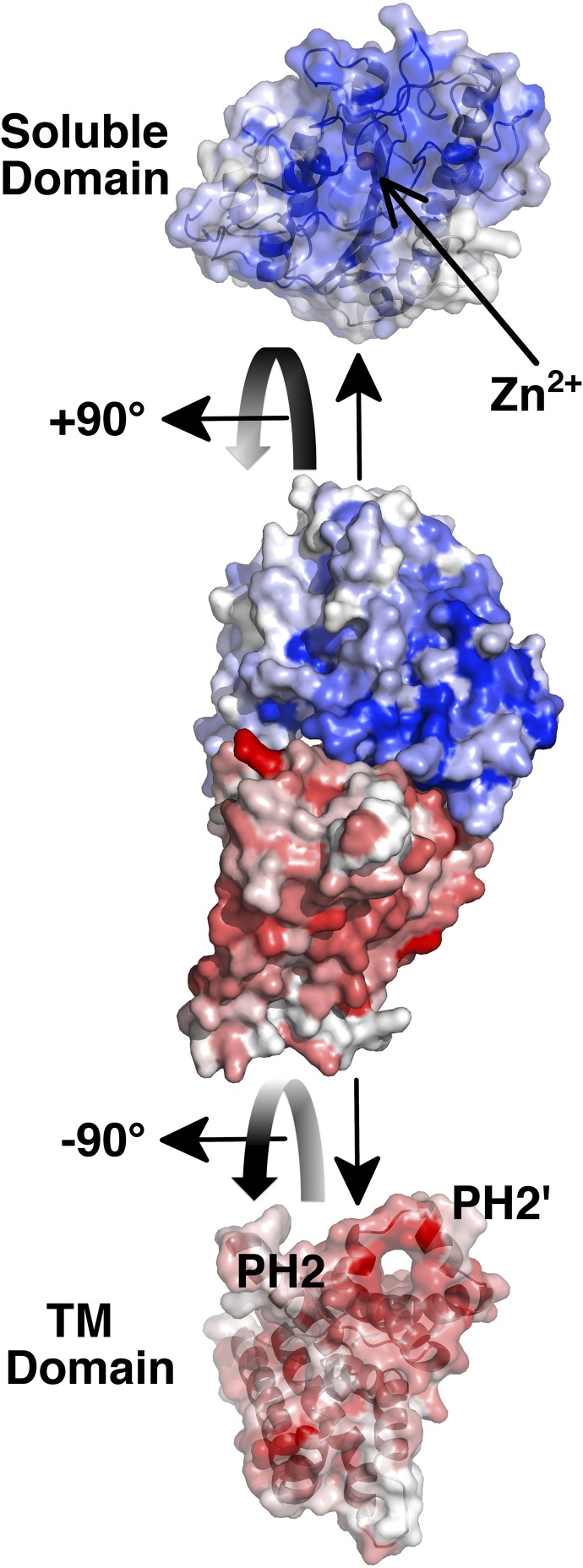
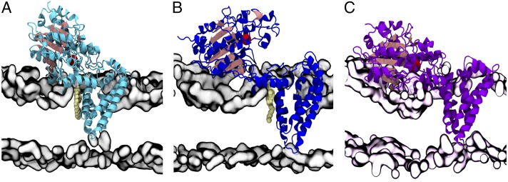
Multidrug-resistant (MDR) gram-negative bacteria have increased the prevalence of fatal sepsis in modern times. Colistin is a cationic antimicrobial peptide (CAMP) antibiotic that permeabilizes the bacterial outer membrane (OM) and has been used to treat these infections. The OM outer leaflet is comprised of endotoxin containing lipid A, which can be modified to increase resistance to CAMPs and prevent clearance by the innate immune response. One type of lipid A modification involves the addition of phosphoethanolamine to the 1 and 4' headgroup positions by phosphoethanolamine transferases. Previous structural work on a truncated form of this enzyme suggested that the full-length protein was required for correct lipid substrate binding and catalysis. We now report the crystal structure of a full-length lipid A phosphoethanolamine transferase from Neisseria meningitidis, determined to 2.75-Å resolution. The structure reveals a previously uncharacterized helical membrane domain and a periplasmic facing soluble domain. The domains are linked by a helix that runs along the membrane surface interacting with the phospholipid head groups. Two helices located in a periplasmic loop between two transmembrane helices contain conserved charged residues and are implicated in substrate binding. Intrinsic fluorescence, limited proteolysis, and molecular dynamics studies suggest the protein may sample different conformational states to enable the binding of two very different- sized lipid substrates. These results provide insights into the mechanism of endotoxin modification and will aid a structure-guided rational drug design approach to treating multidrug-resistant bacterial infections.

Keywords: Neisseria; lipid modification; membrane protein structure; molecular dynamics; multidrug resistance.


Conflict of interest statement

The authors declare no conflict of interest.

Figures

Fig. 1.
Fig. 1.
Molecular structure of NmEptA. (A) Ribbon representation of NmEptA. The amino-terminal TM domain is shown in red, and the carboxyl-terminal soluble domain is shown in blue. The side chains of the three TM domain tryptophan residues (Trp126, Trp148, and Trp207) are shown as yellow spheres. (B) The topology diagram of NmEptA showing the likely positioning of the protein relative to the periplasmic membrane with the coloring as shown in A. (C) Secondary structure of the soluble domain and (D) TM domain with the helical numbering labeled. (E) Electrostatic surface representation of NmEptA, calculated using APBS. The surface is color-contoured from −4 kT/e to +4 kT/e (negative in red, positive in blue). The bound Zn2+ ion in A and C is shown as an orange sphere. To delineate the proposed orientation of the protein within the bilayer, the membrane surface, representing the hydrophobic portion of the bilayer, is drawn as horizontal black lines.
Fig. 2.
Fig. 2.
Active site region of NmEptA. (A) Amphipathic helices PH2 and PH2′, with the polar residues shown in dark green and nonpolar residues shown in pale green. (B) Difference electron density contoured at 3 σ, with a model of DDM included in the density. (C) Ribbon representation of NmEptA showing the tunnel (green) in the structure, as calculated by CAVER. The Zn2+ ion is represented as an orange sphere. In B and C, the TM domain is colored as a red ribbon representation, and the soluble domain is colored as a blue ribbon representation. The active site residues are shown as ball and stick representation colored with white bonds, and the DDM molecule is colored with yellow bonds. The membrane surface is shown by a black line in each panel.
Fig. 3.
Fig. 3.
Surface area representation of NmEptA for the full-length structure and for the soluble domain and the TM domain. The colored shading of the surfaces corresponds to the different domains, as indicated. Each domain has been rotated from the view of the full-length structure to show the interaction surface. The intensities of the red and blue colors correspond to the level of sequence conservation, as indicated in the sequence alignment in SI Appendix, Fig. S1. The position of the PH2 and PH2′ helices in the TM domain and the position of the bound Zn2+ ion in the soluble domain are indicated.
Fig. 4.
Fig. 4.
Molecular dynamics simulations of NmEptA in a DPPE and PE/PG membrane environment. (A) Representative closed conformation of NmEptA embedded in DPPE bilayer obtained after 100-ns simulation at 310 K with a bound DPPE molecule positioned between periplasmic helices PH2 and PH2′. (B) Representative open conformation of NmEptA embedded in DPPE bilayer after 100-ns simulation at 310 K with the bound DPPE lipid. (C) Representative conformation of NmEptA embedded in PE/PG bilayer after 100-ns simulation at 298 K. The bound DPPE molecules are shown in pale yellow spheres, and the Zn2+ ion is shown as an oversized red sphere. The polar region of the bilayer is represented by a gray surface defined by phosphorus and nitrogen atoms for the DPPE bilayer and by a pale pink surface for the mixed PE/PG bilayer.

References

    1. World Health Organization . Antimicrobial resistance: Global report on surveillance. WHO; Geneva: 2014. pp. 1–256.
    1. US Department of Health and Human Services . Antibiotic resistance threats in the United States 2013. Centers for Disease Control and Prevention; Atlanta: 2013. pp. 1–113.
    1. Osei Sekyere J, Govinden U, Bester LA, Essack SY. Colistin and tigecycline resistance in carbapenemase-producing Gram-negative bacteria: Emerging resistance mechanisms and detection methods. J Appl Microbiol. 2016;121(3):601–617. - PubMed
    1. Liu YY, et al. Emergence of plasmid-mediated colistin resistance mechanism MCR-1 in animals and human beings in China: A microbiological and molecular biological study. Lancet Infect Dis. 2016;16(2):161–168. - PubMed
    1. McGann P, et al. Escherichia coli Harboring mcr-1 and blaCTX-M on a Novel IncF Plasmid: First Report of mcr-1 in the United States. Antimicrob Agents Chemother. 2016;60(7):4420–4421. - PMC - PubMed

Publication types

MeSH terms